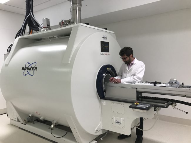
9.4T Bruker Biospec MRI UWA

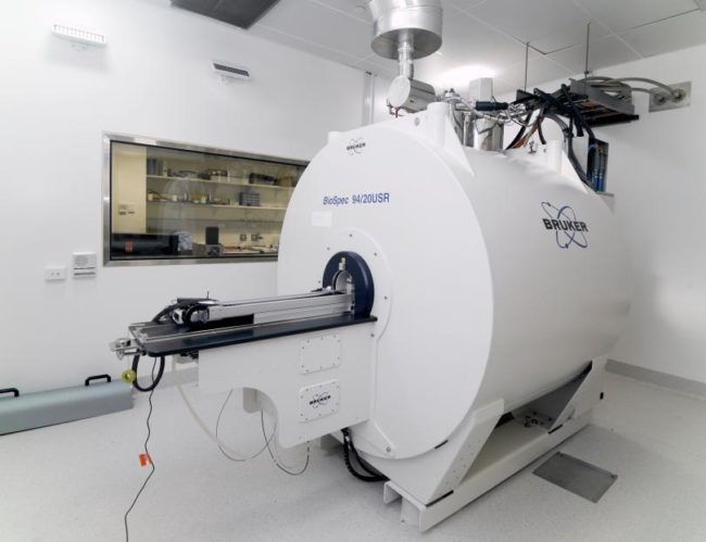
Bruker Biospec 9.4T Preclinical MRI

Kirk Feindel operating the 9.4T MRI

The Bruker BioSpec 9.4T MRI is a high performance multi-nuclear system for non-invasive high-resolution imaging and spectroscopy in preclinical, preserved sample, plant, and materials research.

A range of volume, surface, phased array, and cryogenic imaging coils as well as species specific heated beds are available to accommodate various preclinical needs, in addition to planar surface coils for materials applications. Three sizes and strengths of imaging gradients are available to best tailor the imaging capabilities to the diverse and individual needs of both preclinical and materials researchers.

This instrument has automatic hardware detection, an automatic animal positioning system, and the latest generation of parallel receive and transmit electronics to decrease experiment time without compromising image resolution or contrast.

  • Anatomical imaging studies

  • Preclinical functional MRI (fMRI)

  • Diffusion tensor imaging (DTI)

  • MR spectroscopy

  • Multi-nuclear MRI (H-1, C-13, F-19, Na-23, P-31)

  • Preclinical cardiac imaging

Inquiries

To the Node

Profile photo of Paul Parizel
UWA Node Director
UWA Facility Fellow

To NIF Central

[wpforms html_class = "tatsu-wp-form-rounded-with-underline tatsu-wp-form-submit-rounded" id="4698"]

Biograph MR-PET (large bore) Monash

Biograph MR-PET (large bore)

The Biograph MR-PET is a fully integrated 600mm bore 3T MRI and PET system. This allows for the simultaneous acquisition of PET data with the soft tissue discrimination of MRI. As MRI emits no ionising radiation PET scans can be performed for a longer duration allowing higher accuracy. All data is fully integrated with kinetic and standard reconstructions covering the whole body if required.

  • 40% greater sensitivity then PET-CT

  • High MR image quality for off-centre imaging, fat saturation and spectroscopy

  • 32″ BOLDScreen for stimuli presentation

  • Two CCTV cameras in scan room with wide angle coverage

  • Range of application suites for optimised examinations of all regions

Inquiries

To the Node

Dr Mike de Veer
Monash Facility Fellow

To NIF Central

[wpforms html_class = "tatsu-wp-form-rounded-with-underline tatsu-wp-form-submit-rounded" id="4698"]

BioFLECT Monash

FLECT-CT

The TriFoil FLECT-CT small animal imaging system is located at the Monash Insitute for Pharmaceutical Sciences (MIPS – Parkville). The TriFoil FLECT-CT instrument provides in vivo optical imaging with uniform sensitivity and resolution throughout the object. The system provides images of deep tissue phenomena without loss of image or quantitative accuracy. The X-ray CT scanner provides co-registered images for anatomical correlation that significantly improves the segmentation and resolution of the optical images.

The system is capable of imaging rodent animal models with axial and transaxial fields of view of 38mm and 100mm (mice), and 42 frequency excitation options, up to 16 light filtration options, and is configured with “InVivoScope” control and analysis software.

  • FLECT system capable of imaging in the range of ~500 – 870 nm

  • Individualised animal tissue heterogeneity map for compensation/correction of attenuation of photons based on CT image

  • Deep tissue algorithms for fluorescence recovery based on individualised geometry of each animal

  • Ability to plug-in imaging cells compatible with MRI, and to fuse FLECT, CT, and MRI images

  • Diffusion MRI

  • Simultaneous fMRI and EEG

Inquiries

To the Node

Dr Mike de Veer
Monash Facility Fellow

To NIF Central

[wpforms html_class = "tatsu-wp-form-rounded-with-underline tatsu-wp-form-submit-rounded" id="4698"]

9.4T MRI Small Animal Monash-ARAMBI

9.4T MRI Small Animal

The 9.4T animal magnetic resonance imaging (MRI) system (Bruker) located at the ARA-MBI campus in Prahran is optimised for rodent and high-resolution sample imaging. The ultra high magnetic field coupled with rodent cryocoils provides structural and functional images with higher resolution and signal to noise ratio compared with standard MRI. This scanner can be used for brain, abdominal, cardiovascular, muscular and articular, as well as dynamic molecular imaging.

  • Small animal imaging studies

  • Mouse and rat cryocoils for brain and whole body imaging

  • Mouse and rat cardiac surface coils for heart imaging

  • 40 mm 19F fluorine imaging coil for use with rodents or samples

  • 35 mm volume coil for high-resolution mouse head or sample imaging

  • 86 mm radio frequency transmit and receive coils

  • Suitable isofluorane anaesthesia and animal monitoring system

  • Full range of beds to cater for more species and samples

Inquiries

To the Node

Facility Manager

To NIF Central

[wpforms html_class = "tatsu-wp-form-rounded-with-underline tatsu-wp-form-submit-rounded" id="4698"]

9.4T MRI Small Animal UQ

9.4T MRI Small Animal

Gary and Karine using the MR.

The Bruker Biospec MRI scanner consists of a 9.4 Tesla, 300 mm ultrashield refrigerated 30 cm magnet interfaced to a scalable Bruker Avance III spectrometer that allows unparalleled flexibility for MRI scanning of small and medium sized research animals and other samples.

The system incorporates a cryoprobe, which enables maximum signal and image quality to be achieved for rodent mouse brain imaging. The system also includes 8 parallel RF transmit and receive channels for multi-coil phased array imaging and numerous coils to investigate nuclei other than standard protons or “water” imaging, notably carbon-13 coils to investigate metabolism, sodium-23 coils to investigate cell viability, phosphorus-31 coils to investigate tissue energetics and fluorine-19 coils for labelling of targeted tracers.

  • Cryoprobe for rodent and mouse imaging

  • C-13, Na-23, P-31 and F-19 coils

  • MR Spectroscopy (MRS)

Inquiries

To the Node

Prof Markus Barth
Queensland Node Co-Director
Dr Gary Cowin
QLD Facility Fellow
Research Fellow, CAI

To NIF Central

[wpforms html_class = "tatsu-wp-form-rounded-with-underline tatsu-wp-form-submit-rounded" id="4698"]

9.4T MRI Small Animal UNSW

9.4T MRI Small Animal

The MRI facility at the UNSW’s BRIL is one of the most powerful small-animal MRIs in Australia and is able to provide users with state-of-the-art small animal and sample imaging, and MRI biomarker assessment.

  • Structural, physiological, and metabolic imaging studies

  • Small rodent monitoring, anaesthesia, and positioning systems

  • High power gradient system (wide bore)

  • High power gradient insert (micro-imaging)

  • Various coils; cryo-cooled, standard, x-nucleus

Inquiries

To the Node

Photo of Dr Andre Bongers
UNSW Facility Fellow
UNSW - BRIL Node Director

To NIF Central

[wpforms html_class = "tatsu-wp-form-rounded-with-underline tatsu-wp-form-submit-rounded" id="4698"]

9.4T MRI Small Animal Monash

9.4T MRI Small Animal

9.4T MRI with Tara operating

The 9.4T animal MRI system (Agilent/Bruker) housed at the Clayton site is a flexible and adaptable magnetic resonance imaging scanner for pre-clinical imaging and research. The system runs Paravision 6 and can carry out a wide range of pre-clinical imaging functions with a range of proton coils for various applications. The ultra high magnetic field provides structural and functional images with higher resolution in comparison with clinical MRI. It can be used for brain, abdominal, cardiovascular, muscular and articular, as well as dynamic molecular imaging. 

  • Anatomical imaging studies

  • Animal beds for rodents and marmosets

  • Horizontal bore magnet with 205/120 gradient coils

  • Phased array surface coils for rat and mouse brain and heart

  • 200 mm surface coils for 1H-19F (370-420 MHz), 31P-13C (75-170 MHz), 2H-15N (39-82 MHz)

  • 40 mm 190-F coil for rodents

Inquiries

To the Node

Dr Mike de Veer
Monash Facility Fellow
Photograph of Dr Gang Zheng
Monash Facility Fellow

To NIF Central

[wpforms html_class = "tatsu-wp-form-rounded-with-underline tatsu-wp-form-submit-rounded" id="4698"]

7T MRI Whole Body UQ

7T MRI Whole Body

Aiman operating the 7T MRI with a humna subject. Image courtesy of CAI, UQ.

Key technical features of the Siemens Magnetom 7T MRI system include high-performance gradients and multi-receive and multi-transmit radiofrequency capabilities that further increase sensitivity.

The 7T is utilised across a broad range of applications, including neuroscience, engineering, spectroscopy and theranostics. Alzheimer’s and Parkinson’s diseases, Multiple Sclerosis, epilepsy, musculoskeletal and liver diseases are examples of clinical applications that benefit from the high spatial resolution achievable with this instrument.

  • Anatomical imaging studies

  • Functional MRI (fMRI)

  • High-performance gradients

  • Multi-receive and multi-transmit radio-frequency capabilities

Inquiries

To the Node

Prof Markus Barth
Queensland Node Co-Director
Head of Human & Companion Animal Imaging Operations, CAI

4DX Pulmonary Imaging System LARIF

4Dx Permetium Pulmonary Preclinical Imaging System

The 4Dx preclinical scanner is a small animal research scanner which uses x-rays and computed tomography capabilities to measure respiratory function in small rodents.

  • Preclinical lung imaging system

  • No contrast requirement

  • Cardiac and pulmonary gating capabilities

Inquiries

To the Node

SAHMRI Node Co-Director
Profile photo of Georgia Williams
SAHMRI Facility Fellow

To NIF Central

[wpforms html_class = "tatsu-wp-form-rounded-with-underline tatsu-wp-form-submit-rounded" id="4698"]

3T Skyra MRI (large bore) Monash

3T Skyra MRI (large bore)

Monash 3T MRI in use

The Siemens 3 Tesla Skyra MRI scanner has a 70cm wide bore, a full range of imaging coils including an advanced 32-channel head coil, a 20 channel head and neck coil, a 32 channel spine coil, and an 18 channel body coil.

The scanner console includes advanced reconstruction and analysis software for functional and diffusion neuroimaging, perfusion imaging, flow quantification, cardiovascular, musculoskeletal electronencephalography system and an SR Instruments eye tracking unit.

Scanner uses XQ Gradients and has multinuclear spectroscopy and parallel transmit technology (ZOOMit) applications.

  • Neuroimaging and neuropsychological testing

  • Functional MRI (fMRI)

  • Flow quantification

  • Perfusion MRI

  • Diffusion MRI

  • Simultaneous fMRI and EEG

  • MR-compatible pulse oximeter

Inquiries

To the Node

Dr Mike de Veer
Monash Facility Fellow

To NIF Central

[wpforms html_class = "tatsu-wp-form-rounded-with-underline tatsu-wp-form-submit-rounded" id="4698"]
Privacy Settings
Youtube
Vimeo
Google Maps