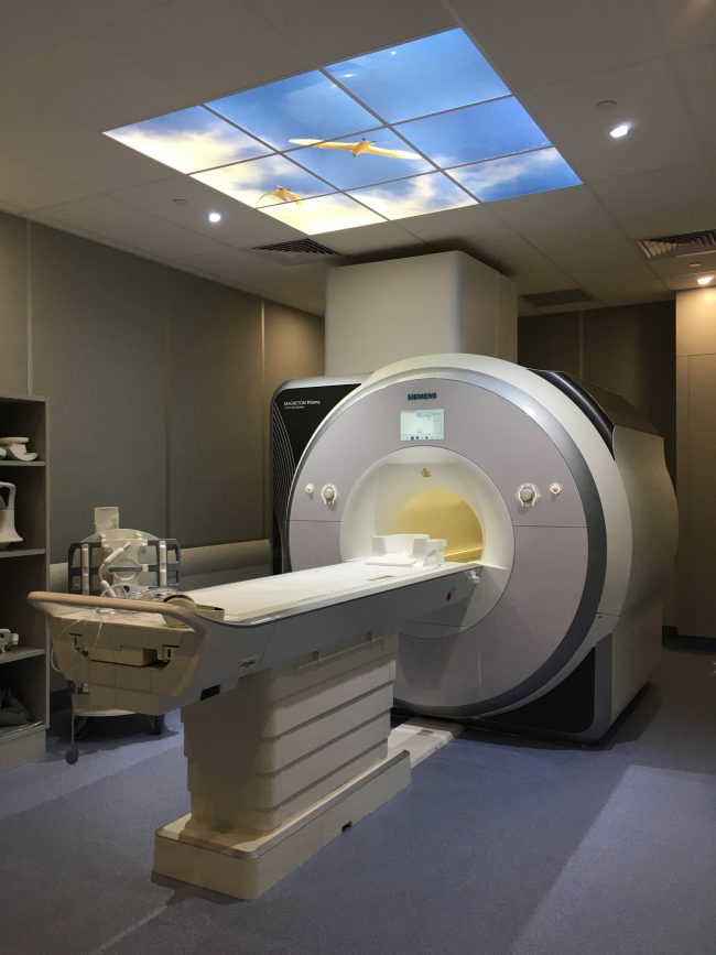
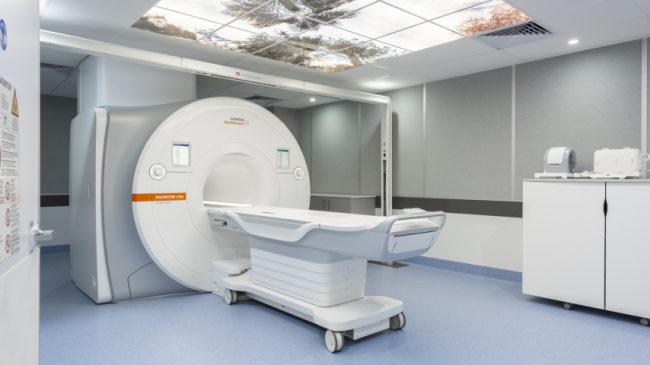
3T Siemens Magnetom Prisma MRI UNSW

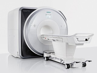
Siemens 3T Magnetom Prisma MRI

The Siemens 3T Magnetom Prisma is a 60 cm bore magnet design with 80mT/m gradients and a maximum slew rate of 200 T/m/s.

  • 64 channel head/ neck coil, 32 channel spine and 18 channel body coils for neuro, cardiac, body and musculoskeletal imaging

  • fMRI, DTI, contrast‐ and arterial spin labelling based perfusion diffusion, cardiac and body imaging, T1 and T2 mapping, vascular imaging

  • Elastography hardware and software

  • Cambridge Research Systems BOLDscreen 32″ LCD display and Lumina response pads for fMRI studies

  • Contrast injector

  • Physiology monitoring and recording, cardiac and respiratory gating

Inquiries

To the Node

Director, Research Imaging NSW
UNSW - BRIL Node Director
UNSW - NeuRA Node Director

To NIF Central

[wpforms html_class = "tatsu-wp-form-rounded-with-underline tatsu-wp-form-submit-rounded" id="4698"]

3T Siemens Magnetom Vida MRI UNSW

3T MRI - Siemens Magnetom Vida

The Siemens 3T Magnetom Vida, located at the RINSW, is a 70 cm wide bore magnet design with a 60 mT and 200 T/m/s slew rate gradient system.

  • Broad range of imaging coils, e.g., 64 & 20 channel head coil, 60 channel body coil, peripheral angiography coils and several MSK coils for neuro, cardiac, body and musculoskeletal imaging

  • fMRI, DTI, perfusion, diffusion, free‐breathing and single breath hold cardiac and body imaging sequences, quantitative

  • Imaging such as T1, T2, iron, fat and stiffness mapping, vascular imaging, spectroscopy etc

  • Laser‐guided patient alignment system, flat table, specialised RF antennae, and sequences for MR guided Radiotherapy treatment planning

  • Cambridge Research Systems BOLDscreen 32″ LCD display and Lumina response pads for fMRI studies

  • Elastography hardware and software

  • Contrast injector and vital sign monitor

Inquiries

To the Node

Director, Research Imaging NSW
UNSW - BRIL Node Director
UNSW - NeuRA Node Director

To NIF Central

[wpforms html_class = "tatsu-wp-form-rounded-with-underline tatsu-wp-form-submit-rounded" id="4698"]

Siemens Biograph mCT FLOW (PET/CT) HIRF

Biograph mCT FLOW (PET/CT)

The PET/CT Scanner provides details of disorders at a molecular level (PET) and a detailed image of the body’s anatomy (CT). PET/CT can be used to diagnose, localise and monitor conditions such as brain disorders, heart disease, brain injury and cancer. With a 78 cm bore, 22 cm axial field of view, LSO scintillators and a 128 slice CT with 0.28 sec rotation time, the Siemens Biograph mCT FLOW offers effective human imaging research capabilities.

  • Co-registration of PET with CT volumes

  • Flat scanning palette for radiotherapy applications

  • Full laser positioning for RT applications

  • Head holders for RT positioning

Inquiries

To the Node

QLD Node Co-Director (HIRF)

To NIF Central

[wpforms html_class = "tatsu-wp-form-rounded-with-underline tatsu-wp-form-submit-rounded" id="4698"]

3T Siemens Magnetom Prisma HIRF

3T MRI - Prisma

The Siemens Magnetom 3T Prisma at the HIRF site of the QLD Node is one of only a few in Australia. This 3T MRI delivers the capacity to conduct cutting-edge human research through neuroimaging and functional connectivity, and structural details of soft body tissues such as the eyes, blood vessels, the female reproductive system, bladder, prostate and joints. The 3T-MRI can be used to detect conditions such as strokes, multiple sclerosis, cancerous tumours and damage to the brain or spinal cord.

  • Anatomical imaging studies

  • Functional MRI (fMRI)

  • Perfusion MRI

  • Diffusion MRI

Inquiries

To the Node

QLD Node Co-Director (HIRF)

To NIF Central

[wpforms html_class = "tatsu-wp-form-rounded-with-underline tatsu-wp-form-submit-rounded" id="4698"]

Biograph mMR (PET/MR 3T) HIRF

Siemens Biograph mMR (PET/MRI)

This ground-breaking PET/MRI Scanner, located at HIRF, is one of a few of its kind in Australia. It provides simultaneous anatomical (MRI) and functional (PET) data to locate tumours and understand disease processes. It offers the most precise early diagnosis of diseases such as brain, ovarian and prostate cancers and monitoring of processes within those cancers. It can also be used to study how drugs and PET tracers (small compounds) are taken up by tumours and surrounding areas. This will provide a more comprehensive understanding of the biology of tumours. The scanner provides high-quality images of organs in motion, such as the liver, and will help researchers to identify the best treatment strategies for patients. The MRI/PET enables organ motion to be tracked and corrected for higher-quality imaging.

  • Anatomical studies

  • Combined PET and MR

  • 12 coils avalable

  • Up to 9 micron resolution

  • Power injector ancillary

Inquiries

To the Node

QLD Node Co-Director (HIRF)

To NIF Central

[wpforms html_class = "tatsu-wp-form-rounded-with-underline tatsu-wp-form-submit-rounded" id="4698"]

What is Ultrasound / Photoacoustic imaging?

Ultrasound / Photoacoustic imaging

Ultrasound imaging employs sound waves with frequencies >20 kHz. These waves are pulsed through internal structures and the resulting ‘echo’ is used to generate an image of the internal structures. Optoacoustic or photoacoustic imaging is an imaging technique which detects ultrasound waves following light absorption from specific molecules. This technique allows molecules with different spectral profile to be detected. Advancements in the development of nanoparticles and infrared fluorescent reporters that are detectable by optoacoustics have also increased the possibilities of molecular imaging.

What is PET / SPECT?

Positron Emission Tomography

PET-CT image of rat

Positron Emission Tomography (PET) takes advantage of emissions from the decay of radioactive materials (e.g., radiotracers) to precisely locate and visualise physiological processes. Single-photon emission computed tomography (SPECT) uses a more direct way of measuring the emitted gamma rays, resulting in a broader range of usable isotopes at the expense of some resolution. Both PET and SPECT are typically paired with CT to co-locate radiotracers with anatomical features.

What is Optical imaging?

Optical imaging

Optical imaging detects wavelengths in the optical, ultraviolet, or infrared spectrum to image whole specimens. For example, specific genes, cells, or organisms can be “tagged” with a gene encoding one of the luciferase enzymes that enable some bacteria, insects, and animals to glow. When the tagged entity is active, it glows. The emitted light corresponds to the number and location of the tagged entities. This information enables scientists to non-invasively observe the spread of disease or the effects of a drug throughout the system.

What is MPI?

Magnetic Particle Imaging

Magnetic Particle Imaging (MPI) is an emerging molecular imaging modality that measures the location and concentration of superparamagnetic nanoparticle tracers in vivo (typically Iron Oxide, SPIOs) by detecting their response to spatially dependent magnetic fields. The technology is used in various research projects with focus on cell tracking, targeted imaging in cancer and neuroscience, angiogenesis/vascularity quantification as well as in the development of MPI tracer technology to leverage the translational potential of the technology.

What is CT?

Computed Tomography

3D rendering of a spine from CT - image courtesy of LARIF

Computed tomography (CT) uses multiple axial scans to generate cross-sectional information or 3-dimensional reconstructions. X-ray CT is the typical mechanism for taking ‘slices’ which are then digitally reconstructed into 3-D volumes.

Privacy Settings
Youtube
Vimeo
Google Maps