Quantum GX MicroCT WSU
Inquiries
To the Node

Western Sydney University Node Director

University of Western Sydney Facility Fellow

Simultaneous use of both Siemens Inveon scanners has been routine, and faster throughput imaging using dual-mice beds designed in-house has been established. Non-invasive methods from image-derived input functions for quantification and kinetic modelling have been developed and validated. In vivo validation of Partial Saturation Approach (PSA) modelling method using 11C-raclopride has also been established. The complete process from radioisotope production by the cyclotron, radiochemical labelling and imaging continues to be delivered, and post-mortem analysis for biodistribution is now routine.

The Siemens Inveon multi-modality PET/CT imaging scanner is capable of providing three dimensional CT and PET images of live mouse and rat as well as fixed samples. The system can deliver high resolution CT images (<10 µM for a field-of-view <20mm) with a maximum field-of-view of 80 mm X 50 mm (with a resolution of approximately 50 µM).
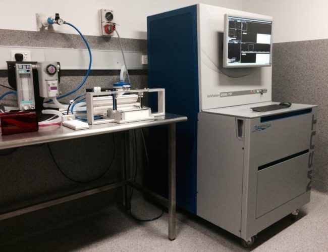

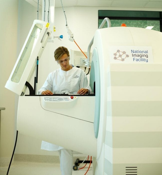
The Multispectral Optoacoustic Tomography (MSOT) inVision256-TF (iThera Medical) was acquired with the help of the Cancer Institute NSW equipment grant 2014. It is the only system capable of multispectral tomographic imaging of an entire mouse.


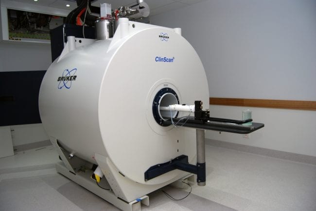

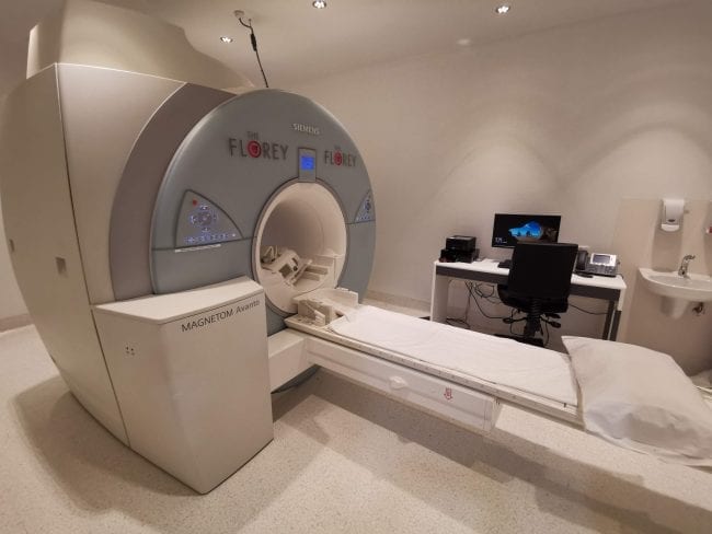
The world’s first Bruker ClinScan MR/PET scanner was commissioned at CAI in late 2012 as part of the NCRIS funded National Imaging Facility UQ node. This system allows simultaneous acquisition of MRI and PET images of an animal or sample. The technology combines the exquisite structural and functional characterisation of tissue provided by MRI with the extreme sensitivity of PET imaging of metabolism and tracking of uniquely labelled cell types or cell receptors. The Clinscan MRI system is comprised of a 7 Tesla, 30 cm bore superconducting magnet, with operating software identical to the Siemens clinical MRI platform.
This enables the most direct translation of research outcomes from animals to humans, benefiting a wide range of biomedical and scientific research. The PET insert has been developed to provide optimal performance in the high magnetic field of the MRI system.

The UNSW Node houses a ‘mock scanner’ to assist preparations with anxious participants, allowing them to experience being inside a small space before going into the real scanner


A mock MRI scanner is a practice MRI scanner that mimics the sounds and equipment of the MRI environment. This scanner is available to researchers accessing the Florey facilities.
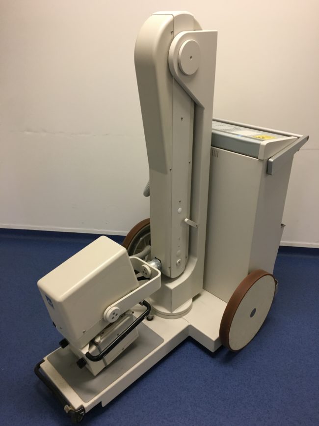

Villa Visitor mobile X-Ray unit
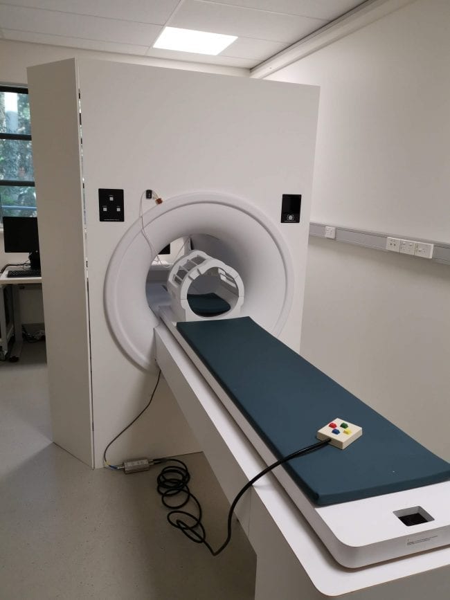
Small animal micro CT Skyscan. This facility is located at the North Tce site of LARIF.
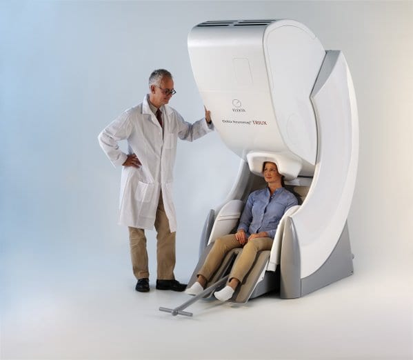
The Neuromag TRIUX, is a magnetoencephalography (MEG) platform from Elekta. It addresses key MEG requirements that are critical for robust functional mapping studies. The system features higher tolerance for magnetic interferences, new user interface features and patient comfort enhancements.
The MEG system at Swinburne Neuroimaging (SNI) has been established with the full range of peripheral technologies and stimuli presentation devices available.


Copyright 2021 @ National Imaging Facility. All rights reserved.
Read our Privacy Policy.
admin@anif.org.au