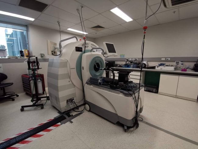
Mediso PET/CT Monash

Mediso PET/CT

The Mediso Nano PET/CT, located at the ARAMBI site, provides a high-precision gantry for exact alignment of PET/CT data. The gantry bore opening is 16 cm and accommodates imaging of mice, rats and rabbits with interchangeable beds and radiation shields. Mediso’s gating package (respiratory & cardiac/ECG) is available for optimal image quality. A complete 360° PET detector ring consisting of 12 modules provides the ultra high resolution and sensitivity, with the reconstructed resolution in the centre of the field of view being less than 1.0 mm.
Image reconstruction is achieved with a fast, iterative and 3D real / post Tera-Tomo™ PET engine using 3D OSEM (Ordered Subset Expectation Maximization) protocols. PET and/or CT scanning can extend up to 300 mm in the axial direction by automated translation of the bed for whole body imaging, with a transaxial range of 45-125 mm. Real time reconstruction of CT data can achieve voxel sizes in the range of 13-37 micron.

  • Co-registration of PET with CT volumes

  • Low-dose protocols (<50 mGy / 3 min scan)

  • Longitudinal rodent studies

  • Imaging chambers with integrated animal support

Inquiries

To the Node

Research Fellow

To NIF Central

[wpforms html_class = "tatsu-wp-form-rounded-with-underline tatsu-wp-form-submit-rounded" id="4698"]

MALDI-TOF Mass Spec UQ

MALDI-TOF Mass Spec

A Bruker Autoflex MALDI-TOF/TOF MSI facility is available for both advanced MSI and protein analysis. MSI enables the spatial distribution of ions to be imaged directly from tissue sections to a resolution of 20-50 µm, and examples of application include biomarker studies in cancer and drug distribution. It can also be used to localize chemically unlabeled molecular markers and/or bioactive peptides in tissue by generating unique ion profiles through the use of isotopic labeling.

The technology is an ideal platform for mass analysis of biomolecules, characterisation of protein folding and sequencing, and a robotic LC-spotter allows full-scale proteomic experimentation.

  • Biomolecule distribution

  • Proteomic characterisation

Inquiries

To the Node

Prof Markus Barth
Queensland Node Co-Director
Imaging Mass Spectrometry Specialist, CAI

To NIF Central

[wpforms html_class = "tatsu-wp-form-rounded-with-underline tatsu-wp-form-submit-rounded" id="4698"]

Magnetic Particle Imaging (MPI) UNSW

Magnetic Particle Imaging (MPI)

image of the mpi equipment

In Q1 2020, UNSW has installed a Magnetic Insight Momentum pre-clinical MPI scanner as the first facility in Australia to provide this novel technology. With its unique set of features MPI supports tracer applications in murine disease models for medical sciences and biology research as well as to the progress of MPI as a new translational imaging modality.  The technology is used in various research projects with focus on cell tracking, targeted imaging in cancer and neuroscience, angiogenesis/vascularity quantification as well as in the development of MPI tracer technology to leverage the translational potential of the technology. 

  • Anatomical imaging studies

  • No nuclear technologies required

  • Spatial resolution ~1 mm

  • High tracer sensitivity (detection limits nano-molar trace concentrations)

Inquiries

To the Node

Photo of Dr Andre Bongers
UNSW Facility Fellow
UNSW - BRIL Node Director
Profile photo of Saeed
UNSW Facility Fellow

To NIF Central

[wpforms html_class = "tatsu-wp-form-rounded-with-underline tatsu-wp-form-submit-rounded" id="4698"]

MPI Monash

Magnetic Insights Magnetic Particle Imaging (MPI)

The MPI at MBI is scheduled to be installed in 2020.

  • Quantitative molecular imaging

  • Integrated CT module

  • HYPER module for heating accuracy and real-time monitoring of therapeutic heat dose

Inquiries

To the Node

Facility Manager

To NIF Central

[wpforms html_class = "tatsu-wp-form-rounded-with-underline tatsu-wp-form-submit-rounded" id="4698"]

Large Animal CT LARIF

Large Animal CT

LARIF is home to a 16-slice Philips Briliance CT scanner. The system has various reconstruction capabilities e. g. volume rendering techniques, multiplanar reconstructions etc. and can be used for a variety of imaging.

  • Large animal imaging

  • Routine CT studies

  • Forensic imaging

  • CT angiography

  • CT-guided fluoroscopy

Inquiries

To the Node

SAHMRI Node Co-Director

To NIF Central

[wpforms html_class = "tatsu-wp-form-rounded-with-underline tatsu-wp-form-submit-rounded" id="4698"]

IVIS Spectrum-CT UNSW

IVIS Spectrum-CT

The IVIS® SpectrumCT pre-clinical in vivo imaging system expands upon the versatility and advanced optical feature sets of the IVIS and Maestro™ platforms integrated with low dose microCT to support longitudinal imaging. The IVIS SpectrumCT enables simultaneous molecular and anatomical longitudinal studies, providing researchers with essential insights into complex biological systems in small animal models. The constant horizontal gantry motion and the flat panel detector provide low-dose imaging and automated optical and microCT integration. The stable revolving animal platform table rotates 360° to acquire full 3D data. Multiple animals can be scanned simultaneously while maintaining an average dose per scan at about 13 mGy, with a scanning and reconstruction time of less than a minute. Optical and microCT modalities can also operate independently. Multispectral fluorescence imaging (400-900nm wavelength) can be performed with the system for 2D or 3D imaging. Deep tissue detection and quantification can be achieved through trans-illumination available on this system. The system is fully equipped with its own isoflurane anaesthetic system and is capable of imaging up to five mice simultaneously.

  • Longitudinal imaging

  • Full 3D data

  • Isofluorane anaesthetic system

  • Up to 5 mice simultaneously

Inquiries

To the Node

UNSW Facility Fellow
UNSW - BRIL Node Director

To NIF Central

[wpforms html_class = "tatsu-wp-form-rounded-with-underline tatsu-wp-form-submit-rounded" id="4698"]

IVIS Lumina Live Imaging UNSW

IVIS Lumina Live Imaging

The Lumina enables real-time, non-invasive visualisation and tracking of bioluminescent or fluorescent cells, proteins or gene activity within a living animal. It can also be used to visualise dishes, plates, and tissue samples. The system integrates a highly sensitive charge-coupled device (CCD) cooled camera capable of detecting exceptionally low light levels, with a light-tight imaging chamber and includes a broad range of filter sets that permits imaging of a wide range of fluorophores.

  • Anatomical imaging studies

  • Isoflurane anaesthetic system

  • Up to 3 mice simultaneously

  • LivingImage software

Inquiries

To the Node

UNSW Facility Fellow
UNSW - BRIL Node Director

To NIF Central

[wpforms html_class = "tatsu-wp-form-rounded-with-underline tatsu-wp-form-submit-rounded" id="4698"]

IVIS Lumina II Imaging System UWA

IVIS Lumina II Pre- clinical small animal In vivo Bioluminescence/ Fluorescence Imaging System

The IVIS Lumina II is a high-sensitivity, low noise, in vivo imaging technology platform that enables noninvasive visualization and tracking of cellular and genetic activity within a living organism, in real time.

The IVIS Lumina II takes very low light level images that can be saved and displayed for subsequent analysis. In addition to bioluminescent imaging, the IVIS Lumina II incorporates the XFO-12 fluorescence equipment that enables fluorescent imaging capabilities for both in vitro and in vivo applications. The sensitive range of the CCD camera sets the wavelength range for fluorescence applications from 400 to 950nm.

  • In-vivo fluorescence imaging

  • In-vivo bioluminescence imaging

  • Autofluorescence correlation

  • Min image pixel resolution 50 um

  • Heated stage and anaesthesia manifold

  • 10 excitation and 4 broad emission filter sets

Inquiries

To the Node

Profile photo of Paul Parizel
UWA Node Director
UWA Facility Fellow

To NIF Central

[wpforms html_class = "tatsu-wp-form-rounded-with-underline tatsu-wp-form-submit-rounded" id="4698"]

Inveon PET/SPECT/CT Monash

Inveon PET/SPECT/CT

PET-SPECT-CT at MBI

The Inveon Multimodality System is a versatile platform for laboratory animal CT, SPECT, and PET studies on a single integrated gantry. The system can be configured for PET-CT, SPECT-CT, PET-SPECT-CT or CT only. The large area CT system has a field of view up to 10 cm x 10 cm and resolution down to 20 microns. The PET system can deliver 1.4mm FWHM spatial resolution, and a maximum field of view of 10cm x 30cm. Whole mouse and rat SPECT studies are possible using gamma rays ranging in energy from 30 to 300 keV, with automated zoom for optimising the field of view.

  • Animal imaging studies

  • Animal support, such as portable isofluorane, warming pallets, and monitoring of temperature, ECG, and respiration

  • Integrated video system for animal positioning and monitoring

  • Fused 3D imaging of static or dynamic volumes

  • Complex volumetric segmentation for quantification

Inquiries

To the Node

Dr Mike de Veer
Monash Facility Fellow

To NIF Central

[wpforms html_class = "tatsu-wp-form-rounded-with-underline tatsu-wp-form-submit-rounded" id="4698"]
Privacy Settings
Youtube
Vimeo
Google Maps