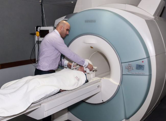
3T MRI Whole Body UQ

3T MRI Whole Body

Aiman operating the 3T Mri. Image courtesy of CAI, UQ

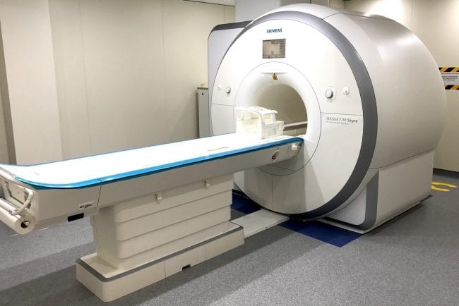
The Siemens PRISMA is a high performance 3T MRI scanner for Neuro-, whole-body and peripheral imaging. The system is complete with a wide range of coils and software for applications in neurology, cardiology, angiography, oncology, orthophaedics and paediatrics, and includes a development environment for new sequence and hardware development.

  • 64-channel head coil

  • 12-channels for body imaging

  • Multi-nuclear capable

  • Functional MRI (fMRI)

  • Musculoskeletal imagingĀ 

Inquiries

To the Node

Prof Markus Barth
Queensland Node Co-Director
Head of Human & Companion Animal Imaging Operations, CAI

To NIF Central

[wpforms html_class = "tatsu-wp-form-rounded-with-underline tatsu-wp-form-submit-rounded" id="4698"]

3T MRI Whole Body UNSW-NeuRA

Philips Ingenia CX 3T MRI scanner

This is a state-of-the art 3T digital MRI scanner with 60 cm bore (110 cm tunnel flare), 50 cm maximum field of view that can accommodate patients weighing up to 250 kg.

  • Anatomical imaging studies

  • Functional MRI (fMRI)

  • MR eye tracking system

  • Broad range of imaging coils for neuro, body, and musculoskeletal imaging

  • Wireless physiology monitoring and recording for cardiac (VCG, PPU) and respiratory gating

Inquiries

To the Node

UNSW-NeuRA Facility Fellow
UNSW - NeuRA Node Director

To NIF Central

[wpforms html_class = "tatsu-wp-form-rounded-with-underline tatsu-wp-form-submit-rounded" id="4698"]

3T MRI Whole Body Swinburne

3T MRI Whole Body

Swinburne Neuroimaging’s (SNI) Siemens Prisma Fit 3T magnet features multiple coils and the full range of peripheral technologies and stimuli presentation devices.

  • Anatomical imaging studies

  • Functional MRI (fMRI)

  • High speed eye tracking (Eyelink 1000)

  • Combined capabilities of MRI, EEG and MEG

Inquiries

To the Node

Dr Matthew Hughes
Swinburne Facility Fellow
Swinburne University of Technology Node Director

To NIF Central

[wpforms html_class = "tatsu-wp-form-rounded-with-underline tatsu-wp-form-submit-rounded" id="4698"]

3T MRI ā€“ Prisma Florey

3T MRI - Prisma

Shawna Farquharson operating the Human MRI

Human imaging facilities at the Florey node includes a Siemens Prisma, a 60 cm bore MRI scanner, with dedicated head coils to enable internationally competitive research. A wide selection of head coils are available, including single-channel transmit-receive up to 64 channel receive. The state of the art scanner is also equipped with specialised MRI coils for body & extremity imaging studies.

  • Anatomical imaging studies

  • Functional MRI (fMRI)

  • MR Spectroscopy (MRS)

  • Perfusion MRI

  • Diffusion MRI

  • Simultaneous fMRI and EEG

Inquiries

To the Node

Assoc Prof David Abbott
Florey Informatics Fellow

To NIF Central

[wpforms html_class = "tatsu-wp-form-rounded-with-underline tatsu-wp-form-submit-rounded" id="4698"]

3T MRI ā€“ Skyra Florey

3T MRI - Skyra

Shawna Farquharson operating the Human MRI

Human imaging facilities at the Florey node includes a Siemens Skyra, a 70cm bore MRI scanner, with dedicated head coils to enable internationally competitive research. A wide selection of head coils are available, including single-channel transmit-receive up to 64 channel receive. The state of the art scanner is also equipped with specialised MRI coils for body & extremity imaging studies.

  • Anatomical imaging studies

  • Functional MRI (fMRI)

  • MR Spectroscopy (MRS)

  • Perfusion MRI

  • Diffusion MRI

  • Simultaneous fMRI and EEG

Inquiries

To the Node

Assoc Prof David Abbott
Florey Informatics Fellow

To NIF Central

[wpforms html_class = "tatsu-wp-form-rounded-with-underline tatsu-wp-form-submit-rounded" id="4698"]

3T MRI LARIF

3T MRI - Skyra

3T MRI at LARIF

LARIFā€™s feature instrument is the high field MRI Siemens ā€˜Skyraā€™ scanner. The Skyra 3T is a 48-channel XQ gradient MRI scanner with 70 cm wide bore. The system comes with 32 Ā¬channel head coil, 20-channel head/neck coil, TxRx head coil, special purpose coil, flex coils, loop coils, 32-channel spine coil, 18-channel body coils and MR contrast injector. It is used for various applications including veterinary imaging, forensic imaging, large animal research and fetal medicine.

The scanner includes advanced imaging and analysis software, including TWIST, TIM whole body suite, TIM planning suite, Syngo Native, Syngo Tissue 4D, MyoMaps, ZOOMit, TimTx TrueShape, QISS, SMS EPI, 3D ASL, and IDEA licensing.

  • 4D Flow

  • Spectroscopy

  • Diffusion tensor imaging

  • Neuro fMRI ready

  • Advanced cardiac

  • Multinuclear ready

Inquiries

To the Node

SAHMRI Node Co-Director
SAHMRI Facility Fellow

To NIF Central

[wpforms html_class = "tatsu-wp-form-rounded-with-underline tatsu-wp-form-submit-rounded" id="4698"]

16.4T MRI Small Animal UQ

16.4T MRI Small Animal

16.7T MRI at UQ. Image courtesy of CAI, UQ

The Bruker Ultrashield Plus 700 WB Avance NMR spectrometer has a vertical open bore diameter of 89 mm. It has an available space of up to 34 mm diameter, which is optimal for live mouse imaging and various ex vivo samples.

The scanner is equipped with three types of gradient inserts: micro5, micro2.5 and mini0.5 which provide gradient strengths of 4.8, 2.5 and 0.5 mT/m/A. To achieve highest sensitivity, the scanner is equipped with new generation SAW and quadrature microimaging coils (5, 10, 15, 20, 25, 28 and 35 mm diameters). The 5-15 mm coils are suitable for imaging of excised (fixed tissue samples) such as brain, spinal cord and biopsy samples. The 20-35 mm coils are suitable for live mouse imaging.

  • Anatomical imaging studies

  • High Resolution Magic Angle Spectroscopy (HR-MAS)

  • Micro-imaging

  • High-resolution diffusion imaging

  • Susceptibility-weighted imaging

Inquiries

To the Node

Prof Markus Barth
Queensland Node Co-Director
Research Fellow, CAI

To NIF Central

[wpforms html_class = "tatsu-wp-form-rounded-with-underline tatsu-wp-form-submit-rounded" id="4698"]

14.1T MRI Small Animal WSU

14.1T MRI Small Animal

The BRUKER AVANCE III 600 MHz Wide Bore NMR/MRI Spectrometer (14.1 T) is equipped with high current gradient amplifiers capable of generating XYZ gradients up to 300 G/cm (k-space and q-space) and a Z gradient 3000 G/cm (q-space). It is equipped with various probes and accessories that enable it to be used for a wide range of NMR experiments such as diffusion, micro-imaging, and high-resolution protein structure determination.

  • Micro-imaging studies

  • High-resolution protein structure determination

  • MR Spectroscopy (MRS)

  • Diffusion MRI

Inquiries

To the Node

Bill Price smiling at the camera in a fetching pink shirt
Western Sydney University Node Director
University of Western Sydney Facility Fellow

To NIF Central

Please email admin@anif.org.au

11.7T MRI Small Animal WSU

11.7T MRI Small Animal

500 MHz MRI at WSU

BRUKER AVANCE II 500 MHz Wide Bore NMR/MRI Spectrometer (11.7 T) is equipped with high current gradient amplifiers capable of generating XYZ gradients up to 300 G/cm (k-space and q-space) and a Z gradient 3000 G/cm (q-space).

It is equipped with various probes and accessories that enable it to be used for a wide range of NMR experiments such as diffusion, micro-imaging, high-resolution MAS, and high-resolution protein structure determination.

  • Micro-imaging studies

  • High-resolution magic-angle spinning (MAS)

  • High-resolution protein structure determination

  • Diffusion MRI

Inquiries

To the Node

Bill Price smiling at the camera in a fetching pink shirt
Western Sydney University Node Director
University of Western Sydney Facility Fellow

To NIF Central

Please email admin@anif.org.au

Privacy Settings
Youtube
Vimeo
Google Maps