Imaging critical to brain cancer treatment

Imaging critical to brain cancer treatment: Olivia Newton-John Cancer Research Institute to collaborate with Telix in ground-breaking new study

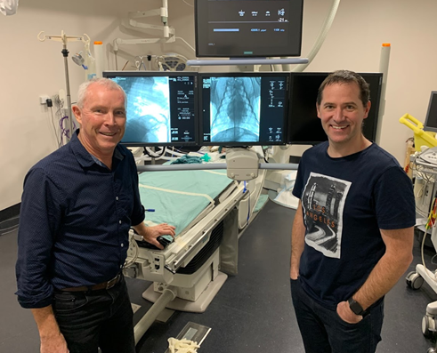
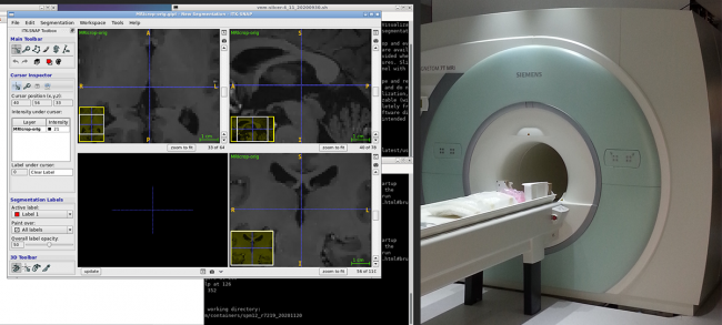
National Imaging Facility (NIF) Node partner, the Olivia Newton-John Cancer Research Institute (ONJCRI) will work with globally recognised biopharmaceutical company Telix to evaluate the use of a novel radiotracer (O-(2-[18F]fluoroethyl)-L-tyrosine or 18F-FET) to image patients with glioblastoma (GBM), a type of brain cancer, with positron emission tomography (PET) (FET-PET).

The collaboration between ONJCRI and Telix will enable a synergistic approach to improving the lives of people with GBM, which is the most common primary brain cancer in adults.

The ONJCRI is a global leader in the development of immunotherapies, targeted therapeutics, and personalised cancer medicine, while Telix is focused on the development of clinical-stage products that address significant unmet medical need in oncology and rare diseases.

The FET-PET in Glioblastoma (FIG) study will recruit up to 210 recently diagnosed adult GBM patients at 10 sites around Australia, aiming to definitively establish the role of FET-PET in the management of patients with GBM. The FIG Study is funded by the Medical Research Future Fund (MRFF), the Australian Brain Cancer Mission (ABCM), and the Cure Brain Cancer Foundation, and also involves the Trans-Tasman Radiation Oncology Group (TROG) and the Australasian Radiopharmaceutical Trials Network (ARTnet). 

The NIF’s LaTrobe University – ONJCRI Node Director, and Clinical Trial Co-Chair, Prof Andrew Scott AM said the study would utilise imaging to bring critical new treatment opportunities to light and have potentially life-saving impacts.

“Imaging is integral to effective diagnosis, staging and determination of the treatment pathway for all cancers, but is vitally important in GBM which is very aggressive and can be difficult to treat,” Professor Scott said.

“This ground-breaking study will use 18F-FET, a new PET tracer which can show us if tumour cells are active. This is a more functional imaging technique compared to magnetic resonance imaging (MRI), the current standard imaging tool, and could potentially provide a powerful imaging biomarker for the management of brain cancer and improve survival rates.”

National Imaging Facility CEO, Prof Wojtek Goscinski said the collaboration was an exciting opportunity to see the life-changing impacts that cutting-edge imaging capabilities can have on people living with debilitating illnesses.

“Medical imaging plays a critical role in the diagnosis, treatment and monitoring of life-threatening diseases like GBM,” Prof Goscinski said.

“It is excellent to see Australian-led research use imaging with the aim to improve the treatment of patients with GBM and save lives.

“It’s exciting for NIF’s LaTrobe University – ONJCRI node to be involved in an industry partnership that has the potential to expand the country’s economic growth, and position Australia as a global leader in cancer research,” he said.

You can read more about the announcement here.

COVID-19 Research at LARIF: Using fluoroscopy for lung ventilation analysis

LARIF has teamed up with Australian biomedical company, 4DMedical, and University of Adelaide scientists Associate Professor David Parsons and Dr Martin Donnelly to address the COVID-19 crisis, through testing a novel ventilator, the now patented 4DMedical ‘XV technology’, and a large animal model of Acute Respiratory Distress Syndrome (ARDS).

Associate Professor David Parsons and Dr Martin Donnelly in the LARIF Cath Lab

The NCRIS-enabled facilities and expertise at the Large Animal Research and Imaging Facility (LARIF) NIF Node, located in the South Australian Health and Medical Research Institute (SAHMRI), were utilised by a consortium of doctors, engineers, and medical researchers as part of the Australian Lung Health Initiative (ALHI).

Read More

PET for Plants

Clinical PET/CT scanners deliver non-invasive, precise anatomical and functional imaging of the human body. Did you know the same systems have been used to investigate plants?

A team of cross-disciplinary researchers at the University of Melbourne, University of Adelaide, and the University of British Columbia have teamed up to demonstrate the utility of clinical PET/CT scanners to image plants.

Read More

Open-Field PET

Understanding the molecular mechanisms that regulate behaviour, memory, and reward-based learning has challenged neuroscientists for decades. Until recently, research into these fundamental mechanisms in the brain has relied on invasive or confounding methodologies, limiting studies of neurological disorders that affect behaviour and learning, such as depression, age-related neurodegeneration, and addiction.

Read More

Changes in the brain related to parenthood

Parents can tell you that having kids changes your life. Recent studies by the Cognitive Neuroimaging Team at Monash Biomedical Imaging have shown just how deep those changes go by analysing brain structure in older adults that have had children. Parenthood was shown to cause life-long changes in both mothers and fathers, with a follow-up study suggesting that motherhood can protect the ageing maternal brain.

Read More

CT and 3D printing improving clinical PPE

Frontline medical workers put themselves at risk during a pandemic to deliver critical health care and save lives. Personal protective equipment (PPE) such as gloves, gowns, and face shields can reduce the risk of infection. To prevent contamination through airborne droplets, healthcare workers can employ an air-purifying respirator to push filtered air into their face shield or hood.

Read More

Development of Microbeam Radiation Therapy

Only two in ten people diagnosed with brain cancer will survive for at least five years, a figure that has barely improved in the last 30 years. Due to the blood-brain barrier and the risk of damaging normal brain tissue, brain cancer treatments are complex and multidisciplinary. Despite advances in surgical techniques, radiotherapy and chemotherapeutics, brain tumours remain challenging to treat. Hence, image-guided microbeam radiation therapy (MRT) with nanoparticle enhancement is being investigated for the treatment of brain cancer in animal models.

Read More

Creating a Lizard Brain Atlas

Until recently, reptilian evolutionary studies lacked an important resource – a lizard brain atlas. As the subject of numerous ecological and behavioural studies, the Australian tawny dragon (Agamidae: Ctenophorus decresii) was an ideal candidate for creating a high-resolution MRI atlas of a representative scaled reptile (squamata). Such data is not only a resource for studies of the genus but also informs environmental decision making through an improved understanding of animal adaptation and evolution.

Read More
Privacy Settings
Youtube
Vimeo
Google Maps