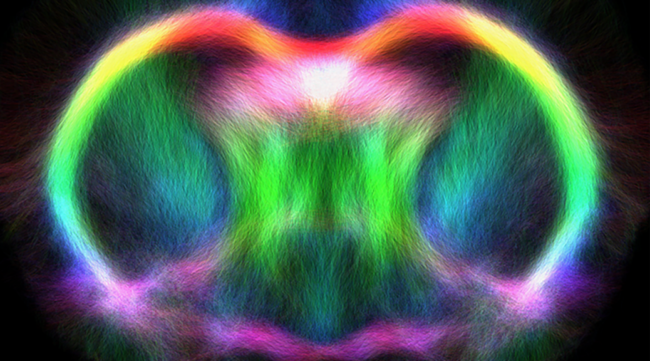
Novel Tractography to Detect Mild Traumatic Brain Injury

A mild traumatic brain injury (mTBI), often referred to as a concussion, rarely has lasting effects and is often presumed to cause only transient disturbances to brain function. However, repeated mTBIs, particularly those occurring in the sports and military settings, have been associated with cumulative and chronic neurological impairments, and the development of neurodegenerative diseases such as chronic traumatic encephalopathy (CTE).

There is evidence that these long-term adverse effects of repeated mTBIs are in part due to the recurring insults occurring before the brain has recovered from the initial mTBI and is in a period of increased cerebral vulnerability (ICV). There is increasing evidence that mTBI triggers complex biological changes including inflammatory, metabolic, neuronal, vascular and axonal abnormalities. It is believed that such changes are responsible for ICV and therefore, the identification of reliable markers that indicate when the brain is no longer in a state of ICV might allow them to be used to guide medical decisions.

The current clinical management of mTBI is largely guided by the presence or absence of neuropsychological symptoms, and typically evaluated by subjective and/or self-reported methods. Symptoms may include physical, cognitive, co-ordination, emotional, and sleep abnormalities. The onset of symptoms, although typically rapid, can take minutes or hours to occur, and symptoms are usually mild, or may even go unrecognized.

Recovery is determined to have occurred after all post-injury symptoms have resolved, at which point patients are commonly cleared to return to pre-injury activity. However, there is now evidence that the resolution of symptoms might not accurately indicate that the brain has recovered from the neuropathophysiological changes induced by mTBI. Therefore, research is required to guide and facilitate more informed medical decisions pertaining to return to pre-injury activity. In particular, it is critical that objective markers sensitive to the brain’s changes and recovery after an mTBI are identified.

Magnetic Resonance Imaging (MRI) and blood proteomics might provide objective measures of pathophysiological changes in mTBI, and indicate when the brain is no longer in a state of ICV. In a collaborative study, the use of MRI, blood proteomics, and behavioral methods as markers to detect changes and estimate recovery after experimental mTBI in rat models was investigated. Rats were given a sham or mild fluid percussion injury (mFPI), and behavioral testing, MRI, and blood collections were conducted up to 30 days post-injury.

There were cognitive impairments for three days post-mFPI, before normalizing by day 5 post-injury. In contrast, advanced MRI (i.e., tractography) and blood proteomics (i.e., vascular endothelial growth factor) detected a number of abnormalities, some of which were still present 30 days post-mFPI.

These findings suggest that MRI and blood proteomics are sensitive measures of the molecular and subtle structural changes following mTBI. Of particular significance, this study identified novel tractography measures that are able to detect mTBI and may be more sensitive than traditional diffusion-tensor measures. Furthermore, the blood and MRI findings may have important implications in understanding ICV and are translatable to the clinical setting.

For more information on this project, contact Mr. David Wright (wrightd@unimelb.edu.au).


Collaborators
Anatomy and Neuroscience, The University of Melbourne
The Florey Institute of Neuroscience and Mental Health
Department of Medicine, The Royal Melbourne Hospital
Department of Anatomy, Physiology, and Genetics, Uniformed Services University of the Health Sciences, USA
Department of Electrical and Electronic Engineering, The University of Melbourne
Centre for Stroke and Brain Injury, The University of Newcastle
School of Health Sciences, The University of Newcastle

Imaging adds a new dimension into student learning

Life presents itself in an intriguing array of three-dimensional structures. Through hands-on examination of specimens, students are presented with a highly valuable resource that aids their understanding of the diversity and complexity of taxa. However, the fragility, rarity and cost restricts the number and diversity of physical specimens that educators can provide to their classes. Additionally, student access to these specimens is often restricted to very narrow time periods. As a consequence, students tend to have life forms visualized to them using two-dimensional formats that include photographs and schematics.

However, high fidelity 3D virtual models are often used in medicine and science to reveal ‘hidden’ properties of organisms to aid diagnose and visualize research data. Using state-of-the-art technologies and computer software, the resulting 3D data are visually highly engaging and intellectually stimulating. These are key qualities that educators are looking for to involve students in subject material, and as such, 3D virtual models of ‘real-life’ biological structures have an enormous potential for utilization in education and student learning.

Recently, an initiative lead by Dr Vera Weisbecker, from the School of Biological Sciences, The University of Queensland has lead the incorporation of such models into undergraduate studies. This has been an expansion from her research into aspect of mammalian cranial development where she has used her expertise in the area of microCT to develop a novel online resource for use in her third year course that explores the diversity of extant and extinct vertebrate taxa. To get microCT scans of many of the specimens, she relies on facilities and expertise of National Imaging Facility at the Centre for Advanced Imaging at UQ node. These scans are processed by the author, who was given free-range to populate the online site and design the layout of the library interface. The library has expanded to incorporate models derived from both microCT scans and digital photogrammetry (the latter is the author’s specialty) in addition to receiving permission to incorporate models from numerous national and international institutions (e.g., Smithsonian Institute, Idaho Museum of Natural History, University of New England). This translates to an increased benefit for enrolled students that have limitless access to every model, which can be viewed during relevant lectures and practicals, or at any other time for revision and exploration.

Despite its recent generation, the 3D virtual library has received positive feedback from students and academics alike. The resource material continues to expand the number and diversity of specimens within the library, as well as new formats to present the information for student interaction (e.g., 3D pdf practical manuals). Perhaps not surprising, additional UQ courses have begun to incorporate visualizations from the site.

For more information, contact Dr Anthony Romilio (a.romilio@uq.edu.au) and visit their website and for details about the microCT and PET-CT facilities at UQ node of National Imaging Facility contact Dr. Karine Mardon (k.mardon@uq.edu.au).

In addition to developing teaching material for several UQ courses, Anthony is currently compiling a 3D digital database of Western Australia’s dinosaur coast, a stretch of 80km coastline where countless fossil footprints occur.


Collaborators
School of Biological Sciences, The University of Queensland
Centre for Advanced Imaging, The University of Queensland

Imaging for a better understanding of Anorexia Nervosa

Anorexia Nervosa (AN) is a serious psychiatric condition characterised by significantly low body weight and a fear of weight gain. A disturbance in the experience of one’s own body weight or shape is a core feature of the illness, which has a mortality rate among the highest of any mental illness. Thus, it is critical to gain a better understanding of the neurobiological basis of the illness which currently remains unclear.

 
The potential neurobiological underpinnings of AN have typically been investigated with the use of functional Magnetic Resonance Imaging (fMRI), in which brain states evoked during an experimental and a control condition are compared, with the aim of elucidating task-specific activations. Recently, however, researchers have begun to investigate synchronous brain activity at rest to examine ‘functional connectivity’ between brain regions. The term ‘functional connectivity’ is used to signify the correlation of activity time courses between brain regions. The examination of functional connectivity at rest provides information about neuronal communication in the brain, and how integration of information may relate to behaviour.

 
A study completed by the collaborators from University of Melbourne, Swinburne University of Technology node, Austin & St. Vincent’s hospitals, and Monash University & the Alfred hospital examined functional connectivity between sensorimotor and visual brain regions in AN. 26 females with AN and 27 healthy controls participated in this study and underwent a resting state functional magnetic resonance imaging scan at the neuroimaging facility at Swinburne University of Technology node. AN patients showed reduced functional connectivity between visual regions and sensorimotor regions, relative to healthy controls. These findings suggest that reduced functional connectivity between somatosensory and early visual regions may be related to visuospatial processing deficits in AN, and their misperception of body size. Gaining a better understanding of how deficits in visuospatial processing and reduced functional connectivity within these networks relate to AN may facilitate the development of more effective treatments in the future, specifically designed to improve these disturbances in the illness.
For more information on this study, contact Andrea Phillipou (ap@unimelb.edu.au).


Collaborators

Department of Optometry & Vision Sciences and Department of Psychiatry, The University of Melbourne
Department of Mental Health, The Austin Hospital
Department of Psychiatry, St Vincent’s Hospital
Faculty of Health Sciences, Australian Catholic University
Brain and Psychological Sciences Research Centre, Swinburne University of Technology
Monash Alfred Psychiatry Research Centre, Monash University and The Alfred Hospital

Visualisation and Characterisation of Feto-placental Vasculature

Proper vascular development of the human placenta is crucial for meeting the metabolic needs of the developing fetus during pregnancy. Maternal environmental stressors such as malnutrition disrupt the elaboration of the feto-placental vasculature that in turn impacts on placental function and results in reduced fetal growth. The ramifications of this are not only on short-term fetal health but also long-term health outcomes. Indeed, distortion in placental shape and size strongly associate with later adult health outcomes such as cardiovascular disease, obesity and cancer.

Dr Caitlin Wyrwoll of the School of Anatomy, Physiology and Human Biology, at The University of Western Australia is leading a multidisciplinary team that is investigating, in rodent models, how common environmental stressors in pregnancy alter feto-placental vascular morphology and placental function. Ultimately the team will seek to identify potential therapeutic targets to enhance placental vascular development and then apply this to experimental models to assess the outcomes on fetal development and adult health.

The research project involves collaboration with the Western Australian nodes1 of the National Imaging Facility and the Australian Microscopy and Microanalysis Research Facility to image, visualise and characterise the geometry of the arterial and venous feto-placental vascular trees using high-resolution X-ray microscopy (ZEISS Xradia 520 Versa). Dexamethasone administration during rodent pregnancy is used as a model to simulate excess placental and fetal glucocorticoid exposure (a known effect of prolonged stress). Control and treated rats are anaesthetised at day 22 of gestation and their uteri collected. The feto-placental units are dissected and fetal anaesthesia induced. The individual feto-placental vascular trees are cleared of blood and perfused with Microfil®, a radio opaque polymer casting compound. Each cast is stabilised in PBS in a plastic vial and imaged using a wide field-of-view of ~13.4 mm, a voltage of 50kV, more than 3000 projections through 360 degrees, and an exposure time of 7s. The ZEISS XMReconstructor software is used to reconstruct an image volume (standard parallel beam backprojection algorithm) with voxels of size ~7.0 µm.

The team have completed a preliminary study involving control and dexamethasone-treated rats and both the venous and arterial feto-placental vasculature trees. A visual comparison of treatment to control indicates that for both types of vascular tree, there is reduced branching in the fine vessels and reduced vessel density. A quantitative comparison indicates reduced total vessel length and total vessel volume.

A methodology is currently being developed for a more comprehensive and automatic quantitative assessment of vasculature morphology and geometry. This includes automatic segmentation, filtering, centre-line extraction and characterisation of the vessel tree in terms of its branching characteristics such as its branching hierarchy and angles, vessel diameters and the tortuosities of vessel segments. Furthermore, these vascular tree images are being used in a world-first study to model placental blood flow using computational fluid dynamics.

For more information on this work, contact Dr. Caitlin Wyrwoll (caitlin.wyrwoll@uwa.edu.au).

Collaborators
School of Anatomy, Physiology and Human Biology, The University of Western Australia
School of Mechanical and Chemical Engineering, The University of Western Australia
Centre for Microscopy, Characterisation and Analysis, The University of Western Australia

Grape split imaging to examine physical changes before and after splitting using diffusion magnetic resonance

Berry split is a condition in which the grape epidermis splits which often occurs during periods of high rainfall and is a significant cause of grape crop loss. In damp conditions there is increased uptake of water via osmosis and decreased water loss from transpiration. The pre-dawn turgor pressure of table grape cultivars lies in the vicinity of 5-38 kPa but prior to berry split can be as high as 1.5-3.7 MPa.
In order to examine and characterise the immediate effect of fruit split on grape, National Wine & Grape Industry and Western Sydney University node of National Imaging Facility have been collaborating on an ongoing project, which investigates the physical changes within the grape berry both before and after splitting using diffusion magnetic resonance imaging. Thirty-six table grapes of the Thompson Seedless variety were involved in the study and assigned to three groups: a control group (12 berries), a group in which they were wrapped in damp tissue (12 berries), and a total immersion group (12 berries). Five axial images (including diffusion tensor images) spaced evenly apart along the length of each berry were acquired simultaneously every hour to create a time-course study of each grape.
For each grape that split within the MRI during the study it was observed that there was an immediate change in the diffusion coefficient in the region of the wound. This region increased in volume over the course of the subsequent scans and correlated with regions of non-vital cells (as determined by fluorescence microscopy). It was determined from the study that grape berries left exposed to standing water after splitting exhibit greater cell death within the vicinity of the split. Therefore, the surface of split berries should be kept dry if possible to reduce further damage.


Collaborators

Nanoscale Organisation and Dynamics Group, University of Western Sydney
School of Medicine, University of Western Sydney
National Wine & Grape Industry Centre
NSW Department of Primary Industries

Sources

Dean, R.J., Bobek, G., Stait-Gardner, T., Clarke, S.J., Rogiers, S.Y. and Price, W.S. (2015), Time-course study of grape berry split using diffusion magnetic resonance imaging. Aust. J. Grape Wine R. doi: 10.1111/ajgw.12184
Dean R.J., Stait-Gardner, T., Clarke, S.J., Rogiers, S.Y., Bobek, G. and Price, W.S. (2014) Use of diffusion magnetic resonance imaging to correlate the developmental changes in grape berry tissue structure with water diffusion patterns. Plant methods 10(1):35


Abnormal brain areas common to the focal epilepsies

A group of scientists at The Florey Institute may soon be able to diagnose a common form of epilepsy after a simple 10-­minute brain scan. The result? Patients will commence immediate treatment and minimize the risk of further damage caused by seizures.

New research published in Brain Connectivity shows that people with focal epilepsy seemingly share characteristic brain network connectivity in three precise regions of the brain, even though the seizure site is in heterogeneous brain regions.

People with focal epilepsy, including all the patients in this new study, have slower psychomotor reflexes, and neuropsychological symptoms such as depression and working memory deficiencies. About one per cent of the population will develop epilepsy at some point in their lifetimes, with childhood and old age being more vulnerable periods. Over half of all diagnosed epilepsies are focal in nature, arising in specific brain regions.

Mangor Pedersen, together with a team led by Professor Graeme Jackson at the Florey node of National Imaging Facility (NIF), used functional Magnetic Resonance Imaging (fMRI) scans obtained on a 3T Siemens Trio located at the Melbourne Brain Centre, Austin Hospital campus. The analysis techniques used in this study may be used to target MR biomarker data allowing patients to be classified as having focal epilepsy, versus other types of epilepsy. The team scanned brains of 14 people with focal epilepsy, and compared them to 14 age-­and sex-matched people without the disease. The group then used two connectivity measures -­ a local network between one voxel and the 27 surrounding voxels (about one cubic centimeter of brain), and a more distributed network from each single voxel connected to all the other voxels in the brain – to show abnormal connectivity in three brain regions of people with focal epilepsy.

The three common brain areas in people with focal epilepsy were both shallow & deep brain regions in the temporal lobe (just in front of and above the ear) and the prefrontal cortex (at the front of the head in between the eyes). What amazed the researchers was that passing the connectivity results through a multivariate pattern analysis (a “machine learning algorithm”) differentiated healthy people from those with focal epilepsy with almost 90 per cent accuracy.

Mangor said of the work, “Focal epilepsy is a disease where seizures originate from different areas of the brain. In this study we tested whether patients had any brain markers in common. We used network connectivity and pattern analysis to classify brain patterns at a single subject level. We hope that this work is a preliminary step towards using network analysis from functional imaging and pattern analysis to detect focal epilepsy biomarkers.”

The work is the culmination of three years’ hard work as part of Mangor’s Ph.D research. However, he is not content to leave it there, saying “we now need to shore up these findings firstly by scanning more patients.” Other future experiments are to scan people with co-morbities, like depression, but not focal epilepsy. This will help assess the reliability of the clinical fMRI classification.


Collaborators

The Florey Institute of Neuroscience and Mental Health, Austin Campus, Melbourne, VIC, Australia.
The University of Melbourne, Florey Department of Neuroscience and Mental Health, Melbourne, VIC, Australia.
Department of Neurology, Austin Health, Melbourne, VIC, Australia

Source

Pedersen, Mangor, et al. “Abnormal brain areas common to the focal epilepsies: Multivariate pattern analysis of fMRI.” Brain connectivity (2015).

Simultaneous scanning of two mice in a small-animal PET scanner

In preclinical Positron Emission Tomography (PET) imaging, several research groups have recently proposed different experimental set ups allowing multiple animals to be simultaneously imaged in a scanner in order to reduce the costs and increase the throughput. Simultaneous scanning of several animals also ensures injections with the same specific activity, which may be otherwise subject to significant variations when animals are injected at different time and possibly with tracers from different productions. In addition, it is well known that the performance of a PET system, in terms of spatial resolution and sensitivity, is optimal at the center of the field of view (FOV) and degrades quickly with increasing distance from the center. In previous studies, the technical feasibility has been demonstrated and the signal degradation caused by additional mice in the FOV characterized, however, the impact of the signal degradation on the outcome of a PET study has not yet been studied.

In a project collaborated by the ANSTO/University of Sydney node of the National Imaging Facility, the collaborators thoroughly investigated, using Monte Carlo simulated [18F]FDG and [11C]Raclopride PET studies, different experimental designs for whole-body and brain acquisitions of two mice and assessed the actual impact on the detection of biological variations as compared to a single mouse setting.

First, the validation of the PET-SORTEO Monte Carlo simulation platform for the simultaneous simulation of two animals was extended. Then, [18F]FDG and [11C]Raclopride input mouse models for the simulation of realistic whole-body and brain PET studies were designed. Simulated studies allowed the scientists to accurately estimate the differences in detection between single- and dual-mode acquisition settings that are purely the result of having two animals in the FOV. Validation results showed that PET-SORTEO accurately reproduced the spatial resolution and noise degradations that were observed with actual dual phantom experiments. The simulated [18F]FDG whole-body study showed that the resolution loss due to the off-center positioning of the mice was the biggest contributing factor in signal degradation at the pixel level and a minimal interanimal distance as well as the use of reconstruction methods with resolution modeling should be preferred. Dual mode acquisition did not have a major impact on ROI-based analysis except in situations where uptake values in organs from the same subject were compared. The simulated [11C]Raclopride study however showed that dual-mice imaging strongly reduced the sensitivity to variations when mice were positioned side-by-side while no sensitivity reduction was observed when they were facing each other.

This is the first study showing the impact of different experimental designs for whole-body and brain acquisitions of two mice on the quality of the results using Monte Carlo simulated [18F]FDG and [11C]Raclopride PET studies. This study validates unique experimental capabilities, which enable researchers to make the most of the radiotracers production and scanners availability.

Collaborators

1 Australian Nuclear Science and Technology Organization (ANSTO), Kirrawee DC, Australia
2 Brain & Mind Centre, National Imaging Facility (NIF), University of Sydney/ANSTO Node, Sydney, Australia
3 CERMEP – Imagerie du vivant, Lyon, France
4 Institut Pluridisciplinaire Hubert Curien, Université de Strasbourg, Strasbourg, France
5 CNRS, UMR7178, 67037 Strasbourg, France

Source

Reilhac, Anthonin, et al. “Simultaneous scanning of two mice in a small-animal PET scanner: a simulation-based assessment of the signal degradation.” Physics in Medicine and Biology 61.3 (2016): 1371.

Enhanced MRI of Preclinical Prostate Cancer


MRI is a useful imaging tool in prostate cancer management. It provides excellent soft tissue contrast and multidimensional information, does not involve exposure to ionizing radiation, and is non-invasive. However, like other imaging modalities such as computed tomography (CT), transurethral ultrasound (TRUS) and nuclear imaging, MRI cannot adequately detect small tumors.

The ability to accurately detect and locate small tumors is necessary for early detection of disease and for assessment of response to therapy in cancer patients. In recent years, the use of biomarker-targeted probes linked with nanoparticle-based contrast agents to enhance these imaging modalities has been a major area of research. Iron oxide magnetic nanopar­ticles (MNPs) are powerful contrast agents for MRI. Their superparamagnetic properties make them effective at reducing transverse (spin-spin) T2-relaxation time, causing negative contrast in magnetic resonance (MR) images. MNP-assisted MRI has the potential to improve the assess­ment of cell surface receptor expression on tumors, liver function (macrophage content and activity), inflammation, degenerative diseases, angiogenesis, perfusion and apop­tosis.

This project evaluates the potential ability of MNPs to enhance MRI of prostate cancer by performing MRI on on mice with pre-established orthotopic LNCaP-luc tumors and intravenously injected with either MNPs alone or J591-MNPs. MR images of tumors from mice that received the J591-MNP conjugates show significant darkening at the prostate region, at the 2- and 24-h post-injection timepoints, as shown in the above image.

These observations have major clinical implications because tumor-targeting MNPs could potentially enable the early detection of tumors confined within the prostate by MRI. Based on its biocompatibility, stability, together with its ability to enhance MRI, PSMA-targeting MNPs have promise to be translated into the clinic to improve the management of prostate cancer.

Research team

Brian Wan-Chi Tse, Gary J Cowin, Carolina Soekmadji, Lidija Jovanovic, Raja S Vasireddy, Ming-Tat Ling, Aparajita Khatri, Tianqing Liu, Benjamin Thierry & Pamela J Russell


Source

Tse, Brian Wan-Chi, et al. “PSMA-targeting iron oxide magnetic nanoparticles enhance MRI of preclinical prostate cancer.” Nanomedicine10.3 (2015): 375-386.

Global BioImaging Project


EuroBioImaging

AMMRF Logo

nif logo

AMMRF and NIF announce their inclusion in EU Horizon 2020-funded Global BioImaging (GBI) project that commences today.

The grant to Euro-BioImaging is built on their existing collaboration with two NCRIS-supported projects, the Australian Microscopy & Microanalysis Research Facility (AMMRF) and the National Imaging Facility (NIF) and with India-BioImaging, around imaging infrastructure.

Euro‐BioImaging is a large‐scale pan‐European research infrastructure project on the European Strategy Forum on Research Infrastructures (ESFRI) Roadmap.

The GBI alliance reflects the current revolution in imaging technologies that supports biomedical science from visualisation of molecules inside cells through to imaging processes occurring in the whole animal. GBI will work to facilitate access to a global network of these imaging platforms; enable exchange of experience in technology development; explore standardisation of access protocols, data formats and processing protocols. Such standardisation, together with the sharing of data, will facilitate transglobal collaborative discovery projects with translation to multi-centre clinical trials.

GBI will also enable the identification and sharing of best practice in facility operation and management, new open access data analysis tools and properly curated image storage systems. Other proposed activities include joint international training and exchange programmes for building staff expertise and the further development of MyScope, AMMRF’s online training tool.

Through this interconnected collaborating infrastructure Australian researchers will have the opportunity to engage with multi-lateral, international biomedical microscopy and imaging facilities.

GBI will establish the foundation for a long-term alliance for mutual benefit between Euro-BioImaging and its international partners with the aim of providing sustainable services in imaging technologies to the international scientific community. Improved international connections will also be able to support Australian industry and the Australian Government’s Innovation Agenda.

The AMMRF’s CEO, Dr Miles Apperley, will represent the Australian partners on the GBI management board as they create the global network of state-of-the-art research infrastructure in imaging.

The Global BioImaging Project relates to the Horizon 2020 topic INFRASUPP-6-2014 – international cooperation for research.


Privacy Settings
Youtube
Vimeo
Google Maps