Imaging adds a new dimension into student learning
Life presents itself in an intriguing array of three-dimensional structures. Through hands-on examination of specimens, students are presented with a highly valuable resource that aids their understanding of the diversity and complexity of taxa. However, the fragility, rarity and cost restricts the number and diversity of physical specimens that educators can provide to their classes. Additionally, student access to these specimens is often restricted to very narrow time periods. As a consequence, students tend to have life forms visualized to them using two-dimensional formats that include photographs and schematics.
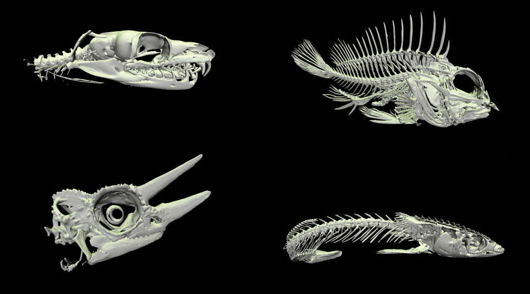
However, high fidelity 3D virtual models are often used in medicine and science to reveal ‘hidden’ properties of organisms to aid diagnose and visualize research data. Using state-of-the-art technologies and computer software, the resulting 3D data are visually highly engaging and intellectually stimulating. These are key qualities that educators are looking for to involve students in subject material, and as such, 3D virtual models of ‘real-life’ biological structures have an enormous potential for utilization in education and student learning.
Recently, an initiative lead by Dr Vera Weisbecker, from the School of Biological Sciences, The University of Queensland has lead the incorporation of such models into undergraduate studies. This has been an expansion from her research into aspect of mammalian cranial development where she has used her expertise in the area of microCT to develop a novel online resource for use in her third year course that explores the diversity of extant and extinct vertebrate taxa. To get microCT scans of many of the specimens, she relies on facilities and expertise of National Imaging Facility at the Centre for Advanced Imaging at UQ node. These scans are processed by the author, who was given free-range to populate the online site and design the layout of the library interface. The library has expanded to incorporate models derived from both microCT scans and digital photogrammetry (the latter is the author’s specialty) in addition to receiving permission to incorporate models from numerous national and international institutions (e.g., Smithsonian Institute, Idaho Museum of Natural History, University of New England). This translates to an increased benefit for enrolled students that have limitless access to every model, which can be viewed during relevant lectures and practicals, or at any other time for revision and exploration.
Despite its recent generation, the 3D virtual library has received positive feedback from students and academics alike. The resource material continues to expand the number and diversity of specimens within the library, as well as new formats to present the information for student interaction (e.g., 3D pdf practical manuals). Perhaps not surprising, additional UQ courses have begun to incorporate visualizations from the site.
For more information, contact Dr Anthony Romilio (a.romilio@uq.edu.au) and visit their website and for details about the microCT and PET-CT facilities at UQ node of National Imaging Facility contact Dr. Karine Mardon (k.mardon@uq.edu.au).
In addition to developing teaching material for several UQ courses, Anthony is currently compiling a 3D digital database of Western Australia’s dinosaur coast, a stretch of 80km coastline where countless fossil footprints occur.
Collaborators
School of Biological Sciences, The University of Queensland
Centre for Advanced Imaging, The University of Queensland


