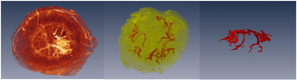
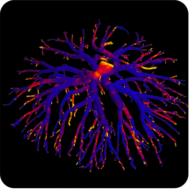
Understanding Feto-Placental Vasculature

Proper vascular development of the human placenta is crucial for meeting the metabolic needs of the developing fetus during pregnancy. Maternal environmental stressors such as malnutrition disrupt the elaboration of the feto-placental vasculature that, in turn, impacts on placental function and results in reduced fetal growth. The ramifications of this are not only on short-term foetal health but also on long-term health outcomes. Indeed, distortion in placental shape and size strongly associate with later adult health outcomes such as cardiovascular disease, obesity and cancer.

Read More

Micro-CT of re-regeneration in lizard tails

Re-regeneration to reduce adverse effects associated with tail loss

Caudal autotomy, the ability to drop and regenerate a portion of the tail, is a widely used anti-predation strategy in many lizard species. Intra-vertebral autotomy planes within a series of the lizard’s caudal vertebrae allow individuals to autotomise a portion of their tail to escape a threat, such as a predator’s grasp. Once autotomised, the tail regenerates with a rigid cartilage rod in place of the original bony vertebrae. Although an effective anti-predation strategy, it has both short and long-term costs to the individual associated with physical tail loss, as well as the energy required for regeneration. Additionally, a regenerated tail lacks autotomy planes, where subsequent autotomy events having to at a more proximal position at a caudal vertebra with an intact autotomy plane.

Read More

Detecting atherothrombosis using targeted MRI

Cardiovascular disease (CVD) is responsible for more than a quarter of all deaths in Australia and remains the global leading cause of death accounting for 17.9 million deaths per year. Of all CVDs, stroke and coronary artery disease account for the majority of deaths. A common underlying cause in these conditions in atherosclerosis, characterised by the build-up of abnormal deposits inside the arteries. Atherosclerotic plaques can rupture and cause thrombosis, or blood clots, resulting in stroke and myocardial infarction. Diagnostic strategies for the detection of thrombi are currently invasive and may not be sensitive to early biomarkers such as localised coagulation and inflammation.

A/Prof Ta, of Griffith University, has teamed up with researchers across Australia and internationally to develop a new form of MRI contrast agent. These ultra-small dual positive and negative contrast iron oxide nanoparticles (DCIONs) provide both T1-positive and T2-negative contrast effects, overcoming the limitations of single modality contrast agents. This duality is particularly important for imaging intravascular thromboses, as current single-contrast nanoparticles results in a black dye against a black artery. Further, the DCIONs are monodisperse, water-soluble, and biocompatible, of critical importance to biomedical applications.

In-vivo MRI of carotid artery thrombus (green arrows) detection after injection with a conjugated DCION. Image from Ta et al 2017.

Using non-invasive MRI at the NIF QLD Node, the application of a DCION conjugated to an enzyme found in activated platelets demonstrated accurate and sensitive detection of intravascular thrombosis. Work is continuing to further optimise the early detection of thrombi, expected to allow for earlier and more effective preventative treatments and improved clinical outcomes for patients at risk of stroke and myocardial infarction.

A/Prof Ta is enthusiastic about the future applications of DCIONs beyond thrombosis diagnoses, stating that “these nanoparticles have the potential to replace traditional gadolinium-based contrast agents due to their stronger T1 contrast effect. Existing alternatives cannot do what these nanoparticles can.”

If you have any concerns about heart disease or atherosclerosis, please talk to your GP and check this website.

Stent-electrode array for cortical neural activity

Minimally invasive endovascular stent-electrode array for high-fidelity, chronic recordings of cortical neural activity

This news has been contributed by Assoc. Prof. Bradford Moffat of the Melbourne Brain Centre Imaging Unit, Department of Radiology and Medicine, The University of Melbourne, Parkville.

National Imaging Facility Fellow, Assoc. Prof. Bradford Moffat collaborated with Dr. Tom Oxley’s group at the University of Melbourne for this high profile publication[1] that appeared in the journal “Nature Biotechnology”.

Read More

MRtrix3

MRtrix3: Advanced tools for the analysis of diffusion MRI data

Diffusion-weighted MRI (dMRI) is a commonly-used medical imaging modality for the investigation of tissue microstructure, exploiting the local hindrance and restriction of water diffusion as indirect probes. The neuroimaging research community utilises this technology extensively for the study of brain white matter in particular, reconstructing structural connectivity pathways and analysing estimated tissue properties.

Read More

MRI investigations of placental structure and function

Preeclampsia is a medical condition affecting up to 3% of pregnant women in Australia. Characterised by high blood pressure and protein in the urine, it is a leading cause of morbidity and mortality in both mothers and infants. Furthermore, preeclampsia has been linked to long-term health consequences for both mother and child.

3-D reconstructions of the placenta from MRI images. (left) Foetal surface view of the placenta. (middle) Maternal surface view of the placenta with an overlay showing maternal vasculature. (right) Side view showing maternal vasculature alone

Hampering early diagnosis, prevention, and treatment efforts is a lack of understanding of preeclampsia pathophysiology.  Currently, the cause of this condition is unknown. Prof Annemarie Hennessy and a team of researchers at Western Sydney University are utilising the WSU NIF Node, in collaboration with NIF Fellow Dr Timothy Stait-Gardner, to learn more about this serious condition.

In this project, high-resolution magnetic resonance imaging is being used to examine placental changes in vivo in mouse models of preeclampsia. In addition to the in vivo studies, high-resolution scans of fixed mouse placentas, normal and abnormal, have been used to create a placental atlas. The creation of a placental atlas and a number of publications have provided important information on mouse models of preeclampsia, including its characterisation and how to differentiate between different models of preeclampsia from T2 maps of the mouse placentas. These works have provided some of the basis for investigations of new treatments of preeclampsia.

Publications:

This story was contributed by the Western Sydney University NIF Node. For further information, please contact Dr Timothy Stait-Gardner.

Local Connectivity Networks Disrupted by Sports-Related Concussion

From https://journals.sagepub.com/doi/10.1177/2059700219861200
Functional local connectivity is decreased in acutely concussed players compared to controls. (a) Statistically significant brain regions at family-wise error rate p < 0.05; 5000 permutations using threshold-free cluster enhancement. Cool-scaled color bar denotes the magnitude of voxel t-values. (b) Individual-level z-normalized fMRI local connectivity values where each grey circle denotes an individual subject; red line is the group mean; red shaded area is the 95th percentile of the mean value; and blue shaded area is one standard deviation from the mean.

Head injuries, including concussion, are taken very seriously in sporting professions. To date, making an accurate diagnosis of acute concussion has been made difficult by the lack of a reliable direct biomarker for injury and recovery. This diagnostic gap can lead to unknown recovery periods and potentially long-term impacts for athletes.


Researchers at the Florey Institute of Neuroscience and Mental Health set out to understand functional brain changes in professional players in the Australian Football League who had been diagnosed with acute sport-related concussion.


The world-first study, published in the Journal of Concussion, utilised NIF infrastructure, the 3T Trio and Skyra MRI scanners, and NIF expertise, Facility Fellows Shawna Farquharson and David Abbott.

 Functional MRI (fMRI) was undertaken to assess functional connectivity alongside anatomical imaging. Although no anatomical damage was observed, the authors described a decreased intrinsic fMRI connectivity within the right frontoparietal regions in acutely concussed footballers. In other words, all 20 concussed athletes showed reduced activity in parts of the brain responsible for executive function, working memory and switching tasks.


“By looking at how the different parts of the brain talk to each other, we can see how these three brain networks are affected, and these changes may help explain the symptoms we see in concussed players.” – Dr Mangor Pedersen, study co-author, from the Florey Institute of Neuroscience and Mental Health.


One interesting finding in this study is that concussion appears to affect particular networks in the brain. These findings are in agreement with some of the typical clinical features of concussion; however, they are based on trends seen as a group. In future, the authors intend to investigate individual brain networks and develop guidelines for personalised treatment and recovery.


This story was contributed by the Florey Institute of Neuroscience and Mental Health NIF Node. For more information, please contact Shawna Farquharson or David Abbott.


More information about concussion is available here, or by speaking to your GP.

Hyperpolarized 129Xe MR imaging of lung

Magnetic Resonance Imaging (MRI) has a range of applications in medical diagnosis, and more than 25,000 scanners are estimated to be in use worldwide. However, lung imaging suffers from some technical challenges, limiting its application in pulmonary disease diagnosis and treatment.


Due to low proton density, movement and high susceptibility difference between air and tissue, conventional proton MRI struggles to image lung tissue and function. These can be partially overcome by the introduction of contrast agents into the lung. Hyperpolarised gases are promising contrast agents for imaging lung structure and function. The two most common gasses are helium (3He) and xenon (129Xe) isotopes. Helium isotopes are challenging and expensive to obtain, and do not provide significant functional readouts. Xenon can be challenging to work with, but promises novel physiological measurements not previously feasible. This resulting technique, termed hyperpolarized 129Xe MR imaging, has revolutionised the field of functional lung imaging.


Five years ago, an Australian Research Council (ARC) grant was awarded to researchers at Monash Biomedical Imaging (MBI) and ANSTO to design and build a machine that could reliably produce hyperpolarized xenon. In this project, Dr Wai Tung Lee and NIF Facility Fellow Dr Gang Zheng have teamed up to develop a new multimodal technology capable of high-quality investigations of the human lungs. While Dr Lee, of ANSTO, was responsible for the construction of the polarizer and production of polarized 129Xe for MR imaging, Dr Zheng, of Monash University, designed the MR experiments and adapted imaging protocols.

Figure 1. Schematic diagram of the polarizer system. Note: Some details omitted for clarity.

Hyperpolarized 129Xe (HP-Xe) gas was generated in a custom designed and constructed SEOP system at Monash Biomedical Imaging (Figure 1 and Figure 2). The system consists of a gas-polarizing unit and a gas injection and recycle unit. The core component of the gas-polarizing unit is an optical pumping cell (OPC) where the gas is polarized. The OPC is placed in an oven to regulate the amount of Rb vapour by maintaining a constant temperature between 60ºC-90ºC. The optical pumping process uses two 240W narrow-bandwidth lasers tuned to the Rb D1 transition wavelength of 794.7nm. Additional optics condition the laser light to circular polarization and shape the laser profile to fully illuminate the OPC. HP-Xe is produced in batches rather than using a continuous-flow process.

Figure 2. The spin-exchange-optical pumping system. A. Gas polarizing unit in Room 1; B. Gas injection and recycling unit in Room 2.

MR experiments were performed on a clinical 3T whole-body system (Skyra, Siemens Medical Solutions, Erlangen, Germany) with a broadband RF amplifier. HP-129Xe MRI was enabled using a bird-cage transmit/receive chest coil (RAPID Biomedical GmbH, Wuerzburg, Germany). A 2D-GRE sequence for X-nuclei MRI was used to image HP-Xe gas in lung. Proton channel images were acquired for the localization of the lung. The resonance frequency of 129Xe was set to 34.09 MHz on the 3T scanner. The human subject first quickly flushed his lung with pure nitrogen, and then immediately inhaled HP-Xe in the Tedlar bag. After inhalation of HP-Xe, the subject held their breath during the scan. The total imaging time was less than 10s.


Dr Zheng outlines the results to date, “We have successfully imaged gas-phase HP-129Xe in a range of phantoms, such as the Tedlar bag, syringe and the glass cell (Figure 3). We repeated the gas-phase imaging in both ex-vivo (Figure 4) and in-vivo lamb lungs (Figure 5). Imaging results supported that our polarizer can provide sufficient polarization for lung imaging. In 2019, we successfully performed a human experiment and a gas signal from the lungs was clearly identified (Figure 6). In addition, all studied showed a clear Rf peak of gaseous and dissolved xenon which should allow us to develop methods to assess lung function. We plan to study human pulmonary disease in the near future and hope to find additional collaborators to help translate this modality into clinical use.”

Gallery Notice : Images have either not been selected or couldn't be found

Construction of the hyperpolarizer is complete; it can routinely produce HP-129Xe for research use. The technique has successfully imaged HP-129Xe in phantoms, ex-vivo and in-vivo lamb lungs and a human lung. If you are considering lung research, please see more on the Monash website, or get in touch with Dr Gang Zheng to discuss your project needs.


Publications:

Zheng G, Lee WT, Tong X, et al., A SEOP Filling Station at the Monash Biomedical Imaging Centre. Conference on polarization in Noble gases (PiNG), 2017.


Zheng G, Lee WT, Tong X, et al., Phase imaging of hyperpolarized 129Xe gas in a human lung. ISMRM 2020, submitted on 6 Nov 2019.


Acknowledgements:

National Imaging Facility, ARC (Grant LE130100035); NHMRC (Grant APP606944); CASS Foundation; Monash Biomedical Imaging, ANSTO


This story was contributed by Dr Gang Zheng of the Monash University NIF Node.


Impact of surgical lymph node removal

The impact of surgical lymph node removal on metastatic disease and the response to immunotherapy

Surgical resection of cancer remains the frontline therapy for millions of cancer patients every year, but disease recurrence after surgery is common with a relapse rate of around 45% for lung cancer. Relapse rates are expected to decline, with new immunotherapies producing extraordinary successes in several solid cancers. Immunotherapy administered after surgery could potentially ‘mop up’ small persisting cancer deposits that lead to disease recurrences. However, uninvolved (tumour-free) draining lymph nodes are the primary ‘factory’ for generating anti-cancer T cell responses; hence, should they be removed, subsequent immunotherapy may be negatively impacted. The aim of this project is to determine in murine models if the response of metastatic disease to immunotherapy is reduced following tumour lymph node resection.

Dr Vanessa Fear of the School of Biomedical Sciences, at The University of Western Australia, is investigating if the response of metastatic disease to immunotherapy is reduced following tumour draining lymph node resection. To do this, the Tumour Immunology Group is using an AB1 Model of metastatic disease. Tumour progression is visualised using IVIS imaging and histology following resection to track the effectiveness of treatment regimes. Ultimately, the team will seek to determine the impact of lymph node removal at the time of tumour resection to subsequent immunotherapeutic outcomes.

Fig 1: IVIS imaging from AB1-HA tumour model. Mice received AB1-HA_LUC i.v. and lung tumour development monitored on the InVivo Imaging System (IVIS, Lumina II imager). At the imaged timepoints mice received intrapertioneal injections of luciferin (150µg/g) and tumour burden was measured on the IVIS in photons/sec (p/s). A, tumour progression day 14 to day 19.

The research project involves collaboration with the Centre for Microscopy, Characterisation and Analysis, the West Australian Node for the National Imaging Facility to image, visualise and characterise the development of lung metastatic disease using the IVIS Lumina II in vivo bioluminescence imager with the help of Living Image Software (Caliper Life Sciences).

Fig 2: IVIS imaging and histology from AB1-HA tumour model.Comparison of IVIS reading with lung H&E staining showing AB1-HA tumour from the same mouse. Tumour volume determined using FIJI software.

The team have completed preliminary studies determining a 55% metastatic disease onset after surgical resection of the primary tumour. Current investigations in tumour resection and lymph node resection indicate temporal changes in onset of metastatic disease compared to mice with intact lymph nodes.

Further investigations into the impact of lymph node resection on immunotherapy are underway. Future investigations will include other models including lung adenocarcinoma, melanoma, and breast cancer.

Collaborators

School of Medicine, the University of Western Australia

School of Biomedical Sciences, University of Western Australia

Centre for Microscopy, Characterisation and Analysis, the University of Western Australia

This story was contributed by the University of Western Australia. For more information, contact Dr Vanessa Fear or Diana Patalwala.

3D printed devices to treat traumatic pelvic fractures

3D printing is increasingly being used in the healthcare industry to customise medical devices to meet patient-specific needs. Currently, device manufacture is lengthy, limiting the application of customised medical devices. The treatment of traumatic injuries requires intervention as quickly as possible, preferably within days post-injury.

This collaborative research project between the Dept. of Biomedical Engineering at the University of Melbourne and the Dept. of Orthopaedics at the Royal Melbourne Hospital aims to assess the feasibility of 3D printing fracture plates to treat traumatic fractures and speed up the production of devices at the point-of-care for a patient. By performing a proof-of-concept experiment on a set of cadaveric pelvis, Dr Dale Robinson and team are evaluating each phase of the 3D printing workflow. Once implanted, a series of computational models and biomechanical experiments will be used to assess whether the 3D printed fracture plate offers an improvement over a traditionally mass-manufactured plate. Paramount to designing customised implants, the anatomy of each pelvis is being characterised using the University of Melbourne’s NIF Node CT with input from PET/CT Facility Fellow Rob Williams and radiographer Rebecca Glarin. After implantation of the fracture plate, CT may assess the effectiveness of the device in terms of stabilising and reducing the fracture.


3D reconstruction of a fractured human pelvis with a custom 3D printed device simulated in blue to promote appropriate healing.

To date, the project has conducted some scans and used this data for preliminary printing of implants. Plates were designed and printed in collaboration of researchers at Johnson and Johnson and the University of Melbourne. The initial study used 3D printed medical-grade titanium and 3D rendering from the NIF facility CT. In developing this method, iterative reconstruction with maximal overlap to printing was used to be consistent with typical medical CT. This was done while still using radiation dosimetry within standard limits.

This project has the potential to improve patient outcomes by enhancing surgical intervention durability, reducing the duration and number of surgeries, and reducing the risk of life-threatening surgical complications (such as pulmonary embolism and infection) through reduced bedtime. Consequently, the effective implementation of customised 3D printed medical devices is expected to reduce healthcare costs through shorter hospital stays and reduced number of surgical interventions.

This story was contributed by the Department of Biomedical Engineering and the Melbourne Brain Centre Imaging Unit at the University of Melbourne, and Johnson & Johnson. For further information, please contact Rob Williams.

Privacy Settings
Youtube
Vimeo
Google Maps