
Micro-CT of re-regeneration in lizard tails
Re-regeneration to reduce adverse effects associated with tail loss
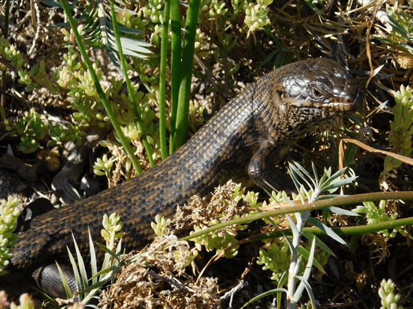
Caudal autotomy, the ability to drop and regenerate a portion of the tail, is a widely used anti-predation strategy in many lizard species. Intra-vertebral autotomy planes within a series of the lizard’s caudal vertebrae allow individuals to autotomise a portion of their tail to escape a threat, such as a predator’s grasp. Once autotomised, the tail regenerates with a rigid cartilage rod in place of the original bony vertebrae. Although an effective anti-predation strategy, it has both short and long-term costs to the individual associated with physical tail loss, as well as the energy required for regeneration. Additionally, a regenerated tail lacks autotomy planes, where subsequent autotomy events having to at a more proximal position at a caudal vertebra with an intact autotomy plane.
Re-regeneration, a regeneration event on an already regenerated tail, has been anecdotally recorded to occur from a result of a physical shearing of the tail, such as struggling free form a predator’s grasp. This would result in a smaller portion of the tail being lost minimising both short and long-term adverse effects on the individual. In this study we investigate (1) the occurrence and use of re-regeneration across three isolated populations of King’s skink (Egernia kingii)that vary in predation risk, (2) assess the internal morphology of re-regeneration using micro-CT technology, and (3) discuss the potential mitigating effects of re-regeneration as well as its use in restoring tail function for lizard ecology.
As re-regeneration has only anecdotally been recorded, James Barr and a team of collaborators at Curtin University and CSIRO created an excellent opportunity to utilise NIF micro-CT technology to provide a highly detailed image of a re-regeneration event. The team assessed the internal morphology of a re-regenerated tail from an E. kingii specimen using the Skyscan 1176 micro-CT scanner located at the Western Australia node of the National Imaging Facility at the Centre for Microscopy, Characterisation & Analysis (CMCA).
Observing re-regeneration using micro-CT
Lizards were captured at the three sites using baited Elliott traps and morphological measurements recorded including tail length, and presence of both regenerated and re-regenerated tissue. An autotomised tail (part of a concurrent study) was reconstructed using CT images. The spinal column was manually selected using a volume of interest (VOI), and 3D models were recreated.
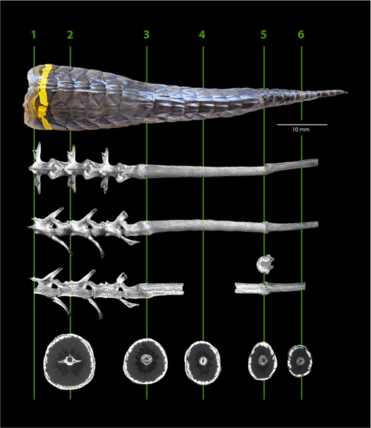
Re-regeneration events were identified as being present in all three populations, with higher occurrences in populations with terrestrial predators. Re-regeneration tissue comprised (mean ± SD) 18.0 ± 14.8% of the total tail length and 38.5% ± 20.6% of the regenerated tail portion. Additional several individuals form the population with higher diversity of terrestrial predators had secondary re-regeneration (regenerated tissue on a re-regenerated tail). Re-regeneration is likely to minimise short and long terms costs associated with caudal autotomy and regeneration. Additionally, as intravertebral autotomy is consciously controlled, the decision to not autotomise their tail but struggle and attempt to cause a shearing event leading to re-regeneration may be a potential alternative anti-predation strategy. The presence of field data provides an ecological context to re-regeneration, which may indicate an alternative means to escape a threat, minimising costs to the individual.
This study provides an enhanced ecological understanding of selective predation pressures and adaptation of defence strategies. Furthermore, regarding cartilage regeneration, this study highlights that the cartilage regeneration process is highly complex, not only involving molecular and genetic variables but also highly influenced by an individual’s natural environment. Ultimately, a greater understanding of cartilage regeneration may lead to therapeutic strategies in humans
For further reading, please see the original publication and other stories generated from this work:
- https://www.nature.com/articles/s41598-019-55231-6
- https://phys.org/news/2019-12-tale-lizard-regrown-tail.html
- https://www.azocleantech.com/news.aspx?newsID=26875
For more information, please contact NIF Facility Fellow Dr Ivan Lozic. This story was contributed by the UWA Node of NIF through James Barr of Curtin University.
This project was funded by the Holsworth Wildlife Research Endowment – Equity Trustees Charitable Foundation & the Ecological Society of Australia. The authors acknowledge the facilities, and the scientific and technical assistance of the National Imaging Facility at the Centre for Microscopy, Characterisation & Analysis, The University of Western Australia, a facility funded by the University, State and Commonwealth Governments. Specifically, the authors would like to mention Diana Patalwala and Jeremy Shaw for their contributions. The authors would like to thank and acknowledge the Department of Biodiversity, Conservation and Attractions (DBCA), Rockingham Wild Encounters and Rottnest Island Authority (RIA) for transportation to, and accommodation on, the islands. Jo Taylor Conservation Officer for the Natural Areas Parks Special Services City of Stirling. JIB was also supported by an RTS scholarship from the Australian government and a CRS scholarship from Curtin University. CAB is supported by a Curtin Research Fellowship.

