#ImagingTheFuture: The world-leading impact of the Australian biomedical imaging community
It’s Chan Zuckerberg Initiative’s #ImagingTheFuture Week, celebrating the remarkable impact of the international biomedical imaging community.
We’re privileged to partner with our NCRIS colleagues at Microscopy Australia to present some of Australia’s impactful imaging projects, supported by our national research infrastructure.
Through open access to state-of-the-art expertise, equipment, tools, data and analysis, we’re proud to empower Australian medical researchers, materials and agriculture scientists to address pressing challenges across research and industry.
The power of advanced imaging technology is driving solutions for Australia’s strategic science and research priorities, fostering innovation, supporting industry and contributing to our health and wellbeing.
Scroll down to read more about some of the impactful and innovative biomedical imaging projects we support through our Preclinical and Frontier Imaging, Advanced Human Imaging, Radiopharmaceuticals, and Imaging Data Collections and Partnerships programs, or head over to our socials and the Microscopy Australia website for more Australian imaging.
Find us on social media:
About Australia’s National Collaborative Research Infrastructure Strategy (NCRIS):
The Australian Government Department of Education helps maintain Australia’s reputation as an established global leader in world-class research by ensuring researchers have access to cutting edge national research infrastructure supported through the NCRIS program. More information: www.education.gov.au/ncris
Better evidence for decision-making in health
Advanced imaging methods and analysis provide critical evidence for decision-making across all aspects of health and clinical science to keep Australia healthy.
Harnessing MEG to model neural dynamics
Partner: Swinburne University of Technology
Imaging expertise:
A/Prof David White
Dr Will Woods
Instrument:
MEG Facility at Swinburne Neuroimaging.
Acknowledgements:
Miao Cao (Swinburne and Peking University), Jiayi Liu (Peking University), Wei Cui (Peking University), Xiongfei Wang (Capital Medical University Sanbo Brain Hospital), Will Woods (Swinburne), David White (Swinburne), Simon Vogrin (Swinburne), Chris Plummer (Swinburne), Changsong Zhou (Hongkong Baptist University), Jia-hong Gao (Peking University)

Image description:
Modelling neural dynamics from non-invasive MEG recordings has the potential to provide crucial insights in the fast-evolving physiological and pathological activity that gives rise to such dynamics. Combining MEG data acquired at the Swinburne NIF node and 3-D velocity field approaches in epilepsy patients, temporospatial patterns in 3-dimensional brain space were obtained and spiral patterns (including singularities) were revealed at seizure onset region, potentially suggesting highly non-linear dynamics and locally unbalanced excitatory-inhibitory neural networks were developed and progressed at seizure onset (from onset up to 1200ms) within the localised brain region. Such developments build on existing work with MEG technology to emphasise the utility of MEG in understanding brain function in health and disease.
Enhancing Low-Field MRI Images through Generative Deep Learning
Partner:
Monash University, The University of Queensland, Herston Imaging Research Facility, SAHMRI, The University of Western Australia
Imaging expertise:
Dr Kh Tohidul Islam
Instrument:
Utilizing paired datasets from 100 healthy participants, images were obtained at Monash Biomedical Imaging facility using Hyperfine Swoop (64mT) and Siemens Biograph mMR (3T) systems. The deep learning model was trained to generate synthetic 3T-like images from 64mT images, and their performance was compared against actual 3T images.
Acknowledgements:
This project is funded by the National Imaging Facility (NIF) and Hyperfine Inc. Special thanks are extended to the NIF at Monash Biomedical Imaging, Monash University, for their facilities and support.
View publication for more information.

Image description:
The image displays MRI scans of a 38-year-old male participant. It includes T1, T2, and FLAIR sequences, each showing four columns: 64mT, 3T, Synthetic 3T, and the difference between 3T and synthetic 3T images. The synthetic images closely resemble the 3T images, demonstrating minimal anatomical variation and highlighting the model’s effectiveness in enhancing 64mT images. This research aims to improve low-field MRI images (64mT) using a Generative Deep Learning method, making them comparable to standard high-field (3T) MRI images.
Better health for the young and older Australians
Imaging studies that look at conditions in younger and older Australians are essential for understanding and promoting healthy development and ageing.
Early detection of biomarker changes in Huntington’s Disease using PET imaging
Partner:
SAHMRI
Imaging expertise:
Dr Muneer Ahamed
Ms Georgia Williams
Instrument:
Large animal PET scanner at SAHMRI
Acknowledgements:
Expertise and help from radiochemistry team and large animal technicians at SAHMRI and contributions from researchers at University of Antwerp, University of Cambridge and University of Warwick drives this project. The authors acknowledge CHDI Foundation, New York for providing the preclinical models.
Find out more information here.

Image description:
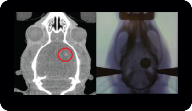
A representative PET image showing [18F]FDOPA uptake in neostriatum in preclinical models.
Huntington’s Disease (HD) is a devastating neurodegenerative condition that causes cognitive, movement and behavioural disturbances, which over time result in progressive disability and eventual death. SAHMRI’s NIF fellows Dr. Muneer Ahamed and Ms. Georgia Williams leading a preliminary feasibility study to validate the use of transgenic preclinical models of HD in PET imaging studies. This study is expected to monitor early-stage (disease onset) and longitudinally evaluate biomarker changes in HD following the slow progression of the HD. This will be first time PET imaging is carried out in this type of HD preclinical model, with the help of NIF’s dedicated large animal PET scanner at SAHMRI.
Growing use of imaging in agriculture and ecology
Imaging is accelerating as an important capability for agricultural and ecological sciences.
Understanding the ecology of coral reefs
Partner:
University of Western Australia
Imaging expertise:
Ms Diana Patalwala
Instrument:
MicroCT
Acknowledgements:
Mr Damian Thomson, CSIRO

Image description:
Micro computerized tomography (μCT) scans of experimental Porites sp. blocks deployed in the lagoon and on the reef slope Ningaloo Reef for 20 months (605 days). Block images show representative scans of two individual blocks (a) pre- and (b) post-deployment in green, and areas of (c) external and (d) internal erosion post-deployment in red.
Porites corals can reveal the past sea conditions by their oxygen isotopes, which reflect the temperature and rainfall of the seawater. This information is useful for studying how the climate and weather patterns have changed over time, and how physical and biological factors influence the distribution and abundance of organisms on the seafloor.
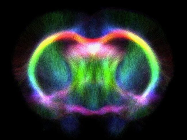
DTI tractogram of a quokka brain: Exploring how marsuipials adapt to different environments
Partner:
Biological Resource Imaging Laboratory (BRIL), UNSW
Imaging expertise:
Dr Andre Bongers
Mr Simone Zanoni
Instrument:
Bruker BioSpec Avance III 94/20 Preclinical MRI
Acknowledgements:
Jyothi Thittamranahalli Kariyappa, Simone Zanoni, Andre Bongers, Lydia Tong, Ken W. S. Ashwell
View publication for more information.


Cultural contributions to materials, engineering and culture
Many varied industrial and research problems— such as chemical processes, materials science, environmental and ecosystems research, security, palaeontology and cultural preservation—are increasingly opening up to the benefits of advanced imaging technologies.
Discovery of a new nasal-emitting trident bat from early Miocene forests in northern Australia
Partner:
Biological Resource Imaging Laboratory (BRIL), UNSW
Instrument:
MILabs U-CT microCT scanner
Acknowledgements:
Suzanne J. Hand, Michael Archer, Anna Gillespie, Troy Myers
View publication for more information.


Understanding equine anatomy: Investigating intestinal muscle structure to prevent rupture
Partner:
Western Sydney University
Instrument:
Diffusion tensor image of a cross-section of equine intestine taken using the 11.7 T MRI at the Biomedical Magnetic Resonance Facility (BMRF) at WSU Campbelltown campus.
Acknowledgements:
Kate Averay1, Denis Verwilghen1, Marianne Keller3, Neil Horadagoda2, Marina Gimeno2
1Camden Equine Centre, University Veterinary Teaching Hospital Camden, Sydney School of Veterinary Science, University of Sydney, Australia
2Pathology Services, University Veterinary Teaching Hospital Camden, Sydney School of Veterinary Science, University of Sydney, Australia
3Sydney School of Veterinary Science, University of Sydney, Australia

Image description:
The research group is looking at equine jejunum rupture and whether there are any anatomical differences in the rupture region. Colour-coding shows the anisotropy of the tissue. The key is on the upper right and shows red is left-right, green is up-down and blue is in and out of the screen. You can clearly see two layers of muscle: the red-green layer wrapping around the intestine and the blue layer running longitudinally.

