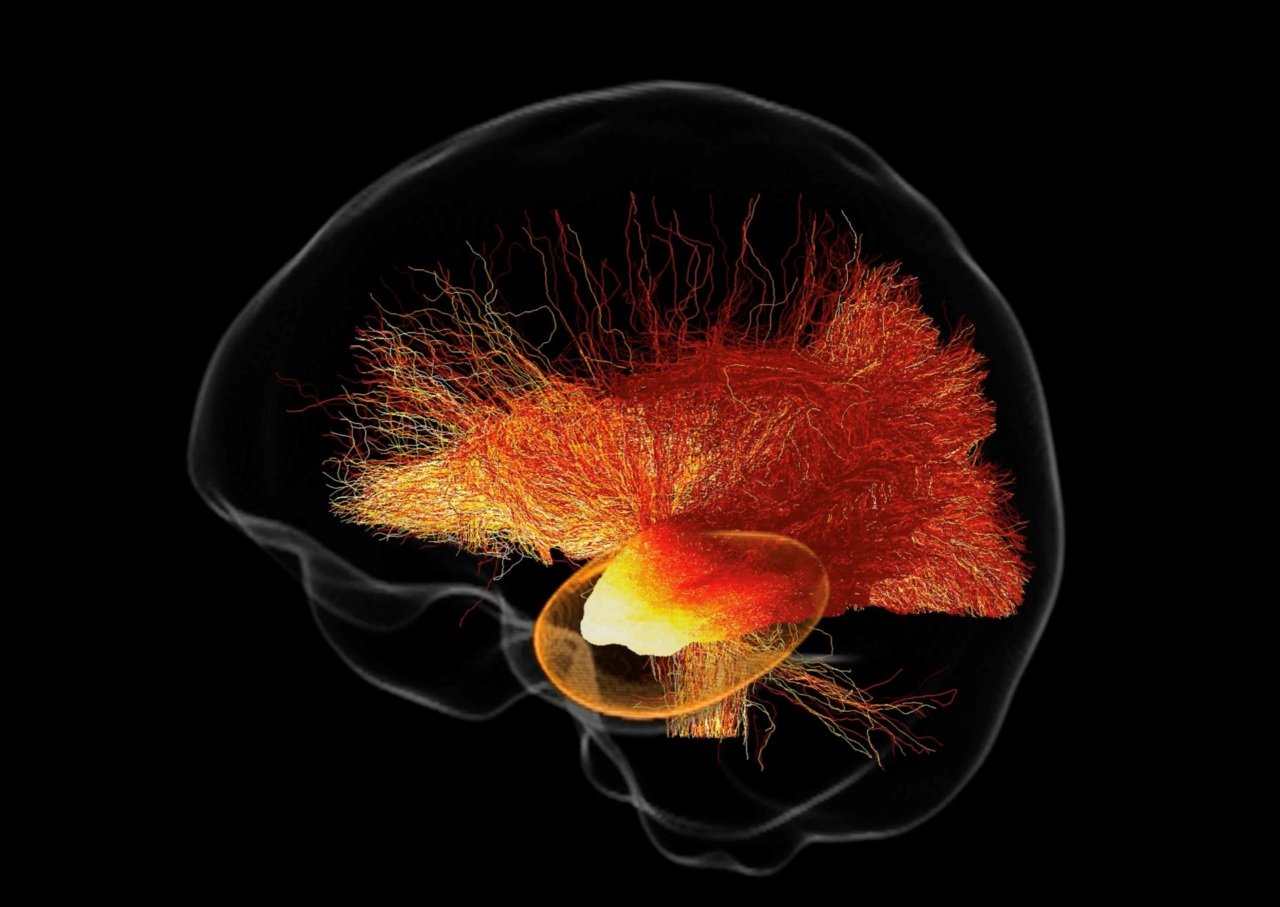
MRtrix3 software maps far-reaching structural effects of surgery in award-winning research Posted on July 1, 2026June 29, 2026 by Sarah Cole
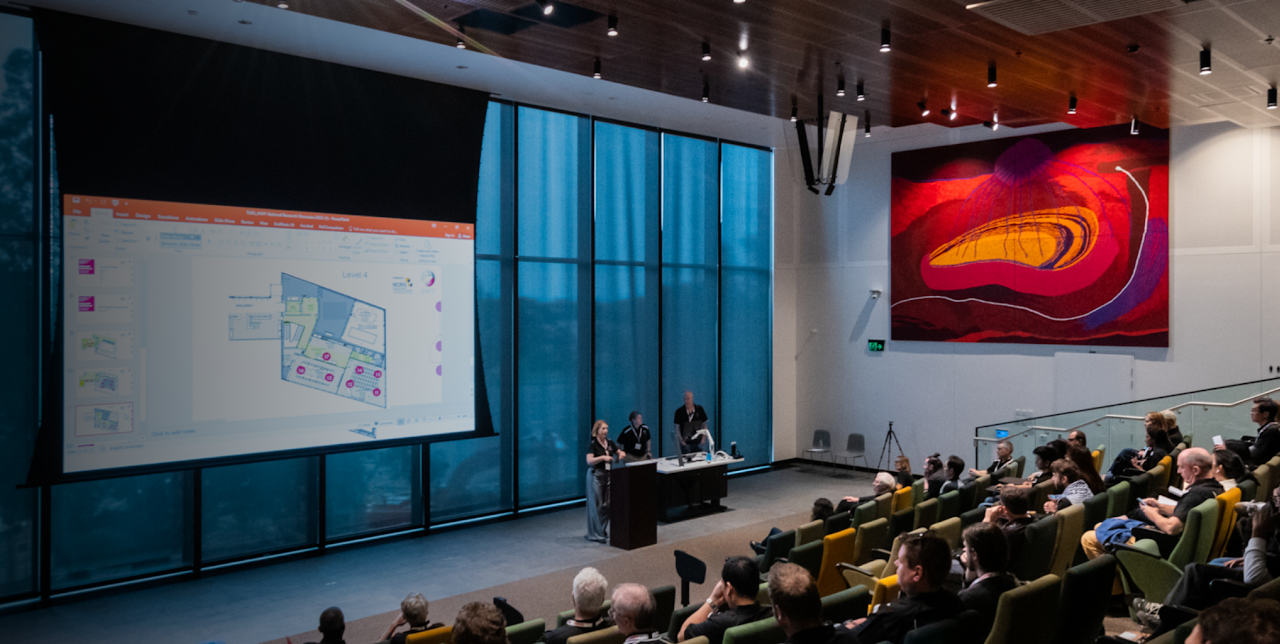
NCRIS: “the best of Australian innovation and dedication to scientific endeavour” Posted on June 30, 2026June 25, 2026 by Sarah Cole
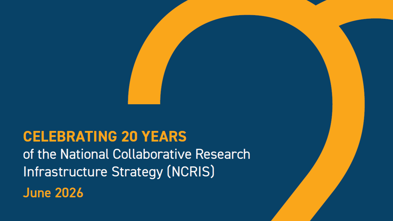
NCRIS and NIF turn 20: decades of building national capability, collaboration and impact Posted on June 28, 2026June 25, 2026 by Sarah Cole
Real-time imaging reveals anti-inflammatory effects of new drug for Parkinson’s disease Posted on June 17, 2026June 17, 2026 by Sarah Cole
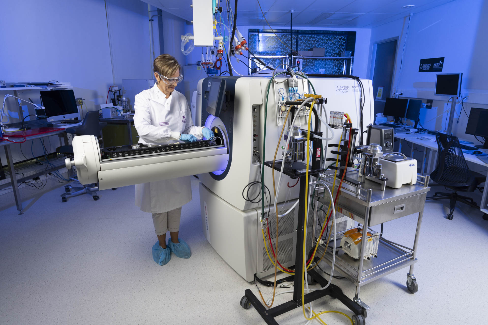
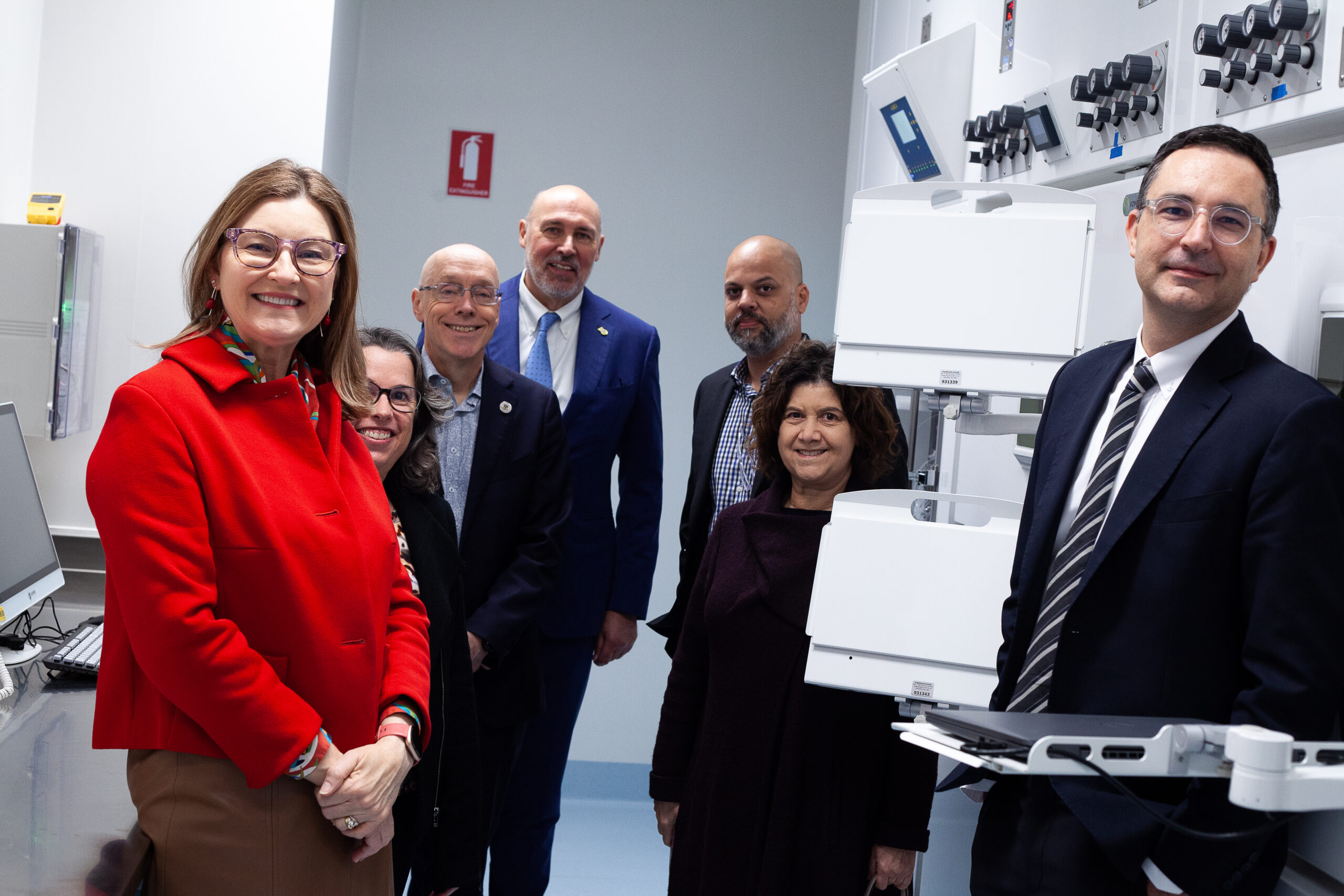
NIF expertise and data use recognised in NCRIS funding announcement Posted on June 2, 2026 by Sarah Cole
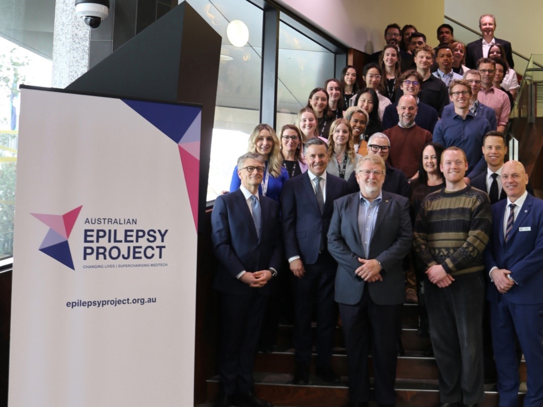
Australian Epilepsy Project awarded $30 million to improve diagnosis and care for Australian patients Posted on May 28, 2026 by Sarah Cole
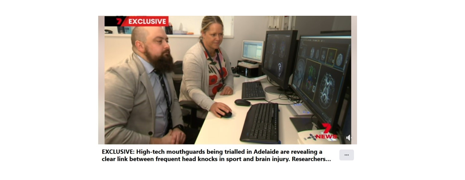
MRIs plus mouthguards aim to transform concussion detection and management in sport Posted on May 12, 2026 by Sarah Cole