Visualisation and Characterisation of Feto-placental Vasculature
Proper vascular development of the human placenta is crucial for meeting the metabolic needs of the developing fetus during pregnancy. Maternal environmental stressors such as malnutrition disrupt the elaboration of the feto-placental vasculature that in turn impacts on placental function and results in reduced fetal growth. The ramifications of this are not only on short-term fetal health but also long-term health outcomes. Indeed, distortion in placental shape and size strongly associate with later adult health outcomes such as cardiovascular disease, obesity and cancer.
Dr Caitlin Wyrwoll of the School of Anatomy, Physiology and Human Biology, at The University of Western Australia is leading a multidisciplinary team that is investigating, in rodent models, how common environmental stressors in pregnancy alter feto-placental vascular morphology and placental function. Ultimately the team will seek to identify potential therapeutic targets to enhance placental vascular development and then apply this to experimental models to assess the outcomes on fetal development and adult health.
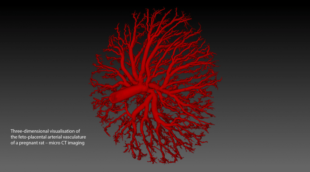
The research project involves collaboration with the Western Australian nodes1 of the National Imaging Facility and the Australian Microscopy and Microanalysis Research Facility to image, visualise and characterise the geometry of the arterial and venous feto-placental vascular trees using high-resolution X-ray microscopy (ZEISS Xradia 520 Versa). Dexamethasone administration during rodent pregnancy is used as a model to simulate excess placental and fetal glucocorticoid exposure (a known effect of prolonged stress). Control and treated rats are anaesthetised at day 22 of gestation and their uteri collected. The feto-placental units are dissected and fetal anaesthesia induced. The individual feto-placental vascular trees are cleared of blood and perfused with Microfil®, a radio opaque polymer casting compound. Each cast is stabilised in PBS in a plastic vial and imaged using a wide field-of-view of ~13.4 mm, a voltage of 50kV, more than 3000 projections through 360 degrees, and an exposure time of 7s. The ZEISS XMReconstructor software is used to reconstruct an image volume (standard parallel beam backprojection algorithm) with voxels of size ~7.0 µm.
The team have completed a preliminary study involving control and dexamethasone-treated rats and both the venous and arterial feto-placental vasculature trees. A visual comparison of treatment to control indicates that for both types of vascular tree, there is reduced branching in the fine vessels and reduced vessel density. A quantitative comparison indicates reduced total vessel length and total vessel volume.
A methodology is currently being developed for a more comprehensive and automatic quantitative assessment of vasculature morphology and geometry. This includes automatic segmentation, filtering, centre-line extraction and characterisation of the vessel tree in terms of its branching characteristics such as its branching hierarchy and angles, vessel diameters and the tortuosities of vessel segments. Furthermore, these vascular tree images are being used in a world-first study to model placental blood flow using computational fluid dynamics.
For more information on this work, contact Dr. Caitlin Wyrwoll (caitlin.wyrwoll@uwa.edu.au).
Collaborators
School of Anatomy, Physiology and Human Biology, The University of Western Australia
School of Mechanical and Chemical Engineering, The University of Western Australia
Centre for Microscopy, Characterisation and Analysis, The University of Western Australia


