PET for Plants
Clinical PET/CT scanners deliver non-invasive, precise anatomical and functional imaging of the human body. Did you know the same systems have been used to investigate plants?
A team of cross-disciplinary researchers at the University of Melbourne, University of Adelaide, and the University of British Columbia have teamed up to demonstrate the utility of clinical PET/CT scanners to image plants.
Using the NCRIS-enabled facilities and expertise at the University of Melbourne NIF Node, the team successfully monitored the temporal dynamics of sodium transport in Barley plants in real-time.
Sodium-ion levels in agricultural soils have a significant impact on crop growth and productivity, hence understanding sodium transport in plants is critical to the agriculture industry.
Although the literature covers a significant number of studies that examine sodium influx, compartmentation, and efflux using 22Na- or 24Na-labeled salts, most of these studies employ destructive methods and lack distribution characteristics in real time, in live plants.
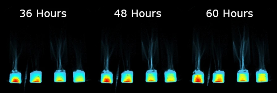
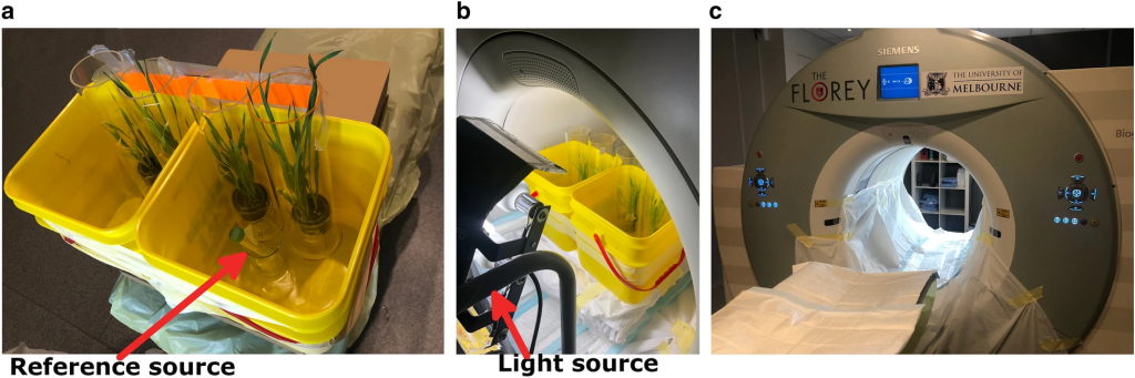
Due to the human-sized 70 cm and 25 cm depth of the Siemens Biograph128 mCT, four whole plants could be simultaneously monitored for up to three days with simulated light and dark (diurnal) conditions.
22Na translocation from roots to shoots was observed, with rates influenced by both nutrient status and channel inhibition.
The PET images showed that plants cultivated in low-nutrient media transported more sodium than plants cultivated in high-nutrient media. In addition, sodium uptake was suppressed in the presence of a cation-channel inhibitor. These results are consistent with cellular studies on ion transport and other radiotracer studies, demonstrating the accuracy of the PET/CT methods described.
In addition, a distinct diurnal pattern of 22Na influx was observed, an effect that was previously only hypothetical. Plants were found to absorb more sodium during the light period and anticipated the change in the light/dark cycle by adjusting the sodium influx rate downward in the dark period.
This work highlights significant potential for plant biology research using existing imaging systems. For example, the study of genetic and treatment effects on crops in saline soil can be observed at the whole-plant level.
One of the technical advantages to using this technology is the CT-based attenuation correction, which is advantageous for better radioactivity quantification in studying plant physiology and CT-based delineation of plant regions of interest (ROIs).
While dedicated plant PET scanners are not accessible to most plant research groups, while clinical PET scanners exist in many research institutions and may be ideal to monitor the movement of ions such as 18F and 11C in whole plants. As demonstrated here, the CT component a clinical PET/CT scanner can be exploited to mark ROIs on PET images to delineate small plant structures for ROI analyses.
This story was contributed by the University of Melbourne NIF Node. For more information, please contact NIF Facility Fellow Mr Rob Williams.

