#IWD2024 | Breaking world records and gender barriers

Ultra-high field MRI is unveiling a picture of endurance, and helping to uncover the physical, social and emotional effects of Associate Professor Donna Urquhart’s incredible Antarctic ultramarathon to empower women and girls.


Harnessing physical and mental resilience to finish an Antarctic ultramarathon

Monash University pain and musculoskeletal researcher, A/Prof Donna Urquhart is pushing past physical limitations, unveiling the intricate connection between mind and body, with a crucial mission to empower girls and women in sports.

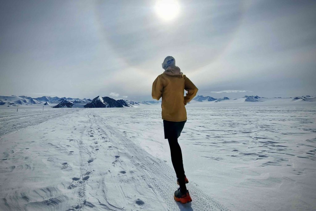
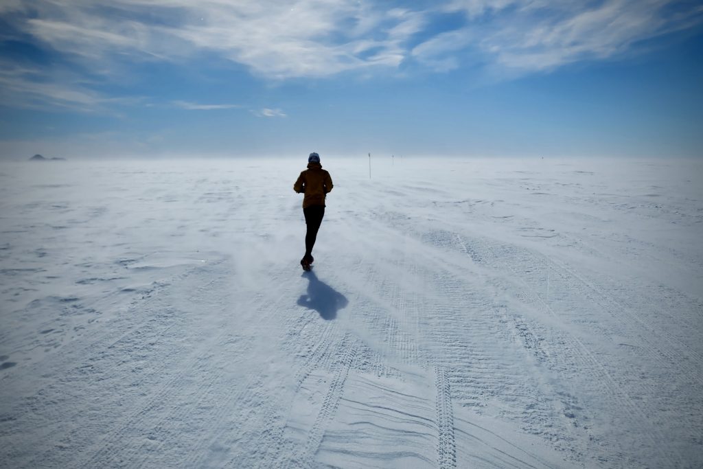
Donna made international headlines in January, after crossing the finish line of the longest ultramarathon in a polar region in history.

As the first woman to conquer this challenge, she ran 1,402km over the space of 28 days at the Union Glacier Camp in Antarctica – the coldest, windiest and driest desert on earth.

Donna said she wanted her run to be more than a record, using the attempt to launch a mission to empower young girls and women to love and discover what’s possible for them in sport, and break down the barriers to participation.

“The alarming statistics show that girls can find sport stressful, and by the age of 15 only 32% are still playing, leaving so few to benefit from the enormous physical, social and emotional aspects of sport,” Donna said.

It took a team of volunteers and two years of training, but Donna said pushing her physical limitations was only half of the battle, emphasising the crucial role of psychological strength in overcoming challenges – with the start of her run posing the most significant hurdle.

“When I first arrived in Antarctica, it was overwhelming. I didn’t know if I could live in a polar region and ultimately run 1,300km,” Donna said. 

She had set a goal to run 50km per day, but by the third day in, the enormity of this challenge hit hard and suddenly, with Donna forging through minus 20-degree 80km per hour winds and limited visibility.

Donna said the physical risks presented by these conditions such as stumbling and falling, hypothermia, polar thigh, frost bite and knee and foot pain, significantly impacted her confidence at the start of the ultramarathon. 

“That first week was so intense – it was a steep learning curve, and I had the realisation that my own initial lack of confidence mirrored the uphill battle women and girls face in sports,” Donna said.

“By pushing my own physical boundaries in this world-record attempt I want to show young girls and women what’s possible when you harness the power of the body and mind.”

“Confidence and self-belief were two emotions that I struggled with early, but this world record run taught me strategies that helped me push on, and I can’t wait to pass this knowledge onto other young women,” Donna said.


Ultra-high field functional imaging is charting new territories in extreme endurance and physical exercise

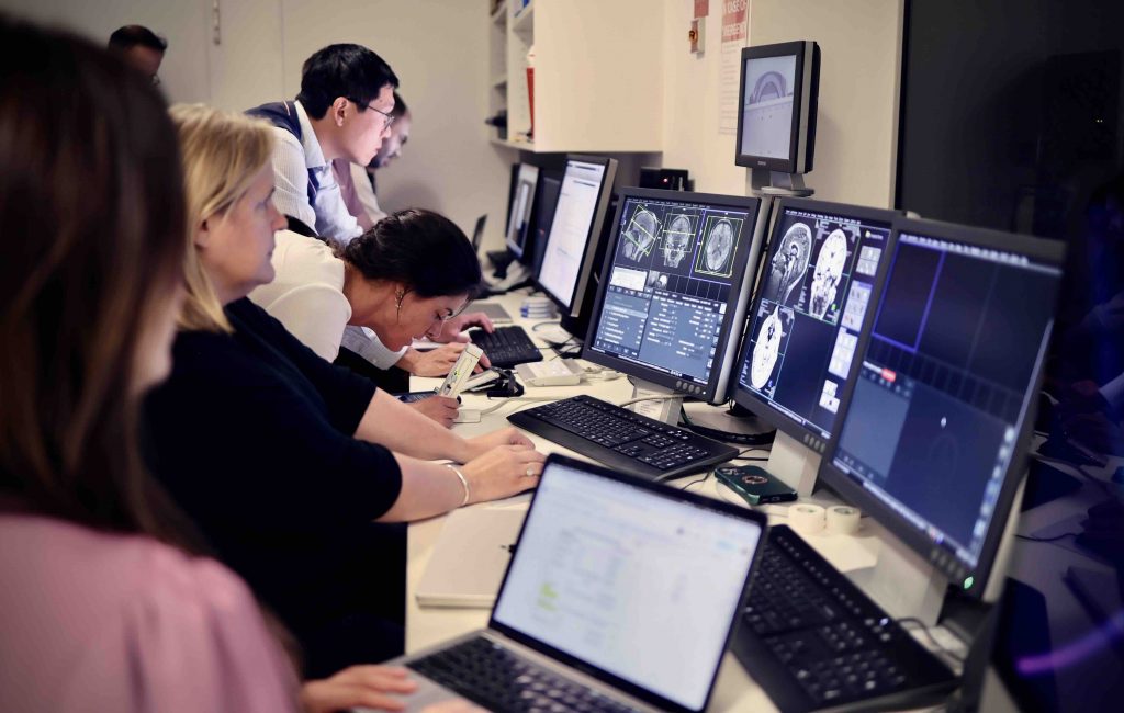
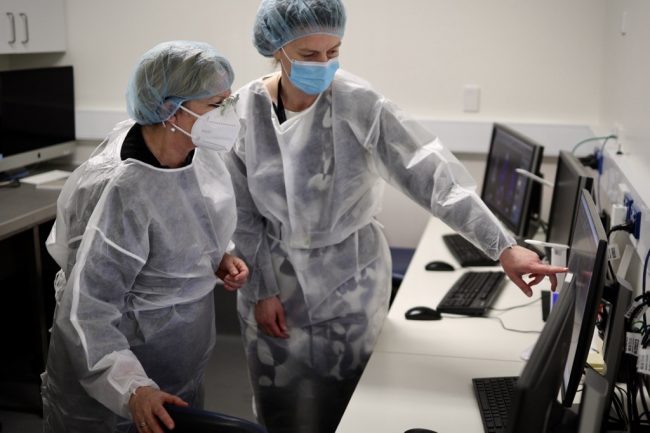
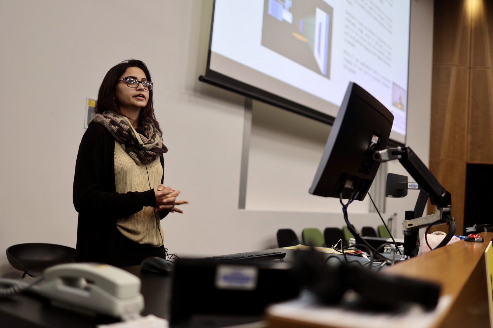
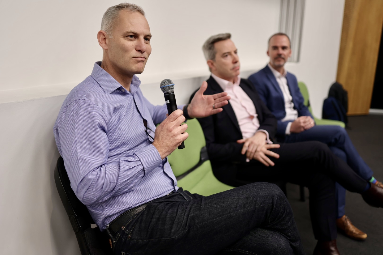
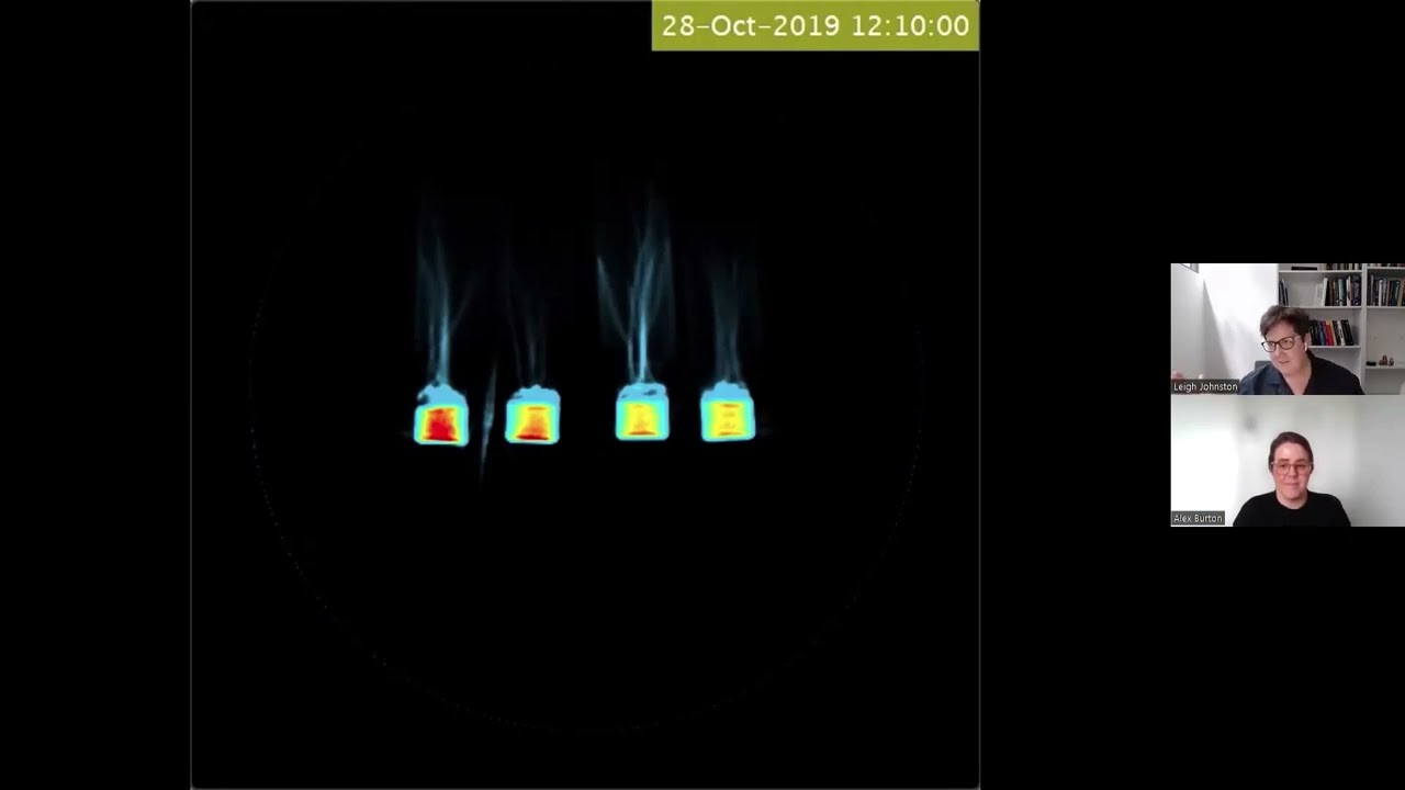
[Pictured: The multi-disciplinary imaging research team, including A/Prof Valentina Lorenzetti, Dr Chao Suo, Dr Hannah Thomson, Ethan Murphy, Anastasia Paloubis, and Arush Arun, with the assistance of University of Melbourne Lead MRI Radiographer Rebecca Glarin, are using imaging to examine the neurological and psychological effects on Donna during this exceptional feat.]

A multi-disciplinary research team is using imaging to examine the neurological and psychological effects on Donna as a result of this exceptional feat, led in collaboration between Donna and A/Prof Valentina Lorenzetti, Lead of the Neuroscience of Addiction and Mental Health Program and Deputy Director of Australian Catholic University’s Healthy Brain and Mind Research Centre, and Dr Karyn Richardson, Wilson Foundation-Brain Park Research Fellow from Monash University.

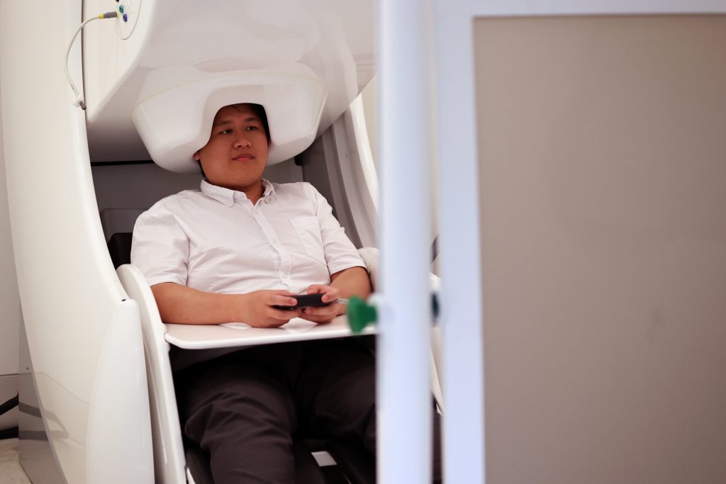
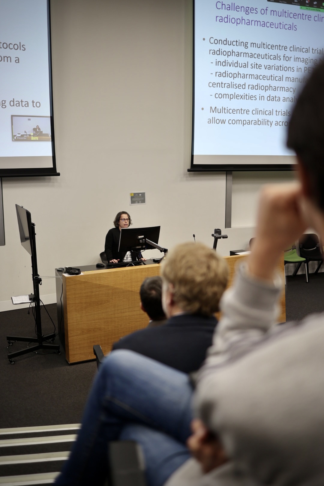
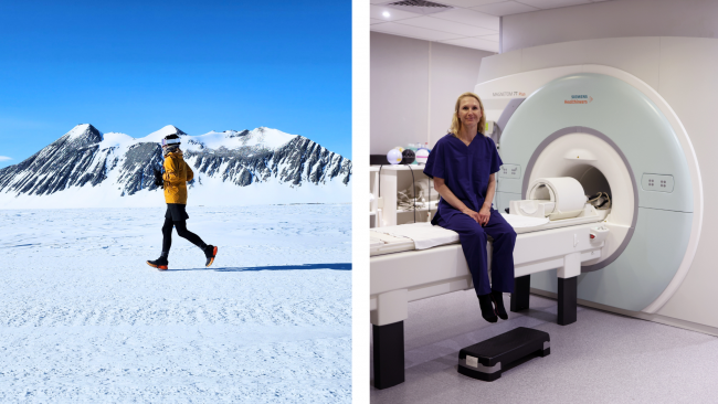
Valentina leads the research team scanning Donna in collaboration with the University of Melbourne Brain Centre Imaging Unit (MBCIU)  on NIF’s ultra-high field Siemens Magnetom 7T MRI.

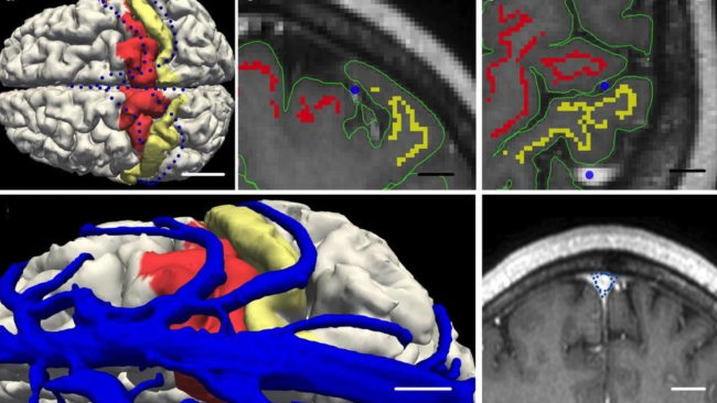
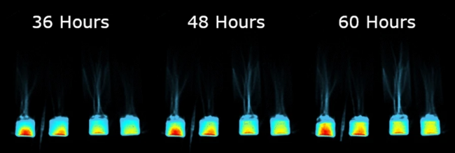
Donna’s exceptional achievement provides an exclusive opportunity for researchers to examine how her brain has changed pre-to-post polar ultramarathon; the 7T MRI can image brain function at a higher resolution than what could be captured on a traditional MRI scan.

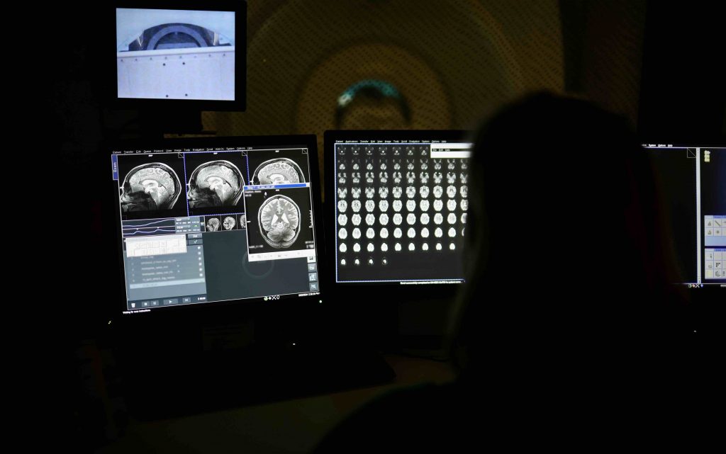
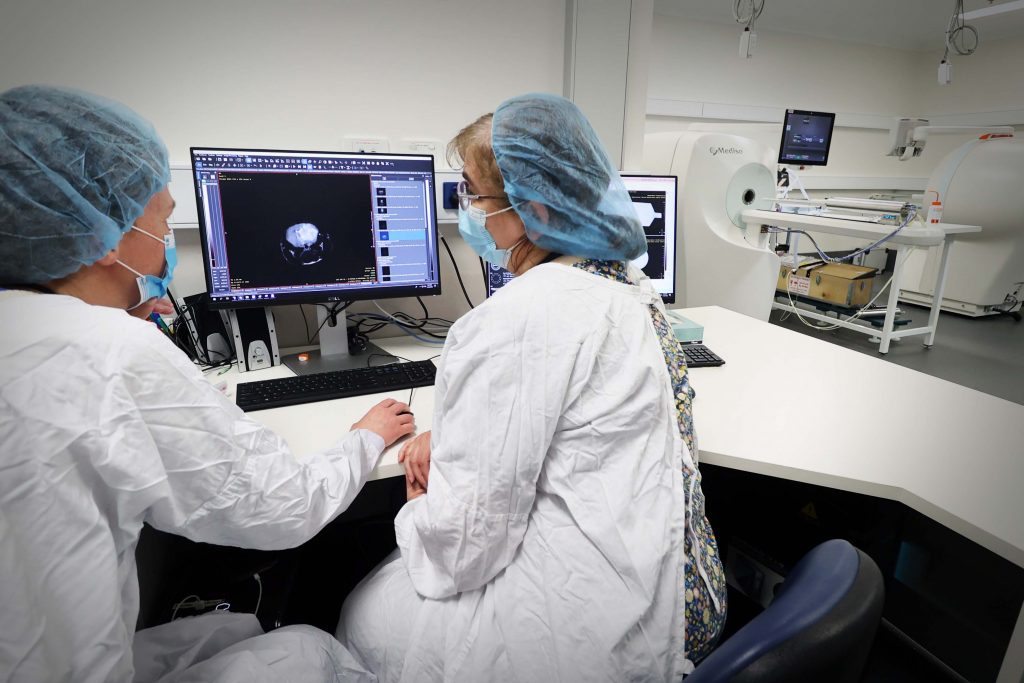
[Pictured: Donna’s brain was scanned with the highest resolution anatomical sequences.]

Donna was scanned twice before the ultramarathon to account for measurement errors, and twice after the ultramarathon (about 2 weeks and 4 weeks), with the highest resolution anatomical sequences, combined with a state-of-the-art functional MRI scan to map the brain networks during rest.

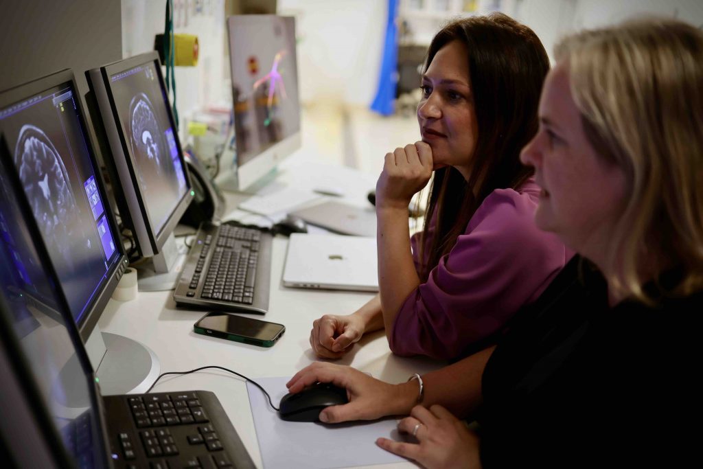
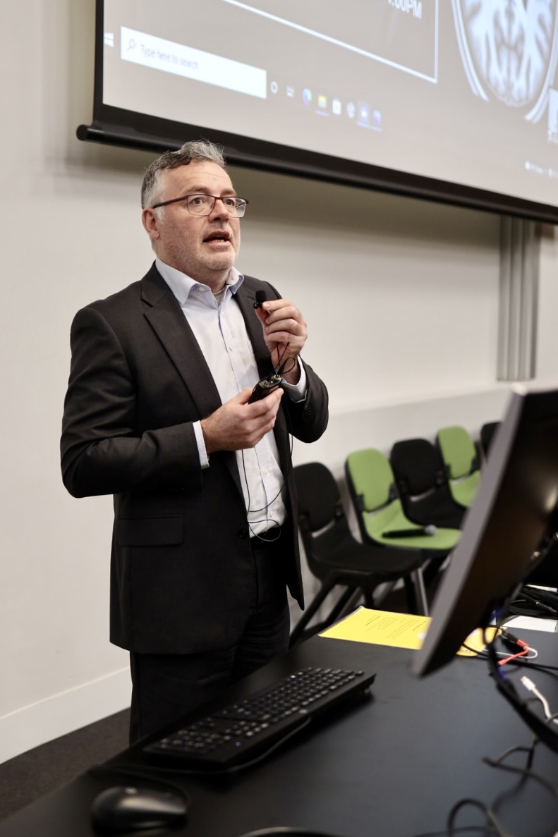
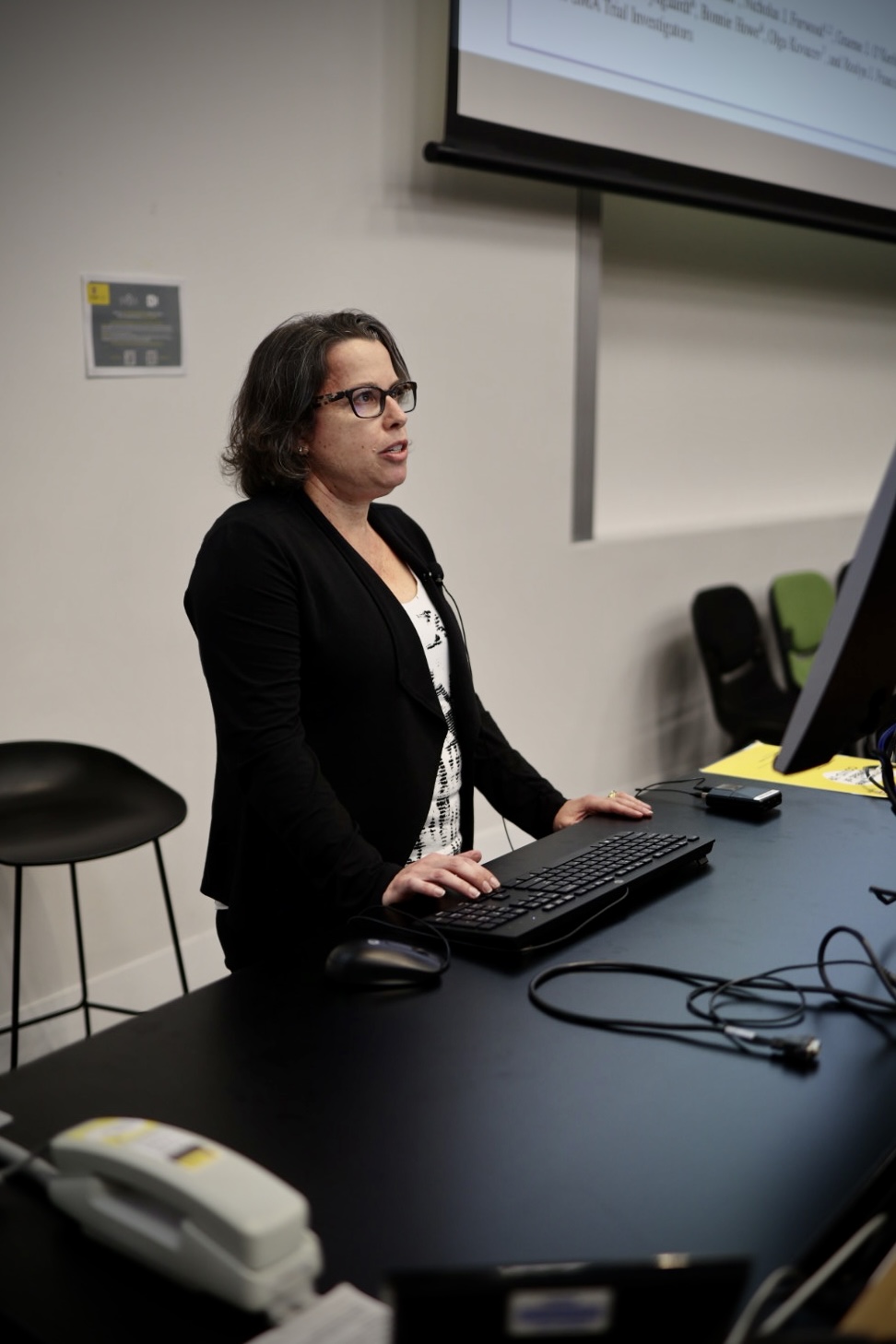
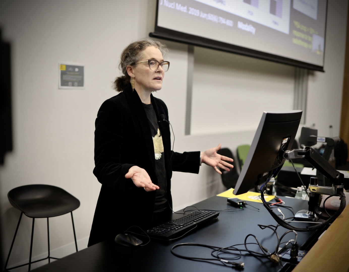
[Pictured: Research imaging team lead A/Prof Valentina Lorenzetti and NIF University of Melbourne Lead MRI Radiographer, Rebecca Glarin]

Valentina said this series of scans can help to provide preliminary evidence on the brain changes that accompany extreme efforts when pushing the body and mind during an ultramarathon in an extreme climate.

“What Donna has done is amazing, and we need examples like this. Everyone knows that exercise is good for you – and now we have the opportunity to showcase the MRI brain images to help convey the message effectively,” Valentina said.


Bridging the mind-body gap: Understanding neurological and psychological insights

Complementary to the imaging study, researcher Dr Karyn Richardson led a mental health and wellbeing assessment to explore how Donna’s mental state changed before and after the event, and how these factors impacted her performance.

Karyn said mental state plays a powerful role in sport, exercise, and endurance and this was a wonderful opportunity to explore these relationships.

Donna completed a variety of questionnaires before and after the ultramarathon to document her general wellbeing, stress and anxiety, coping and confidence.

She also rated a number of factors including pain, confidence, wellbeing, resilience and perceived stress daily in Antarctica, which helped her team to provide additional support during the challenge.

“The data mirrors Donna’s recount of the experience, with a significant period of adjustment over the first week of the run before she acclimatised to the environment and her body adjusted to the daily challenges,” Karyn said.

Karyn said the more confident Donna felt before her run, the quicker she completed the 50km for the day.

“Research has shown that resilience, belief in yourself, and the ability to push through physical challenges are incredibly important for these types of activities, and this is precisely what Donna has done.”

“Donna’s achievement is truly remarkable and her passion to use her experience to inspire young women in sport has blown me away – she is an incredible role model in- and outside of sport,” Karyn said.


Redefining sport for women and girls: Embracing a holistic narrative beyond physicality

The Run Antartica initiative is creating social impact by raising awareness about females’ experience of sport, providing education on a holistic approach to sport, and raising funds for their charity partners, including in disadvantaged communities. Research shows that girls can find sport ‘stressful’ and that 50% stop participating during adolescence (aged 15-19 years). The reasons teenage girls withdraw from sport are multifactorial, however, one of the major contributors is having a lack of confidence.

“This research adds another layer to highlight that sport is about your body, but also about how you train your mind to address the cognitive and emotional components of sport and activity,” Donna said.

“We really see this as a pivotal time in history to evaluate the model we use to look at sport, and change the conversation from focusing on physicality to encompassing a holistic view.”

“We require a comprehensive framework, with a focus on educating young females in the development of cognitive, emotional and social skills in a sporting environment. Our professional athletes are provided with this approach – why aren’t our young people?” she said.

Donna’s mission to empower women and girls in sports aims to disrupt these historical trends which have contributed to dropout rates. In navigating this extraordinary feat, Donna is reshaping the narrative by which we view community-based sport in Australia, empowering the next generation of female athletes to develop a love of sport, and experience the important health and education benefits.

For more information about Donna’s mission or to donate, visit https://www.runantarctica.com/


This research was conducted using NIF expertise and 7T MRI facilities at the Melbourne Brain Centre Imaging Unit at the University of Melbourne.


Victorian collaboration raises over $50m investment in critical imaging capabilities

[Image: La Trobe University – Olivia Newton-John Cancer Research Institute NIF Fellow, Dr Ingrid Burvenich with Minister Tierney]

The Victorian Government has invested $14.83m in National Imaging Facility’s (NIF) research infrastructure in Victoria, in partnership with the Victorian Biomedical Imaging Capability (VBIC), equating to a boost of just over $50m through collaborative co-investment. 

[Image: Victorian Minister for Higher Education, the Hon Gayle Tierney MP]

Victorian Minister for Higher Education, the Hon Gayle Tierney MP visited the Olivia Newton-John Cancer Research Institute on Tuesday, to highlight the impact of the collaboration and the State Government’s investment of through the Victorian Higher Education State Investment Fund (VHESIF) initiative.  

“Collaborative projects such as this demonstrate how our government is supporting higher education and industry to become international leaders in their field,” Minister Tierney said.

[Image: Victorian Minister for Higher Education, the Hon Gayle Tierney MP toured the facilities at the Olivia Newton-John Cancer Research Institute and the Austin hospital]

The funding is supporting the upgrade and expansion of imaging capabilities across NIF’s research facilities in Victoria, including The Florey, La Trobe University and the Olivia Newton-John Cancer Research Institute, Monash University, Swinburne University of Technology, and University of Melbourne in partnership with Peter MacCallum Cancer Centre and the Austin Hospital.  

Critical medical research in areas of national priority such as dementia cancer and epilepsy, as well as agriculture research will be enabled by the co-investment, which includes $26.7m from NIF through the Australian Government’s National Collaborative Research Infrastructure Strategy (NCRIS) funding scheme. 

Infrastructure funded under the collaboration includes human magnetic resonance imaging (MRI) capabilities at Swinburne University of Technology and The Florey, where state-of-the-art high-intensity focused ultrasound will support the development of new treatments for essential tremor and tremor-dominant Parkinson’s disease. 

In addition to this, the University of Melbourne upgraded their ultra-high-field 7T MRI (one of only two in Australia), and acquired a new human PET-CT. 

Preclinical capabilities including PET/MRI and PET/CT to support important drug discovery and testing have been installed at the Olivia Newton-John Cancer Research Institute/La Trobe University and Monash University. 

[Image: NIF preclinical capabilities at the Olivia Newton-John Cancer Research Institute]

A new research cyclotron at Monash (the Australian Precision Radiopharmaceutical Facility APRF) will enable the production of radioisotopes under GMP standards, and enhance Australia’s sovereign capability to produce therapeutics and diagnostics. Complementary to this, radiochemistry hotcell infrastructure upgrades at Monash, the Austin Hospital, and Peter MacCallum Cancer Centre will support the design and development of novel cancer treatments.  

[NIF radiochemistry capabilities at the Austin hospital/Olivia Newton-John Cancer Research Institute]

The funding also enables upgrades to the magnetoencephalography (MEG) at Swinburne, one of only two systems in Australia, supporting the study of brain function. 

[Image: NIF MEG at Swinburne University of Technology]

Development of specialised plant imaging capabilities at the University of Melbourne will underpin research into the effect of climate change on crops and soil, and strategies and applications for agricultural improvements to support Australia’s standing as a world leader in food and beverage production. 

NIF Chief Executive Officer, Prof Wojtek Goscinski said the co-contributed investment underpins transformational initiatives in a number of national priority areas including precision medicine, molecular imaging, drug discovery, diagnostics and plant soil imaging. 

“It’s a privilege for NIF to partner with the Victorian Government and VBIC to support Australia’s strategic science and research priorities” Prof Goscinski said. 

“These capabilities will support Australia as a world-leader in applying advanced imaging technology, resulting in better healthcare, better products, and important discoveries.”

Read the Victorian Government’s announcement here. 

#ImagingTheFuture Week: Enabling breakthroughs in biomedical science and technology

Chan Zuckerberg Initiative’s (CZI) Imaging the Future Week puts a spotlight on the importance of imaging science in biomedicine, and the value of the global imaging community in translating health research.

Imaging is unlocking solutions to the world’s biggest challenges across commercial, clinical and research fields and has helped innovate in bioengineering, biology, medical technology and science, pharmaceutical and non-pharmaceutical therapies.

National Imaging Facility (NIF) supports the Imaging the Future Week initiative, and the 2023 event is focused on highlighting advances in technology and the impact this has on our understanding of health and disease.

As we continue to meet the evolving needs of modern research, NIF is accelerating new technology, enabling experts to develop protocols, tools, imaging data, and the application of imaging to solve complex problems – scroll on to find out more.


Better evidence for decision-making in health

Advanced imaging methods and analysis provide critical evidence for decision-making across all aspects of health and clinical science to keep Australia healthy.


Australia’s largest investment in molecular imaging
Australia’s first open access research Total Body Positron Emission Tomography scanner is NIF’s largest investment to date, and it will deliver a transformative understanding of complex health problems. Next-generation molecular imaging and radiopharmaceuticals are revolutionising how we see biological processes, paving the way for better diagnosis and treatment of chronic, systemic adult and childhood diseases. The instrument will produce high quality data at lower doses of radiation. It can be used to capture information from all body organs simultaneously to build a better picture of complex processes such as ageing, metabolism, brain signalling, behaviour, cognition and drug interactions.

Multidisciplinary collaboration to improve epilepsy outcomes
MRI imaging technology, AI, machine learning and data analysis are helping improve the lives of 150,000 Australians with epilepsy. The Australian Epilepsy Project will combine neuroimaging with cognitive and genetic data, and integrate them using AI, to develop predictive tools that will guide diagnosis and highlight opportunities for precision treatment. Expertise from the Florey Institute of Neuroscience and Mental Health, the University of Melbourne, Monash University and Austin Health drives the project, aiming to reduce seizure frequency and the risk of injury or death.


Better health for the young and older Australians

Imaging studies that look at conditions in younger and older Australians are essential for understanding and promoting healthy development and ageing.


Understanding the development of cerebral palsy
NIF is contributing to valuable data assets, including the first collection to show the way that muscles grow in children with cerebral palsy. The MUGgLE Study is the first longitudinal study comparing muscle growth in the development of children with cerebral palsy and typically developing children. The study is a partnership between Neuroscience Research Australia, the University of NSW and the Cerebral Palsy Alliance Research Institute. Imaging is being used to study muscle tightening and shortening as it happens, with high-resolution measurements of the architecture of whole muscles, giving researchers detailed, anatomically accurate, three-dimensional reconstructions to understand disordered muscle growth. The project has included the development of imaging methods and algorithms to be able to study this, adapting the acquisition protocols as well as the imaging analysis techniques to accommodate measurement of the specific features of muscles.

Brain-computer interface restoring independence after paralysis
An implant the size of a paperclip is allowing people who are paralysed to operate technological devices using their thoughts without open brain surgery. NIF expertise and the 7T MRI at the University of Melbourne enabled early developments of the device which can translate brain signals from the inside of a blood vessel into commands on a computer.

The Synchron Stentrode is a world first brain-computer interface designed to restore functional independence in patients with paralysing conditions like ALS. The device was named one of TIME Magazine’s best inventions of 2021, and is currently undergoing expanded human clinical trials in preparation for submission to the FDA.


Equitable regional and rural health

Crucial to societal equity and research quality, delivering a geographically distributed network of advanced imaging to support research and personalised medicine, and taking part in medical trials, is a major national challenge.


Bringing health equity to regional and rural Australia
NIF is deploying four low-field portable MRI scanners to remote and regional sites to help researchers apply this affordable imaging technology in rural areas. The national mobile magnetic resonance (MR) network will be the first project of its kind world-wide and is a collaboration with partners including Monash University, University of Queensland, South Australian Health and Medical Research Institute (SAHMRI), the Alfred Hospital, Royal Perth Hospital, University of Western Australia and MedTech company, Hyperfine. These portable scanners will be used to understand how this fast-developing technology can help diagnose stroke, traumatic brain injury, and other conditions after testing in research laboratories at NIF nodes to build the usability of low-field MR, including developing techniques to maximise data quality and improve image processing.

Imaging mobilises ground-breaking field ventilator for deployment in the COVID-19 crisis
NIF provided critical support in preclinical testing to mobilise the now commercialised ventilator, 4DMedical ‘XV technology’ at the LARIF multipurpose fluoroscopy laboratory. A team of Australian collaborators, including biomedical company 4DMedical and University of Adelaide scientists created the ground-breaking, simple to use ‘field ventilator’ that can be locally produced at a low cost from easily acquired parts. It was developed in response to the global COVID-19 crisis, which identified potential shortages in essential medical equipment.

NIF collaborators and users funded and recognised among top researchers

NIF is enabling research that has attracted national recognition and funding, with collaborators listed among last year’s top medical researchers in The Australian, and as recipients of competitive Federal Government support. 

Top researchers in health and medical sciences 

In case you missed it, NIF collaborators have been listed in The Australian among the top researchers in 2022, covering expertise in areas including neurodegenerative disease, neuropsychiatry, theranostics and nuclear medicine. 

The list features researchers in 250 fields of academic endeavour, spanning the sciences, the social sciences through to the humanities and the arts. It aims to shine a light on what they do and the benefits they bring to the country. 

The weight of a researcher’s contribution was judged through citations from other researchers in their publications. 

NIF collaborators and users included: 

  • University of Melbourne Professor Christopher Rowe, for research in gerontology and geriatric medicine, covering dementia research, patient care and leadership in molecular imaging research at Austin Health and at the Florey Department of Neuroscience and Mental Health
  • University of NSW Professor Perminder Sachdev, recognised for work in neurology, with a focus on conditions such as drug-induced movement disorders, Tourette syndrome, secondary psychosis, healthy brain ageing and dementia, in particular Alzheimer’s disease and vascular dementia, and research into new interventions such as brain stimulation for neuropsychiatric disorders
  • Professor Louise Emmett, a keynote speaker at National Imaging Facility (NIF) Scientific Symposium, listed for work in nuclear medicine, radiotherapy and molecular imaging at the University of NSW and St Vincent’s Hospital in Sydney.

Australian Research Council support for Centre of Excellence 

Professor Michelle Watt from the University of Melbourne is part of a collaboration that has secured $35 million in funding as part of a new Australian Research Council Centre of Excellence (CoE) in Plants for Space. 

The centre aims to create on-demand, zero-waste, high-efficiency plants for better sustainability for space habitation, with the team covering aspects such as process and systems engineering, law, policy and psychology. 

It will make use of imaging equipment at the University of Melbourne Brain Imaging Centre Unit, where NIF support and work from NIF Node Director Professor Leigh Johnston and NIF Fellow Dr Edward Green have contributed to a Functional Plant Imaging Capability, in collaboration with Professor Watt. 

Professor Watt combines imaging and sensor technologies with modelling to understand how roots function in increasingly dynamic climates – and how to increase productivity and decrease environmental impacts. 

ISMRM and ISMRT ANZ Chapters’ Annual Meetings shine a light on national imaging expertise and infrastructure

[Image: Presentation award winners at ISMRM ANZ, Honours student, Arunan Srirengan, Dr Ed Green, Dr Gwen Schroyen and Dr Myrte Strik. Photo credit: Dr Adam Clemente]

The Australian and New Zealand Chapters of the International Society for Magnetic Resonance in Medicine (ISMRM) and the International Society for Magnetic Resonance Radiographers and Technologists (ISMRT) held their Annual Meetings in Sydney last month, highlighting the work of leading national researchers and clinicians, including members of the NIF network.

NIF enables coordinated open access to magnetic resonance expertise and infrastructure to support leading national researchers and clinicians, and proudly supported the events.

ISMRM ANZ Joint Chapter Annual Meeting 9-10 Nov

ISMRM ANZ hosted sessions on revolutionising MRI technology, advances in neuroimaging, and clinical applications of advanced MRI, in addition to keynote speakers neurologist and leader in stroke medicine, Prof Mark Parsons and Director of the Institute of Medical Physics at the University of Sydney, Prof Annette Haworth.

Dr Zhaolin Chen was a key speaker in the Revolutionising MRI technology session, presenting the NIF Point-of-Care project, a collaboration between NIF, Australian hospitals, and US medical device manufacturer, Hyperfine, to build the usability of low-field MRI and bring critical imaging to remote Australia and deploy imaging in challenging clinical environments such as COVID wards.

[Image: Dr Zhaolin Chen presenting the NIF Point-of-Care Magnetic Resonance project at ISMRM ANZ]

A number of other NIF users spoke at ISMRM ANZ, including:

  • Rebecca Glarin from the University of Melbourne, presenting findings from her PhD on ‘Optimising functional brainstem imaging of sympathetic drive with ultra-high field MRI’.
  • Dr Shahrzad Moinian from the University of Queensland Centre for Advanced Imaging, presenting ‘In vivo microstructural border delineation between areas of the human cerebral cortex using magnetic resonance fingerprinting (MRF) residuals’.
  • Honours student Arunan Srirengan presenting ‘Early identification of cerebral small vessel disease in obstructive sleep apnoea patients using magnetic resonance spectroscopy: a pilot study’, featuring data obtained on the NIF 3T MRI at NeuRA. This session was awarded second prize in the oral presentation awards.
  • Dr Myrte Strik from the University of Melbourne, presenting ‘Altered network topology in patients with visual snow syndrome: a resting-state 7 Tesla MRI study’, winning the award for best Early Career Researcher Data Blitz presentation.

[Image: Dr Shahrzad Moinian from the University of Queensland Centre for Advanced Imaging. Photo credit: Dr Adam Clemente]

Congratulations to University of Melbourne NIF Fellow, Prof Brad Moffatt as ANZ ISMRM Chapter President on the success of the 2022 meeting hosted at UNSW.

ISMRT ANZ Joint Chapter Annual Meeting 12-13 Nov

The ISMRT ANZ 2022 joint meeting program theme was MRI: Past, Present and Future, and featured a range of internationally renowned speakers demonstrating future technologies and cutting-edge imaging techniques.

Keynote presenters included Medical physicist and human brain imaging academic researcher Dr Samantha Holdsworth, Chief of the Quantitative Medical Imaging Laboratory, USA National Institute of Biomedical Imaging and Bioengineering, Dr Carlo Pierpaoli, and founding member of the Society of Cardiovascular Magnetic Resonance and Principal Investigator for the Cardiac Atlas Project, Prof Alistair Young.

NIF Senior Manager and Senior Research Scientist – National Magnetic Resonance Capability, Dr Shawna Farquharson was a key speaker at the Diffusion Weighted Imaging (DWI) Forum, presenting on ‘DWI: Principles and practical applications’.

[Image: Dr Shawna Farquharson, National Imaging Facility]

NIF users showcased at ISMRT ANZ included:

  • Prof Lynne Bilston from NeuRA, presenting ‘Brain Elastography’.
  • Sarah Daniel from the University of Queensland Centre for Advanced Imaging, presenting ‘Image quality enhancement using deep learning for in vivo human kidney MRI’.

[Image: Ms Sarah Daniel from the University of Queensland Centre for Advanced Imaging]

Congratulations to all presenters at ISMRM and ISMRT ANZ.

Victorian imaging network meets to map out innovative future

[Pictured: VBIC Annual Network Meeting guest speakers Prof Lindy Fitzgerald and Prof Amy Brodtmann] 

Innovation, industry partnerships and commercialisation will be among the topics discussed at a meeting bringing together NIF’s capabilities from around Victoria on November 24. 

The annual Victorian Biomedical Imaging Capability (VBIC) network meeting will attract researchers, clinicians and industry partners to Monash Biomedical Imaging in Melbourne. 

Participants will represent NIF nodes at the University of Melbourne, Monash, Swinburne, the Florey, Olivia Newton John Cancer Research Institute and La Trobe University. 

Neurologist Prof Amy Brodtmann will draw on her interests in imaging, stroke and dementia to present research findings from the Cognitive Health Initiative at Monash and Alfred Health  

Guest speaker Prof Melinda Fitzgerald from Curtin University and the Perron Institute will present on a national initiative she heads as CEO, called Connectivity, the Mission for Traumatic Brain Injury.  The initiative includes use of MRI and clinical biomarkers in a national trial to improve the diagnosis and prognosis of traumatic brain injury. 

Sessions at the meeting will also cover the work of imaging experts in research programs employing ultra-high field MRI, CT and PET; as well as presentations from experts in industry partnering and commercialisation. 

NIF Chief Executive Officer Prof Wojtek Goscinski said the meeting would provide a platform for early career researchers and emerging leaders, a showcase for new-generation imaging, and opportunities for important in-person networking. 

It would also enable discussion on the NIF Imaging Roadmap, including supporting innovation and ensuring Australia’s international comparative advantage, Prof Goscinski said. 

The roadmap will add to the substantial impact and jobs that VBIC and NIF have already delivered, with a recent report estimating more than $350 million in economic activity for Victoria. 

VBIC nodes have grown to employ more than 150 FTE imaging staff, and partner with more than 90 organisations, including Austin Health, CSIRO, Melbourne Health, Mental Health Research Institute and the Peter MacCallum Cancer Centre. 

Capital investments have reached $37 million, providing access to human and preclinical MRI and PET-CT, preclinical DEXA scanners and confocal endomicroscopy, as well as magnetoencephalography and nuclear scintigraphy. 

A massive $235 million in major grants has been secured, enabling new research projects in cancer, infection and inflammation, brain function, epilepsy, dementia and even long-term aspirin use. 

Click here for more information about the VBIC network meeting and full programme.

Inaugural NIF Scientific Symposium kicks off #NationalScienceWeek

Leading researchers, clinicians and industry attended the inaugural National Imaging Facility (NIF) Scientific Symposium on 12 August.

The event kicked off National Science Week for NIF, highlighting the critical role of collaboration in translating research challenges to benefit industry and keep Australians healthy, with the theme ‘National partnerships for innovation and impact’.

NIF CEO Prof Wojtek Goscinski said the Symposium was an excellent opportunity to highlight ground-breaking work from Australia’s world-class imaging community.

“It was a privilege to host experts from across Australia, including keynote speakers Prof Graeme Jackson, Prof Louise Emmet and Prof Gemma Figtree, whose work is at the leading edge of imaging globally,” Prof Goscinski said.

“I’d also like to extend my thanks to the presenters who delivered an excellent Technology Showcase session, and Health and Medical Translational Challenges session.

“A particular highlight was hearing from our industry partners, including Telix Pharmaceuticals, Clarity Pharmaceuticals, Cochlear and Nyrada, who discussed the way they engage with national imaging research infrastructure.

“NIF is privileged to have a strong network of world-leading expertise at our fingertips and it was an honour to bring some of these people together to present their work and share ideas at the 2022 Symposium,” he said.

Keynote presentations of the Symposium included:

  • ‘The Australian Epilepsy Project’, Prof Graeme Jackson
  • From mouse to Medicare: the PSMA story in Australia’, Prof Louise Emmett
  • Coronary artery imaging to inform the next Frontier of heart attack prevention’, Prof Gemma Figtree

The Technology Showcase session highlighted NIF’s latest capabilities, including tools for processing and interpreting data, and applications of imaging to solve complex problems, including:

  • ‘Ultra-high field magnetic resonance imaging’, Prof Leigh Johnston and Prof Markus Barth
  • ‘Bringing imaging to rural Australia with a national network of low field mobile MR scanners’, Dr Zhaolin Chen
  • ‘Australian Imaging Service: The national platform for trusted data management and analysis’, Dr Ryan Sullivan
  • ‘Magnetic Particle Imaging’, Dr Andre Bongers
  • An insight into MicroCT imaging: recent advances, applications and impact on research and innovation’, Ms Diana Patalwala
  • Preclinical Research: The Crucial Step in Medical Advancements’, Dr Chris Christou

The Health and Medical Translation Challenges session provided an opportunity for attendees to hear from clinicians and researchers about their journey to making translational impact, including:

  • Neuroimaging in clinical trials: Perspectives of a clinician-researcher’, A/Prof Sylvia Gustin
  • The Australasian Radiopharmaceutical Trials network (ARTnet)’, A/Prof Ros Francis

The Industry Discussion Panel opened up conversation on how imaging accelerates and underpins innovation and future opportunities, with speakers:

  • Dr David Cade, Chief Executive Officer, Telix Pharmaceuticals Asia Pacific
  • Dr Matt Harris, Chief Scientific Officer, Clarity Pharmaceuticals
  • Dr Zachary Smith, Director, Algorithms and Applications, Cochlear
  • Dr Jasneet Parmar, Neuroscience Researcher, Nyrada Inc

#WorldHealthDay: Imaging unlocking research to keep people healthy

#WorldHealthDay: As Australia’s advanced imaging network, we’re focused on addressing national science and research priorities to help keep people healthy. Our expertise, equipment and services are critical to Australia’s ability to translate health discoveries, undertake clinical trials and commercialise medical products.

The importance of protecting Australians from health threats is critical, as is Australia’s strong medical research capability and reputation for quality and standards.

The National Imaging Facility is unlocking solutions to the world’s biggest imaging challenges across commercial, clinical and research fields. We have helped Australians innovate in fields such as bioengineering, clinical science, biology, medical technology, pharmaceutical and non-pharmaceutical therapies.

Thousands of scientists, doctors, and professionals across hundreds of Australian institutions, companies and research organisations use our work to help answer their medical research questions. We also work with engaged volunteers and patients who make a valuable contribution to health and discovery by being part of research.

We’ve included some examples of the medical projects we’re proud to have partnered with to keep people healthy below:

Dr Ciara Duffy from Western Australia’s Harry Perkins Institute of Medical Research imaging the investigation of honeybee venom to treat breast cancer cells at the University of Western Australia’s Centre for Microscopy, Characterisation and Analysis in collaboration with Microscopy Australia

Associate Professor David Parsons and Dr Martin Donnelly performing preclinical testing of a ground-breaking and simple to use ‘field ventilator’ that can be locally produced at a low cost from easily acquired parts at SAHMRI, in collaboration with 4DMedical, and the University of Adelaide

Supporting Australian trials of Biogen’s Aducanumab (Aduhelm), the first disease modifying therapy for Alzheimer’s disease approved by the United States Food and Drug Administration (FDA) with the University of Melbourne, Herston Imaging Research Facility, the Hunter Medical Research Institute, Australian Imaging Biomarkers and Lifestyle Study of Ageing at The Florey Institute of Neuroscience and Mental Health and Austin Health

#IWD | PODCAST: In conversation with Professor Leigh Johnston

The United Nations International Women’s Day (IWD) is an opportunity to reflect on progress made, call for change and celebrate acts of courage and determination by ordinary women who have played an extraordinary role in their communities. 

The National Imaging Facility’s (NIF) mission is to make cutting-edge imaging capabilities accessible to Australian researchers, and we envision a society that provides equal opportunity for people of all genders to learn, work and engage in science, technology, engineering, and mathematics (STEM). 

Today we highlight the exceptional work of women leading the way in these fields and thank them for the impacts of their life-changing research. 


Professor Leigh Johnston is the NIF Node Director at the Melbourne Brain Centre Imaging Unit within the Department of Medicine and Radiology, and is also the Head of the Department of Biomedical Engineering, at the University of Melbourne.  

Professor Johnston started out as an Engineer, but a unique skill set, passion for collaboration, and drive to pursue challenges have led her to have a great impact on the imaging community. 

Listen to our podcast here.


Professor Johnston talks us through some standout imaging projects: 

The baby mummy 

PET for Plants

Clinical PET/CT scanners deliver non-invasive, precise anatomical and functional imaging of the human body. Did you know the same systems have been used to investigate plants?

A team of cross-disciplinary researchers at the University of Melbourne, University of Adelaide, and the University of British Columbia have teamed up to demonstrate the utility of clinical PET/CT scanners to image plants.

Read More
Privacy Settings
Youtube
Vimeo
Google Maps