NIF is enabling the Muscle Growth in the Lower Extremity (MUGgLE) Study, the first longitudinal study comparing muscle growth in children with cerebral palsy and typically developing children.
The project is a collaboration between Neuroscience Research Australia (NeuRA), the University of NSW (UNSW) and the Cerebral Palsy Alliance Research Institute.
The National Health and Medical Research Council-funded study is using magnetic resonance imaging (MRI) to compare muscle growth between typically developing children and children with cerebral palsy, using high-resolution measurements of the architecture of whole muscles.
Researcher Dr Bart Bolsterlee said the longitudinal study will see the lower legs of over 300 children scanned, between the ages of 0-3 months and 5-14 years.
“They will be scanned three times, with one-and-a-half years in between scans. We analyse the images to look at the individual muscles and how they change in size and structure over time,” Dr Bolsterlee said.
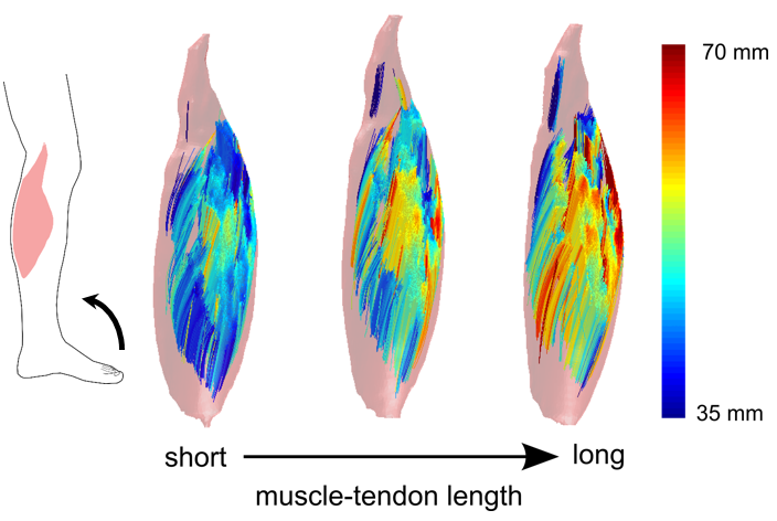
“The key measures we are getting out of this study are not just the volume of muscles, but also the orientations and lengths of their muscle fibres, which is a key determinant of the function of a muscle.
“We also look at the fat content which is a compositional feature of muscles that is quite different between diseased muscles and healthy muscles.
The impacts of this research have real implications for children growing up in Australia, with one-in-seven hundred babies born with cerebral palsy.
“This is very much a fundamental research study – we don’t have any direct clinical outcomes that we are assessing – but what we do know about children with cerebral palsy, the leading cause of childhood physical disability in the western world, is that outcomes can be pretty poor,” Dr Bolsterlee said.
“One-in-three children with cerebral palsy cannot walk independently, and we know this has got something to do with disordered muscle growth.
“It’s obvious from cross-sectional studies that there are quite some differences between the muscles of children with cerebral palsy and their typically developing peers, but nobody has actually studied this longitudinally, so we don’t know when these changes occur.
“We believe that information is necessary to develop new treatments.”
Currently there is no cure for cerebral palsy, and often children undergo severe interventions including complex surgical procedures with drugs to improve daily functioning. These interventions can change muscle growth, but how that affects musculoskeletal function is poorly understood.
Dr Bolsterlee is part of the team developing imaging methods and algorithms to be able to study this, and they are now generating the first data to give a comprehensive picture of how muscles develop typically – and how they develop in children with cerebral palsy.
“Many of the tools that are out there were developed for the brain – I’d say 99% of diffusion imaging software is used to reconstruct the neuronal architecture of the brain. We had to adapt the acquisition protocols as well as the imaging analysis techniques to accommodate measurement of the specific features of muscles we are interested in,” Dr Bolsterlee said.
In addition to configuring the imaging software to analyse data for the muscles, Dr Bolsterlee said there was a lot to consider when optimising scanning protocols to get the best images possible, while scanning children within a limited time.
“I’ve been working at NeuRA for the better part of eight years on this – and it’s really nice to see the first proof of principle demonstrations being taken to large-scale research – and hopefully to clinical practice as well.
“We’ve developed algorithms that several groups around the world are now using,” Dr Bolsterlee said.
This research into muscle imaging has grown the understanding of the architecture of muscles globally.
“Most anatomical knowledge comes from textbooks that are based on dissections of cadaver legs, and these are usually from older people who’ve donated their bodies to science.
“We have a rough understanding of the fibre structure within muscles and how they sit between muscles, but it’s been very difficult to get any information from living human muscles.
“Muscle is one of the most adaptable human tissues in the human body – when you exercise, they get bigger and when you’re lying in bed for too long, they get smaller very quickly.
“So, it’s very important if you want to understand how muscles respond to various stimuli, to have in-vivo imaging methods – or methods that can be applied to living humans,” Dr Bolsterlee said.
Previously, researchers were limited to ultrasound in living patients, which was 2D and only able to capture muscles superficial to the skin because the ultrasonic waves have limited penetration depth.
The MRI diffusion imaging technique allows researchers to look at whole human muscles in 3D, which has led to discoveries in the complex fibre structure of muscles and how it changes when they contract, lengthen or are diseased.
Listen to our podcast with Dr Bolsterlee and NIF Fellow, Dr Michael Green from NeuRA