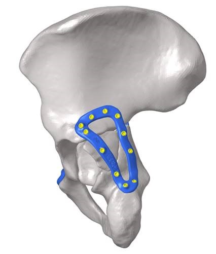
CT and 3D printing improving clinical PPE

Frontline medical workers put themselves at risk during a pandemic to deliver critical health care and save lives. Personal protective equipment (PPE) such as gloves, gowns, and face shields can reduce the risk of infection. To prevent contamination through airborne droplets, healthcare workers can employ an air-purifying respirator to push filtered air into their face shield or hood.

Read More

National Preclinical PET QA

The NIF Molecular Imaging & Radiochemistry (MIR) Theme is a group of NIF Fellows, Directors, and users of NIF facilities that focus on state-of-the-art radiochemistry and molecular imaging applications using PET, SPECT, and MRI.

Integrating preclinical PET systems into a national resource requires the development of defined QA programs to monitor and integrate the data from individual systems. Hence, the MIR Theme initiated a national quality assurance (QA) program for the NIF preclinical PET instruments.

Read More

Stent-electrode array for cortical neural activity

Minimally invasive endovascular stent-electrode array for high-fidelity, chronic recordings of cortical neural activity

This news has been contributed by Assoc. Prof. Bradford Moffat of the Melbourne Brain Centre Imaging Unit, Department of Radiology and Medicine, The University of Melbourne, Parkville.

National Imaging Facility Fellow, Assoc. Prof. Bradford Moffat collaborated with Dr. Tom Oxley’s group at the University of Melbourne for this high profile publication[1] that appeared in the journal “Nature Biotechnology”.

Read More

3D printed devices to treat traumatic pelvic fractures

3D printing is increasingly being used in the healthcare industry to customise medical devices to meet patient-specific needs. Currently, device manufacture is lengthy, limiting the application of customised medical devices. The treatment of traumatic injuries requires intervention as quickly as possible, preferably within days post-injury.

This collaborative research project between the Dept. of Biomedical Engineering at the University of Melbourne and the Dept. of Orthopaedics at the Royal Melbourne Hospital aims to assess the feasibility of 3D printing fracture plates to treat traumatic fractures and speed up the production of devices at the point-of-care for a patient. By performing a proof-of-concept experiment on a set of cadaveric pelvis, Dr Dale Robinson and team are evaluating each phase of the 3D printing workflow. Once implanted, a series of computational models and biomechanical experiments will be used to assess whether the 3D printed fracture plate offers an improvement over a traditionally mass-manufactured plate. Paramount to designing customised implants, the anatomy of each pelvis is being characterised using the University of Melbourne’s NIF Node CT with input from PET/CT Facility Fellow Rob Williams and radiographer Rebecca Glarin. After implantation of the fracture plate, CT may assess the effectiveness of the device in terms of stabilising and reducing the fracture.


3D reconstruction of a fractured human pelvis with a custom 3D printed device simulated in blue to promote appropriate healing.

To date, the project has conducted some scans and used this data for preliminary printing of implants. Plates were designed and printed in collaboration of researchers at Johnson and Johnson and the University of Melbourne. The initial study used 3D printed medical-grade titanium and 3D rendering from the NIF facility CT. In developing this method, iterative reconstruction with maximal overlap to printing was used to be consistent with typical medical CT. This was done while still using radiation dosimetry within standard limits.

This project has the potential to improve patient outcomes by enhancing surgical intervention durability, reducing the duration and number of surgeries, and reducing the risk of life-threatening surgical complications (such as pulmonary embolism and infection) through reduced bedtime. Consequently, the effective implementation of customised 3D printed medical devices is expected to reduce healthcare costs through shorter hospital stays and reduced number of surgical interventions.

This story was contributed by the Department of Biomedical Engineering and the Melbourne Brain Centre Imaging Unit at the University of Melbourne, and Johnson & Johnson. For further information, please contact Rob Williams.

Unwrapping the mystery of ancient Egyptian mummies

Reviving an ancient Egyptian Mummy sounds like something out of a science fiction movie, but researchers at the University of Melbourne have done the next best thing. In a multidisciplinary project with the Faculty of Medicine, Dentistry and Health Sciences headed by Dr Varsha Pilbrow, the head of Meritamun – a young Egyptian woman who lived more than 2000 years ago – has been imaged using CT and reconstructed using 3D-printing technology.


On the left, CT reconstructions of a baby mummy (still being studied). On the right, the CT reconstruction of the head of the mummy named Meritamun from the University of Melbourne’s collection within the School of Biomedical Sciences


Without disturbing the rare specimen and adhering to the controls and procedures of the Human Tissue Act 1982, Meritamun was imaged using the Siemens Human PET/CT in the University of Melbourne National Imaging Facility (NIF) Node. Digital and volumetric displays of tomographic data were acquired and reconstructed for 3D printing to create a skull replica.


The scanning of a mummy with no adverse affects


Biomedical science Masters Student Stacey Gorski has used the CT data to diagnose Meritanum with anaemia, and hypothesizes a cause of death due to parasitic infection.  “The fact that she lived to adulthood suggests that she was infected later in life,” says Gorski. She and supervisor Dr Pilbrow are continuing the investigation, hoping to learn more about the life and death of the ancient Egyptian using forensic pathology.


NIF Facility Fellow Mr Rob Williams facilitating access and providing expertise to the Human PET/CT scanner


In addition to learning more about the pathology and environments of population groups of 2000 years ago, the capability to replicate body parts and organs from CT imaging of specimens offers an opportunity for students to interact with old rare samples without damage to the original.


This story was contributed by the University of Melbourne. Acknowledgements go to Varsha Pilbrow, Julietta Capodistrias, Nina Sellars, Quentin Fogg, Michelle Gough, Gavan Mitchell, Peter Mayal, and Natalie Langowski.

For more information, please contact Rob Williams.

Closing the acquisition time gap – a new sequence scheme for optimised Sodium MRI

Magnetic Resonance (MR) opens a window for the non-invasive investigation of MR-observable nuclei, i.e. nuclei with a non-zero spin. Common nuclei in organic substances, like Carbon-12 and Oxygen-16; however, possess a zero-spin consequently rendering Hydrogen-1 (commonly referred as ‘proton‘ due to the single proton in the nucleus) the first and most frequently imaged nuclei.

Despite the much lower signal regime, also the potential of imaging ‘other nuclei’, so-called x-Nuclei MRI was approached in the early years of MRI. In 1985, only a few years after the first proton-based images were published, Sodium-23 was the second nucleus in the human body that was imaged non-invasively using MRI by Hilal and coworkers [1]. In spite of these early beginnings, x-Nuclei imaging in general and Sodium MRI, in particular, did not see the same high-paced progression as proton-based MRI techniques. This downturn was mostly caused by significant SNR limitations resulting from the low in vivo concentration and complicated NMR-signal characteristics of Sodium nuclei.

While SNR issues are more and more overcome through hardware improvements, particularly the recent advent of high field systems, the challenging signal characteristics continue to encourage the development of dedicated acquisition methods. Sodium possesses a 3/2-spin and as such exhibits a fast bi-exponential signal decay. Consequently, Sodium MRI sequences are commonly performed in 3D with k-space sampled via centre-out trajectory schemes resulting in inherent drawbacks for sampling efficiency and SNR. While previous research in Sodium MRI method development focused on the optimisation of sampling trajectories, we present a new acquisition method that tackles Sodium image quality improvements via a sequence timing optimisation.

Recently, we introduced zero-gradient excitation ramped hybrid encoding (zGRF-RHE) Sodium MRI, a timing optimisation that utilises the sequence dead-time delay (time-gap between transmit and receive mode) for gradient pre-ramping. This concept improves encoding time across k-space and in so doing alleviates signal decay effects during the acquisition. It utilises a hybrid sequence encoding approach where the central k-space gap, resulting from gradient activity before receiver onset, is filled with Cartesian single point acquisitions. A sequence diagram of zGRF-RHE is displayed in Figure 1.


Figure 1: Radial zero‐gradient‐excitation ramped hybrid encoding (zGRF‐RHE) sequence diagram (A) and 2D illustration of hybrid k‐space encoding scheme (B). Gradient‐free excitation is followed by an immediate gradient ramping, data sampling commences after dead‐time. Central k-space is measured separately as single points with the same gradient pre‐ramping condition. TRO, readout duration; td, dead‐time. Figure taken from [2].


Hybrid imaging techniques, like PETRA or its generalised concept RHE, are frequently used in short-T2 proton imaging. Their application to low SNR problems like Sodium was, however, hampered by the employed gradient modulation during signal excitation. In that regard, we establish zero-gradient excitation for an artefact-free excitation profile maximizing image quality. The gradient-free excitation supports high flip angle acquisition, an essential requirement for low SNR imaging like Sodium, but also provides the opportunity to employ advanced RF pulse shapes without introducing a gradient-based excitation bias. Additionally, it should be highlighted that the zGRF-RHE sequence approach essentially describes a timing concept that is not just independent of the RF-excitation but also is not restricted to a particular readout-scheme.

An investigation of 3D-radial zGRF-RHE with standard 3D-radial ultra-short echo time (UTE) imaging can be found in our recent publication [2]. Compared to simulations, phantoms and in-vivo human brain acquisitions confirmed that our proposed sequence timing concept improves on image quality independent of the readout bandwidth and shows reduced image blurring and higher SNR. Figure 2 displays results from an in vivo human brain experiment highlighting improved image sharpness.


Figure 2: (A) Cross sections through matched slices in in vivo experiments at TRO of 2 ms UTE and zGRF‐RHE. zGRF‐RHE shows sharper details around the brain stem (red arrow). (B) Three consecutive transverse slices of CSF masks with contour lines of normalized UTE and zGRF‐RHE at TRO of 2 ms. zGRF‐RHE provides better delineation between lateral ventricles (red arrow). Figure taken from [2].


In summary, this work introduces an enhanced sequence timing concept with particular applicability to challenging low SNR-problems. Ultimately, this approach is expected to be of use for a wider range of x-Nuclei applications with spin > ½.

For further information, the interested reader is pointed to our full publication which can be accessed at https://onlinelibrary.wiley.com/doi/abs/10.1002/mrm.27484.

References
[1] Hilal, S. K., Maudsley, A. A., Ra, J., Simon, H. E., Roschmann, P., Wittekoek, S., Cho, Z., and Mun, S. (1985). In vivo NMR imaging of sodium-23 in the human head. Journal of Computer Assisted Tomography, 9(1):1–7.
[2] Blunck Y, Moffat BA, Kolbe SC, Ordidge RJ, Cleary JO, Johnston LA. Zero-Gradient-Excitation Ramped Hybrid Encoding (zGRF-RHE) Sodium MRI. Magnetic Resonance in Medicine 2018. https://doi.org/10.1002/mrm.27484.


This story was contributed by Yasmin Blunck of the Melbourne Brain Centre Imaging Unit and Department of Biomedical Engineering, University of Melbourne, Parkville

The First Australian Multi-Centre Study of Dementia using Ultra-High Field MRI

Australia is at the forefront of dementia research with world leading studies such as the Australian Imaging and Lifestyle study of Ageing (AIBL) led by a consortium of Australia’s leading Dementia centres, and the recently started Prospective Study of Ageing (PISA) led by the QIMR Berghofer.

The installation of the first human ultra-high field MRI scanner in the southern hemisphere at the Centre for Advance Imaging, the Qld node of the National Imaging Facility, in 2014 opened up a new era of imaging research. The Siemens 7T whole-body MRI scanner brought Australia to the forefront of ultra-high field research enabling examination of the human brain with an unprecedented level of detail.

Subsequently, a second 7T scanner was installed at the Melbourne Brain Centre providing a unique opportunity for a national multi-centre collaboration in ultra-high field MRI and the capability to explore new imaging biomarkers for diagnosis of neurodegenerative disease. A major project is underway, led by the Brisbane-based CSIRO eHealth Research Centre, co-funded by the CRC for Mental Health and in collaboration with the QIMR Berghofer, University of Melbourne and Florey Institute for Neuroscience with the broad aim of characterising new bioimaging biomarkers of neurodegeneration in the aging population. A suite of MRI methods is being applied at both sites on large cohorts of healthy aging subjects and patients diagnosed with fronto-temporal dementia. The scanning part of the project has been successfully completed with superb image quality obtained using state of the art sequences. A significant effort is now underway to analyse this valuable data which may contain a wealth of diagnostic information not otherwise available.


This story was contributed by The University of Queensland

Feature image: (Left) 3D MP2RAGE 0.9mm isotropic showing exceptional tissue contrast, (centre) example of a Quantitative Susceptibility Mapping (QSM), a mechanism for useful chemical identification and quantification of specific biomarkers, and (right) T2W TSE using coronal accquisition for hippocampus subfield examination. 

Privacy Settings
Youtube
Vimeo
Google Maps