#ImagingTheFuture Week: Enabling breakthroughs in biomedical science and technology
Chan Zuckerberg Initiative’s (CZI) Imaging the Future Week puts a spotlight on the importance of imaging science in biomedicine, and the value of the global imaging community in translating health research.
Imaging is unlocking solutions to the world’s biggest challenges across commercial, clinical and research fields and has helped innovate in bioengineering, biology, medical technology and science, pharmaceutical and non-pharmaceutical therapies.
National Imaging Facility (NIF) supports the Imaging the Future Week initiative, and the 2023 event is focused on highlighting advances in technology and the impact this has on our understanding of health and disease.
As we continue to meet the evolving needs of modern research, NIF is accelerating new technology, enabling experts to develop protocols, tools, imaging data, and the application of imaging to solve complex problems – scroll on to find out more.
Better evidence for decision-making in health
Advanced imaging methods and analysis provide critical evidence for decision-making across all aspects of health and clinical science to keep Australia healthy.

Australia’s largest investment in molecular imaging
Australia’s first open access research Total Body Positron Emission Tomography scanner is NIF’s largest investment to date, and it will deliver a transformative understanding of complex health problems. Next-generation molecular imaging and radiopharmaceuticals are revolutionising how we see biological processes, paving the way for better diagnosis and treatment of chronic, systemic adult and childhood diseases. The instrument will produce high quality data at lower doses of radiation. It can be used to capture information from all body organs simultaneously to build a better picture of complex processes such as ageing, metabolism, brain signalling, behaviour, cognition and drug interactions.

Multidisciplinary collaboration to improve epilepsy outcomes
MRI imaging technology, AI, machine learning and data analysis are helping improve the lives of 150,000 Australians with epilepsy. The Australian Epilepsy Project will combine neuroimaging with cognitive and genetic data, and integrate them using AI, to develop predictive tools that will guide diagnosis and highlight opportunities for precision treatment. Expertise from the Florey Institute of Neuroscience and Mental Health, the University of Melbourne, Monash University and Austin Health drives the project, aiming to reduce seizure frequency and the risk of injury or death.
Better health for the young and older Australians
Imaging studies that look at conditions in younger and older Australians are essential for understanding and promoting healthy development and ageing.

Understanding the development of cerebral palsy
NIF is contributing to valuable data assets, including the first collection to show the way that muscles grow in children with cerebral palsy. The MUGgLE Study is the first longitudinal study comparing muscle growth in the development of children with cerebral palsy and typically developing children. The study is a partnership between Neuroscience Research Australia, the University of NSW and the Cerebral Palsy Alliance Research Institute. Imaging is being used to study muscle tightening and shortening as it happens, with high-resolution measurements of the architecture of whole muscles, giving researchers detailed, anatomically accurate, three-dimensional reconstructions to understand disordered muscle growth. The project has included the development of imaging methods and algorithms to be able to study this, adapting the acquisition protocols as well as the imaging analysis techniques to accommodate measurement of the specific features of muscles.

Brain-computer interface restoring independence after paralysis
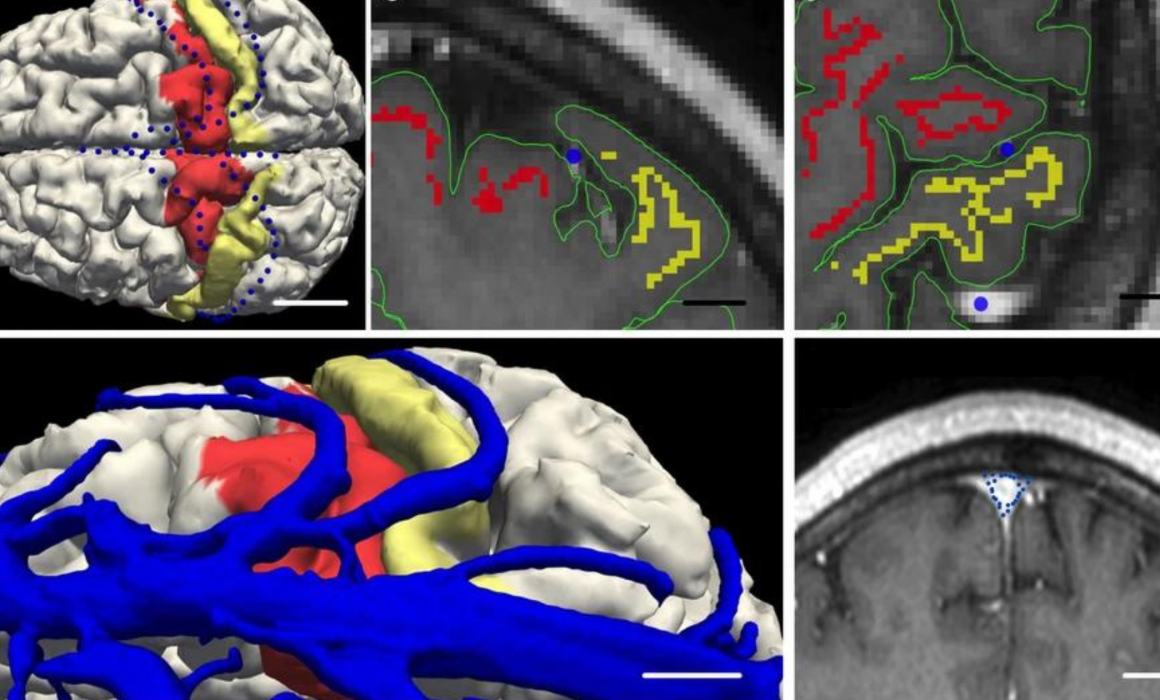
An implant the size of a paperclip is allowing people who are paralysed to operate technological devices using their thoughts without open brain surgery. NIF expertise and the 7T MRI at the University of Melbourne enabled early developments of the device which can translate brain signals from the inside of a blood vessel into commands on a computer.
The Synchron Stentrode is a world first brain-computer interface designed to restore functional independence in patients with paralysing conditions like ALS. The device was named one of TIME Magazine’s best inventions of 2021, and is currently undergoing expanded human clinical trials in preparation for submission to the FDA.
Equitable regional and rural health
Crucial to societal equity and research quality, delivering a geographically distributed network of advanced imaging to support research and personalised medicine, and taking part in medical trials, is a major national challenge.

Bringing health equity to regional and rural Australia
NIF is deploying four low-field portable MRI scanners to remote and regional sites to help researchers apply this affordable imaging technology in rural areas. The national mobile magnetic resonance (MR) network will be the first project of its kind world-wide and is a collaboration with partners including Monash University, University of Queensland, South Australian Health and Medical Research Institute (SAHMRI), the Alfred Hospital, Royal Perth Hospital, University of Western Australia and MedTech company, Hyperfine. These portable scanners will be used to understand how this fast-developing technology can help diagnose stroke, traumatic brain injury, and other conditions after testing in research laboratories at NIF nodes to build the usability of low-field MR, including developing techniques to maximise data quality and improve image processing.

Imaging mobilises ground-breaking field ventilator for deployment in the COVID-19 crisis
NIF provided critical support in preclinical testing to mobilise the now commercialised ventilator, 4DMedical ‘XV technology’ at the LARIF multipurpose fluoroscopy laboratory. A team of Australian collaborators, including biomedical company 4DMedical and University of Adelaide scientists created the ground-breaking, simple to use ‘field ventilator’ that can be locally produced at a low cost from easily acquired parts. It was developed in response to the global COVID-19 crisis, which identified potential shortages in essential medical equipment.


