#ImagingTheFuture Week: Enabling breakthroughs in biomedical science and technology

Chan Zuckerberg Initiative’s (CZI) Imaging the Future Week puts a spotlight on the importance of imaging science in biomedicine, and the value of the global imaging community in translating health research.

Imaging is unlocking solutions to the world’s biggest challenges across commercial, clinical and research fields and has helped innovate in bioengineering, biology, medical technology and science, pharmaceutical and non-pharmaceutical therapies.

National Imaging Facility (NIF) supports the Imaging the Future Week initiative, and the 2023 event is focused on highlighting advances in technology and the impact this has on our understanding of health and disease.

As we continue to meet the evolving needs of modern research, NIF is accelerating new technology, enabling experts to develop protocols, tools, imaging data, and the application of imaging to solve complex problems – scroll on to find out more.


Better evidence for decision-making in health

Advanced imaging methods and analysis provide critical evidence for decision-making across all aspects of health and clinical science to keep Australia healthy.


Australia’s largest investment in molecular imaging
Australia’s first open access research Total Body Positron Emission Tomography scanner is NIF’s largest investment to date, and it will deliver a transformative understanding of complex health problems. Next-generation molecular imaging and radiopharmaceuticals are revolutionising how we see biological processes, paving the way for better diagnosis and treatment of chronic, systemic adult and childhood diseases. The instrument will produce high quality data at lower doses of radiation. It can be used to capture information from all body organs simultaneously to build a better picture of complex processes such as ageing, metabolism, brain signalling, behaviour, cognition and drug interactions.

Multidisciplinary collaboration to improve epilepsy outcomes
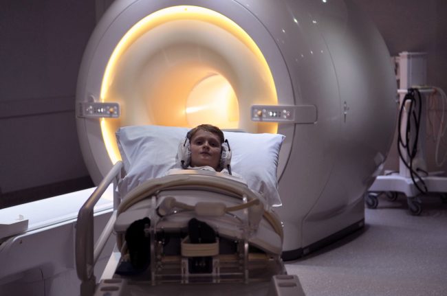
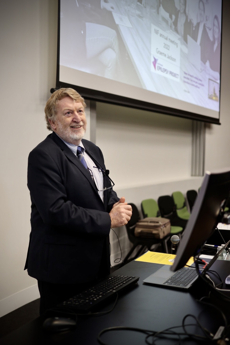
MRI imaging technology, AI, machine learning and data analysis are helping improve the lives of 150,000 Australians with epilepsy. The Australian Epilepsy Project will combine neuroimaging with cognitive and genetic data, and integrate them using AI, to develop predictive tools that will guide diagnosis and highlight opportunities for precision treatment. Expertise from the Florey Institute of Neuroscience and Mental Health, the University of Melbourne, Monash University and Austin Health drives the project, aiming to reduce seizure frequency and the risk of injury or death.


Better health for the young and older Australians

Imaging studies that look at conditions in younger and older Australians are essential for understanding and promoting healthy development and ageing.


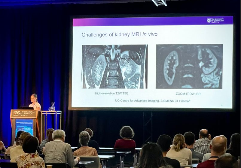
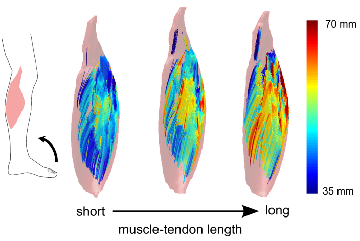
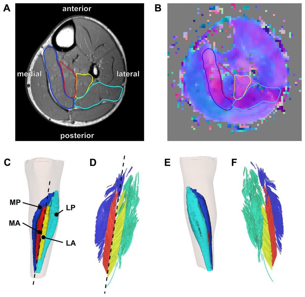
Understanding the development of cerebral palsy
NIF is contributing to valuable data assets, including the first collection to show the way that muscles grow in children with cerebral palsy. The MUGgLE Study is the first longitudinal study comparing muscle growth in the development of children with cerebral palsy and typically developing children. The study is a partnership between Neuroscience Research Australia, the University of NSW and the Cerebral Palsy Alliance Research Institute. Imaging is being used to study muscle tightening and shortening as it happens, with high-resolution measurements of the architecture of whole muscles, giving researchers detailed, anatomically accurate, three-dimensional reconstructions to understand disordered muscle growth. The project has included the development of imaging methods and algorithms to be able to study this, adapting the acquisition protocols as well as the imaging analysis techniques to accommodate measurement of the specific features of muscles.

Brain-computer interface restoring independence after paralysis
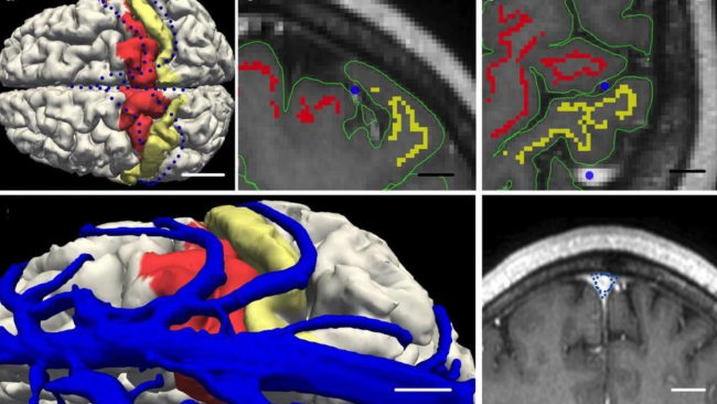
An implant the size of a paperclip is allowing people who are paralysed to operate technological devices using their thoughts without open brain surgery. NIF expertise and the 7T MRI at the University of Melbourne enabled early developments of the device which can translate brain signals from the inside of a blood vessel into commands on a computer.

The Synchron Stentrode is a world first brain-computer interface designed to restore functional independence in patients with paralysing conditions like ALS. The device was named one of TIME Magazine’s best inventions of 2021, and is currently undergoing expanded human clinical trials in preparation for submission to the FDA.


Equitable regional and rural health

Crucial to societal equity and research quality, delivering a geographically distributed network of advanced imaging to support research and personalised medicine, and taking part in medical trials, is a major national challenge.


Bringing health equity to regional and rural Australia
NIF is deploying four low-field portable MRI scanners to remote and regional sites to help researchers apply this affordable imaging technology in rural areas. The national mobile magnetic resonance (MR) network will be the first project of its kind world-wide and is a collaboration with partners including Monash University, University of Queensland, South Australian Health and Medical Research Institute (SAHMRI), the Alfred Hospital, Royal Perth Hospital, University of Western Australia and MedTech company, Hyperfine. These portable scanners will be used to understand how this fast-developing technology can help diagnose stroke, traumatic brain injury, and other conditions after testing in research laboratories at NIF nodes to build the usability of low-field MR, including developing techniques to maximise data quality and improve image processing.

Imaging mobilises ground-breaking field ventilator for deployment in the COVID-19 crisis
NIF provided critical support in preclinical testing to mobilise the now commercialised ventilator, 4DMedical ‘XV technology’ at the LARIF multipurpose fluoroscopy laboratory. A team of Australian collaborators, including biomedical company 4DMedical and University of Adelaide scientists created the ground-breaking, simple to use ‘field ventilator’ that can be locally produced at a low cost from easily acquired parts. It was developed in response to the global COVID-19 crisis, which identified potential shortages in essential medical equipment.

NIF collaborators and users funded and recognised among top researchers

NIF is enabling research that has attracted national recognition and funding, with collaborators listed among last year’s top medical researchers in The Australian, and as recipients of competitive Federal Government support. 

Top researchers in health and medical sciences 

In case you missed it, NIF collaborators have been listed in The Australian among the top researchers in 2022, covering expertise in areas including neurodegenerative disease, neuropsychiatry, theranostics and nuclear medicine. 

The list features researchers in 250 fields of academic endeavour, spanning the sciences, the social sciences through to the humanities and the arts. It aims to shine a light on what they do and the benefits they bring to the country. 

The weight of a researcher’s contribution was judged through citations from other researchers in their publications. 

NIF collaborators and users included: 

  • University of Melbourne Professor Christopher Rowe, for research in gerontology and geriatric medicine, covering dementia research, patient care and leadership in molecular imaging research at Austin Health and at the Florey Department of Neuroscience and Mental Health
  • University of NSW Professor Perminder Sachdev, recognised for work in neurology, with a focus on conditions such as drug-induced movement disorders, Tourette syndrome, secondary psychosis, healthy brain ageing and dementia, in particular Alzheimer’s disease and vascular dementia, and research into new interventions such as brain stimulation for neuropsychiatric disorders
  • Professor Louise Emmett, a keynote speaker at National Imaging Facility (NIF) Scientific Symposium, listed for work in nuclear medicine, radiotherapy and molecular imaging at the University of NSW and St Vincent’s Hospital in Sydney.

Australian Research Council support for Centre of Excellence 

Professor Michelle Watt from the University of Melbourne is part of a collaboration that has secured $35 million in funding as part of a new Australian Research Council Centre of Excellence (CoE) in Plants for Space. 

The centre aims to create on-demand, zero-waste, high-efficiency plants for better sustainability for space habitation, with the team covering aspects such as process and systems engineering, law, policy and psychology. 

It will make use of imaging equipment at the University of Melbourne Brain Imaging Centre Unit, where NIF support and work from NIF Node Director Professor Leigh Johnston and NIF Fellow Dr Edward Green have contributed to a Functional Plant Imaging Capability, in collaboration with Professor Watt. 

Professor Watt combines imaging and sensor technologies with modelling to understand how roots function in increasingly dynamic climates – and how to increase productivity and decrease environmental impacts. 

ISMRM and ISMRT ANZ Chapters’ Annual Meetings shine a light on national imaging expertise and infrastructure

[Image: Presentation award winners at ISMRM ANZ, Honours student, Arunan Srirengan, Dr Ed Green, Dr Gwen Schroyen and Dr Myrte Strik. Photo credit: Dr Adam Clemente]

The Australian and New Zealand Chapters of the International Society for Magnetic Resonance in Medicine (ISMRM) and the International Society for Magnetic Resonance Radiographers and Technologists (ISMRT) held their Annual Meetings in Sydney last month, highlighting the work of leading national researchers and clinicians, including members of the NIF network.

NIF enables coordinated open access to magnetic resonance expertise and infrastructure to support leading national researchers and clinicians, and proudly supported the events.

ISMRM ANZ Joint Chapter Annual Meeting 9-10 Nov

ISMRM ANZ hosted sessions on revolutionising MRI technology, advances in neuroimaging, and clinical applications of advanced MRI, in addition to keynote speakers neurologist and leader in stroke medicine, Prof Mark Parsons and Director of the Institute of Medical Physics at the University of Sydney, Prof Annette Haworth.

Dr Zhaolin Chen was a key speaker in the Revolutionising MRI technology session, presenting the NIF Point-of-Care project, a collaboration between NIF, Australian hospitals, and US medical device manufacturer, Hyperfine, to build the usability of low-field MRI and bring critical imaging to remote Australia and deploy imaging in challenging clinical environments such as COVID wards.

[Image: Dr Zhaolin Chen presenting the NIF Point-of-Care Magnetic Resonance project at ISMRM ANZ]

A number of other NIF users spoke at ISMRM ANZ, including:

  • Rebecca Glarin from the University of Melbourne, presenting findings from her PhD on ‘Optimising functional brainstem imaging of sympathetic drive with ultra-high field MRI’.
  • Dr Shahrzad Moinian from the University of Queensland Centre for Advanced Imaging, presenting ‘In vivo microstructural border delineation between areas of the human cerebral cortex using magnetic resonance fingerprinting (MRF) residuals’.
  • Honours student Arunan Srirengan presenting ‘Early identification of cerebral small vessel disease in obstructive sleep apnoea patients using magnetic resonance spectroscopy: a pilot study’, featuring data obtained on the NIF 3T MRI at NeuRA. This session was awarded second prize in the oral presentation awards.
  • Dr Myrte Strik from the University of Melbourne, presenting ‘Altered network topology in patients with visual snow syndrome: a resting-state 7 Tesla MRI study’, winning the award for best Early Career Researcher Data Blitz presentation.

[Image: Dr Shahrzad Moinian from the University of Queensland Centre for Advanced Imaging. Photo credit: Dr Adam Clemente]

Congratulations to University of Melbourne NIF Fellow, Prof Brad Moffatt as ANZ ISMRM Chapter President on the success of the 2022 meeting hosted at UNSW.

ISMRT ANZ Joint Chapter Annual Meeting 12-13 Nov

The ISMRT ANZ 2022 joint meeting program theme was MRI: Past, Present and Future, and featured a range of internationally renowned speakers demonstrating future technologies and cutting-edge imaging techniques.

Keynote presenters included Medical physicist and human brain imaging academic researcher Dr Samantha Holdsworth, Chief of the Quantitative Medical Imaging Laboratory, USA National Institute of Biomedical Imaging and Bioengineering, Dr Carlo Pierpaoli, and founding member of the Society of Cardiovascular Magnetic Resonance and Principal Investigator for the Cardiac Atlas Project, Prof Alistair Young.

NIF Senior Manager and Senior Research Scientist – National Magnetic Resonance Capability, Dr Shawna Farquharson was a key speaker at the Diffusion Weighted Imaging (DWI) Forum, presenting on ‘DWI: Principles and practical applications’.

[Image: Dr Shawna Farquharson, National Imaging Facility]

NIF users showcased at ISMRT ANZ included:

  • Prof Lynne Bilston from NeuRA, presenting ‘Brain Elastography’.
  • Sarah Daniel from the University of Queensland Centre for Advanced Imaging, presenting ‘Image quality enhancement using deep learning for in vivo human kidney MRI’.

[Image: Ms Sarah Daniel from the University of Queensland Centre for Advanced Imaging]

Congratulations to all presenters at ISMRM and ISMRT ANZ.

World’s first longitudinal muscle study grows understanding of cerebral palsy development

NIF infrastructure is enabling the Muscle Growth in the Lower Extremity (MUGgLE) Study, the first longitudinal study comparing muscle growth in children with cerebral palsy and typically developing children.

The project is a collaboration between Neuroscience Research Australia (NeuRA), the University of NSW (UNSW) and the Cerebral Palsy Alliance Research Institute.

The National Health and Medical Research Council-funded study is using magnetic resonance imaging (MRI) to compare muscle growth between typically developing children and children with cerebral palsy, using high-resolution measurements of the architecture of whole muscles.

Researcher Dr Bart Bolsterlee said the longitudinal study will see the lower legs of over 300 children scanned, between the ages of 0-3 months and 5-14 years.

“They will be scanned three times, with one-and-a-half years in between scans. We analyse the images to look at the individual muscles and how they change in size and structure over time,” Dr Bolsterlee said.

“The key measures we are getting out of this study are not just the volume of muscles, but also the orientations and lengths of their muscle fibres, which is a key determinant of the function of a muscle.

“We also look at the fat content which is a compositional feature of muscles that is quite different between diseased muscles and healthy muscles.

The impacts of this research have real implications for children growing up in Australia, with one-in-seven hundred babies born with cerebral palsy.

“This is very much a fundamental research study – we don’t have any direct clinical outcomes that we are assessing – but what we do know about children with cerebral palsy, the leading cause of childhood physical disability in the western world, is that outcomes can be pretty poor,” Dr Bolsterlee said.

“One-in-three children with cerebral palsy cannot walk independently, and we know this has got something to do with disordered muscle growth.

“It’s obvious from cross-sectional studies that there are quite some differences between the muscles of children with cerebral palsy and their typically developing peers, but nobody has actually studied this longitudinally, so we don’t know when these changes occur.

“We believe that information is necessary to develop new treatments.”

Currently there is no cure for cerebral palsy, and often children undergo severe interventions including complex surgical procedures with drugs to improve daily functioning. These interventions can change muscle growth, but how that affects musculoskeletal function is poorly understood.

Dr Bolsterlee is part of the team developing imaging methods and algorithms to be able to study this, and they are now generating the first data to give a comprehensive picture of how muscles develop typically – and how they develop in children with cerebral palsy.

“Many of the tools that are out there were developed for the brain – I’d say 99% of diffusion imaging software is used to reconstruct the neuronal architecture of the brain. We had to adapt the acquisition protocols as well as the imaging analysis techniques to accommodate measurement of the specific features of muscles we are interested in,” Dr Bolsterlee said.

In addition to configuring the imaging software to analyse data for the muscles, Dr Bolsterlee said there was a lot to consider when optimising scanning protocols to get the best images possible, while scanning children within a limited time.

“I’ve been working at NeuRA for the better part of eight years on this – and it’s really nice to see the first proof of principle demonstrations being taken to large-scale research – and hopefully to clinical practice as well.

“We’ve developed algorithms that several groups around the world are now using,” Dr Bolsterlee said.

This research into muscle imaging has grown the understanding of the architecture of muscles globally.

“Most anatomical knowledge comes from textbooks that are based on dissections of cadaver legs, and these are usually from older people who’ve donated their bodies to science.

“We have a rough understanding of the fibre structure within muscles and how they sit between muscles, but it’s been very difficult to get any information from living human muscles.

“Muscle is one of the most adaptable human tissues in the human body – when you exercise, they get bigger and when you’re lying in bed for too long, they get smaller very quickly.

“So, it’s very important if you want to understand how muscles respond to various stimuli, to have in-vivo imaging methods – or methods that can be applied to living humans,” Dr Bolsterlee said.

Previously, researchers were limited to ultrasound in living patients, which was 2D and only able to capture muscles superficial to the skin because the ultrasonic waves have limited penetration depth.

The MRI diffusion imaging technique allows researchers to look at whole human muscles in 3D, which has led to discoveries in the complex fibre structure of muscles and how it changes when they contract, lengthen or are diseased.

For more information, listen to our podcast with Dr Bolsterlee and NIF Fellow, Dr Michael Green from NeuRA: The MUGgLE Study: Imaging to understand how muscles grow.

Inaugural NIF Scientific Symposium kicks off #NationalScienceWeek

Leading researchers, clinicians and industry attended the inaugural National Imaging Facility (NIF) Scientific Symposium on 12 August.

The event kicked off National Science Week for NIF, highlighting the critical role of collaboration in translating research challenges to benefit industry and keep Australians healthy, with the theme ‘National partnerships for innovation and impact’.

NIF CEO Prof Wojtek Goscinski said the Symposium was an excellent opportunity to highlight ground-breaking work from Australia’s world-class imaging community.

“It was a privilege to host experts from across Australia, including keynote speakers Prof Graeme Jackson, Prof Louise Emmet and Prof Gemma Figtree, whose work is at the leading edge of imaging globally,” Prof Goscinski said.

“I’d also like to extend my thanks to the presenters who delivered an excellent Technology Showcase session, and Health and Medical Translational Challenges session.

“A particular highlight was hearing from our industry partners, including Telix Pharmaceuticals, Clarity Pharmaceuticals, Cochlear and Nyrada, who discussed the way they engage with national imaging research infrastructure.

“NIF is privileged to have a strong network of world-leading expertise at our fingertips and it was an honour to bring some of these people together to present their work and share ideas at the 2022 Symposium,” he said.

Keynote presentations of the Symposium included:

  • ‘The Australian Epilepsy Project’, Prof Graeme Jackson
  • From mouse to Medicare: the PSMA story in Australia’, Prof Louise Emmett
  • Coronary artery imaging to inform the next Frontier of heart attack prevention’, Prof Gemma Figtree

The Technology Showcase session highlighted NIF’s latest capabilities, including tools for processing and interpreting data, and applications of imaging to solve complex problems, including:

  • ‘Ultra-high field magnetic resonance imaging’, Prof Leigh Johnston and Prof Markus Barth
  • ‘Bringing imaging to rural Australia with a national network of low field mobile MR scanners’, Dr Zhaolin Chen
  • ‘Australian Imaging Service: The national platform for trusted data management and analysis’, Dr Ryan Sullivan
  • ‘Magnetic Particle Imaging’, Dr Andre Bongers
  • An insight into MicroCT imaging: recent advances, applications and impact on research and innovation’, Ms Diana Patalwala
  • Preclinical Research: The Crucial Step in Medical Advancements’, Dr Chris Christou

The Health and Medical Translation Challenges session provided an opportunity for attendees to hear from clinicians and researchers about their journey to making translational impact, including:

  • Neuroimaging in clinical trials: Perspectives of a clinician-researcher’, A/Prof Sylvia Gustin
  • The Australasian Radiopharmaceutical Trials network (ARTnet)’, A/Prof Ros Francis

The Industry Discussion Panel opened up conversation on how imaging accelerates and underpins innovation and future opportunities, with speakers:

  • Dr David Cade, Chief Executive Officer, Telix Pharmaceuticals Asia Pacific
  • Dr Matt Harris, Chief Scientific Officer, Clarity Pharmaceuticals
  • Dr Zachary Smith, Director, Algorithms and Applications, Cochlear
  • Dr Jasneet Parmar, Neuroscience Researcher, Nyrada Inc

Neuro Imaging to examine high rates of dementia in older Aboriginal Australians

Early life stress (ELS) has been linked to abnormalities in brain structure and function and may contribute to increased risk of cognitive decline and dementia later in life. ELS has also been associated with the high prevalence of dementia observed in older Aboriginal Australians.

A study at NIF’s UNSW Node, NeuRA Imaging is engaging the Australian Aboriginal community to investigate structural and pathological brain changes that underlie in high rates of dementia and cognitive decline in older Aboriginal Australians.

This will be the first study that investigates neuroimaging in cognitive impairment in older Aboriginal Australians and will inform dementia prevention, diagnosis and policy. It will also contribute to the wider literature on vascular risk in the pathogenesis of Alzheimer’s disease and associated biomedical and social risk factors.

After extensive community engagement with partnering Aboriginal communities including La Perouse, NSW, the initial consultation stage of NeuRA’s Koori Growing Old Well Study indicated that neuroimaging should be included in future dementia studies (Lavrencic et al., 2020, Int Psychogeriatr). Led by NeuRA’s, researchers including Dr Kylie Radford, Professor Tony Broe AM and Dr Louise Lavrencic, the Koori Growing Old Well Study included a community planning survey, pilot MRI study and guidance from an Aboriginal and Torres Strait Islander Steering Committee.

“NIF’s capabilities are allowing this study to investigate underlying brain changes and pathology in ageing and dementia in partnership with Aboriginal communities. The study will give greater detail and is using sophisticated and novel MRI techniques. By having the facility in-house at NeuRA it also means we can ensure a culturally safe and welcoming environment for our participants. With a rapidly ageing population and high rates of dementia, we hope that this ground breaking study will shed light on important ways to promote healthy brain ageing with Aboriginal and Torres Strait Islander peoples,” said Dr Kylie Radford, Senior Research Scientist and Group Leader, Neuroscience Research Australia.

The neuroimaging sub-study is a prospective, cross-sectional non-interventional study where participants will first complete a comprehensive interview and diagnostic assessment as part of the Koori Growing Old Well study. Consenting participants (200) aged 55+ will undergo MR scans with an expected study completion by 2023.

The outcome analyses will include identifying associations between cognitive impairment and hippocampal atrophy/volume and vascular indices on MR. Vascular pathology will be examined for cases of possible or probable Alzheimer’s disease compared to a cognitively intact control group. Correlations between MR measures and early life stress, adult risk and protective factors, cognitive function, and clinically diagnosed cognitive impairment will be investigated.

Diffusion Tensor Imaging of the lower leg: Learnings for muscle contracture and cerebral palsy

Diffusion tensor imaging (DTI) is a magnetic resonance imaging (MRI) technique that exploits the movement of water molecules to reveal microscopic details about tissue architecture. DTI is commonly used in brain imaging studies, used to track neural tracts through the brain. The technique is also ideal for investigating the 3D architecture of muscles, as DTI can be used to obtain detailed, quantitative measurements of the anatomy of complex skeletal muscles in living humans. Prof Robert Herbert’s group at NeuRA utilised the 3T MRI located in the UNSW Node of NIF to take a first look at the compartmentalised soleus muscle to provide reference values for further modelling.

CT and DTI slices through leg muscle with regions highlighting the front back side and rear of the leg followed by 3D reconstructions of fibre muscles coloured to indicate the same regions as shown in the slices above

Reconstruction of the architecture of the human soleus muscle using MRI and DTI, taken from ref., showing (A) the MRI slice (midway between ankle and knee) and (B) the corresponding DTI slice taken on a healthy child participant, with (C – F) showing the 3D reconstruction of the surface of all muscle compartments based on the outlines on the anatomical scan.


The human soleus muscle is particularly difficult to study using conventional techniques, such as ultrasound, due to the depth of the anterior and proximal compartments and difficulty in accurate orientation. Hence, DTI is an ideal method to quantify the macroscopic arrangement of muscle fibres of the soleus and help develop comprehensive, quantitative atlases of human muscle architecture.

Prof Herbert’s team have recently used the method to investigate the leg muscles of children with cerebral palsy. Measurements of the medial gastrocnemius muscles were obtained from structural MRI and DTI scans of 20 children with unilateral spastic CP and 20 typically developing children. The study showed that children with unilateral spastic cerebral palsy had reduced range and muscle volume in the calf on the more affected side compared to typically developing children.

The calf plays a vital role in standing and walking, and the differences detected here provide insight into the pathophysiology of muscle contractures and functional impairments in children with cerebral palsy. This knowledge is essential for orthopaedic surgeons and physiotherapists supporting affected children in learning to walk independently.


For further information, please contact NIF Fellow Dr Michael Green.

This story was contributed by NeuRA.

National Network of Trusted Data Repositories

During 2017 the National Imaging Facility (NIF) nodes at the University of Western Australia (UWA), University of Queensland (UQ), University of New South Wales (UNSW) and Monash University collaborated on a national project to enhance the quality, durability and reliability of data generated by NIF through the Trusted Data Repository project.

●        Quality pertains to a NIF user’s expectation that an animal, plant or material can be scanned and from that data reliable outcomes/characterisations can be obtained (e.g. signal, volume, morphology) over time and across NIF sites.

●        Durability refers to guaranteed long-term availability of the data.

●        Reliability means that the data is useful for future researchers, i.e. stored in one or more open data formats and with sufficient evidential metadata.

The Project, Delivering durable, reliable, high-quality image data, was jointly funded by the Australian National Data Service (ANDS) and Research Data Services (RDS). It was motivated both by NIF’s desire to enhance the quality of the data associated with the use of its facilities, and the desire of ANDS/RDS to facilitate the establishment of Trusted Data Repositories that enable access to data for at least 10 years and includes metadata that documents both the quality of the data and its provenance.

A trusted data repository service is essential for sharing data and ensures that project data created and used by researchers is “managed, curated, and archived in such a way to preserve the initial investment in collecting them” and that the data “remain useful and meaningful into the future” (https://www.coretrustseal.org).

The scope of the Project was limited to MRI data with the understanding that the developed requirements and trusted data repository services could be adapted to, or serve as a basis for other instruments/modalities.

The key outcomes from the Project include:

  1. The NIF agreed process for acquiring trusted data (NAP) – Lists the requirements that must be satisfied to obtain high-quality data, i.e. NIF-certified data, suitable for ingestion in a NIF trusted data repository service. They cover provisioning of a unique instrument identifier, instrument registration with Research Data Australia (https://researchdata.ands.org.au), quality control (QC), quality assurance measures, requisite metadata (including cross-reference to the QC data),  the process by which data is moved from the instrument to the digital repository service and the format(s) of the data.
  2. The NIF requirements for a trusted data repository service – Provides a platform-agnostic checklist of requirements that a basic NIF trusted data repository service should satisfy, including: identification of data by a unique Project identifier, ingestion of data from NIF-compliant instruments, authentication via the Australian Access Federation (https://aaf.edu.au), interoperability and easy deployment across NIF nodes.
  3. Implementations of trusted data repository services for two exemplars:
    1. Preclinical MRI data (with mouse brain data as an example) acquired across three NIF nodes—UNSW, UQ and UWA—using a Bruker BioSpec 9.4T MRI. The services have been implemented using the open source MyTardis/ImageTrove (https://www.mytardis.org) platform.
    2. Clinical ataxia MRI data acquired using a Siemens Skyra 3T MRI scanner in support of a Monash-proposed International Ataxia Imaging Repository (IAIR). The service has been Implemented using the open source XNAT (https://www.xnat.org) platform.

Software developed to support the implementation of the repository services includes: Docker (https://www.docker.com) Compose scripts to permit easy deployment at differents sites, client-side scripts for uploading NIF-certified data to ImageTrove/MyTardis and an XNAT plugin for uploading non-DICOM files.

  1. Assessments of the resulting trusted data repository services against a relevant international metric, the CoreTrustSeal (https://www.coretrustseal.org) Core Trustworthy Data Repositories Requirements.

For NIF users and the broader imaging research community the benefits and impact of this Project include:

  • Reliable and durable access to data
  • Improved reliability of research outputs and the provenance associated with it
  • Making NIF data more FAIR (Findable, Accessible, Interoperable, Reusable – https://www.ands.org.au/working-with-data/the-fair-data-principles)
  • Easier linkages between publications and data
  • Stronger research partnerships

For research institutions they include:

  • Enhanced reputation management
  • A means by which to comply with the Australian Code for the Responsible Conduct of Research
  • Enhanced ability to engage in multi-centre imaging research projects

For NIF they include

  • Improved data quality
  • Improved international reputation
  • The ability to run multi-centre trials

The transition plan post-funding includes: maintenance of existing services for 10 years; the integration of additional instruments; creation of a project web portal; planned new national and international service deployments; refinements and improvements; and CoreTrustSeal certification.

Project documents have been archived in the NIF Customer Relationship Management (CRM) system (accessible by NIF staff). Project software is hosted on GitHub and is freely available for download here: https://github.com/NIF-au/TDR. For further information please contact either the national Project Manager or NIF.

Project Manager and UWA lead: Andrew Mehnert (NIF Informatics Fellow, Centre for Microscopy, Characterisation and Analysis).
NIF lead – Graham Galloway (Chief Executive Officer, NIF)
UQ lead – Andrew Janke (NIF Informatics Fellow, Centre for Advanced Imaging)
UNSW lead – Marco Gruwel (Senior Research Associate, Mark Wainwright Analytical Centre)
Monash lead – Wojtek Goscinski (Associate Director, Monash eResearch Centre)

National network of trusted data repositories establish standard for the future

Imaging equipment such as MRI, PET and CT scanners are capable of producing vast amounts of valuable research data. In order to maximise research outcomes, data must be stored securely, have its quality verified, and should be accessible to the wider research community.

Informatics fellows from around Australia have combined their expertise to build a series of Trusted Data Repositories (TDR’s) to provide researchers with a secure location to store, share and curate their data.

This national project, Delivering durable, reliable, high-quality image data, jointly funded by the Australian National Data Service (ANDS) and Research Data Services (RDS), guarantees the storage of data for at least 10 years for use in future research.

Led by the National Imaging Facility (NIF), the project brought together researchers and informatics specialists from UQ’s Centre for Advanced Imaging (CAI), Monash Biomedical Imaging (MBI), Monash eResearch Centre, the University of Western Australia, RCC (Research Computing Centre, UQ) and the University of NSW. Together, the team has established best practices for TDR’s to store imaging data nationally, through the NIF network.

To read the full article, please click on the following link:

https://cai.centre.uq.edu.au/article/2017/12/national-network-trusted-data-repositories-establish-standard-future

Privacy Settings
Youtube
Vimeo
Google Maps