Brain research supercharged by new ‘control panel’ accessible worldwide

The flood of data from brain research worldwide has now been channelled into an easy-to-use, open-access processing tool built to supercharge our understanding of the human brain.  

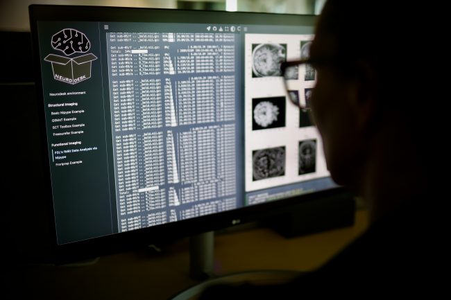
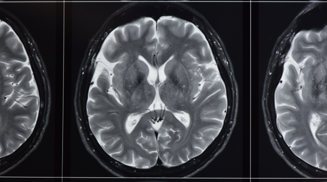
Neurodesk – a platform for processing, analysing and sharing massive datasets – has solved critical issues in the search for answers about brain disorders and diseases such as epilepsy, dementia, schizophrenia and traumatic brain injury. Neuroimaging data comes from modalities such as MRI, PET and MEG/EEG scans. 

Neurodesk was proposed at, led by and supported by National Imaging Facility partners including the University of Queensland, Swinburne University of Technology, and the Australian Imaging Service through University of Sydney.  

“It has removed a significant bottleneck that has hindered our ability to explore the complexities of the human brain, streamlining the processing of vast amounts of neuroimaging data,” says Dr Steffen Bollmann, project lead at the University of Queensland. 
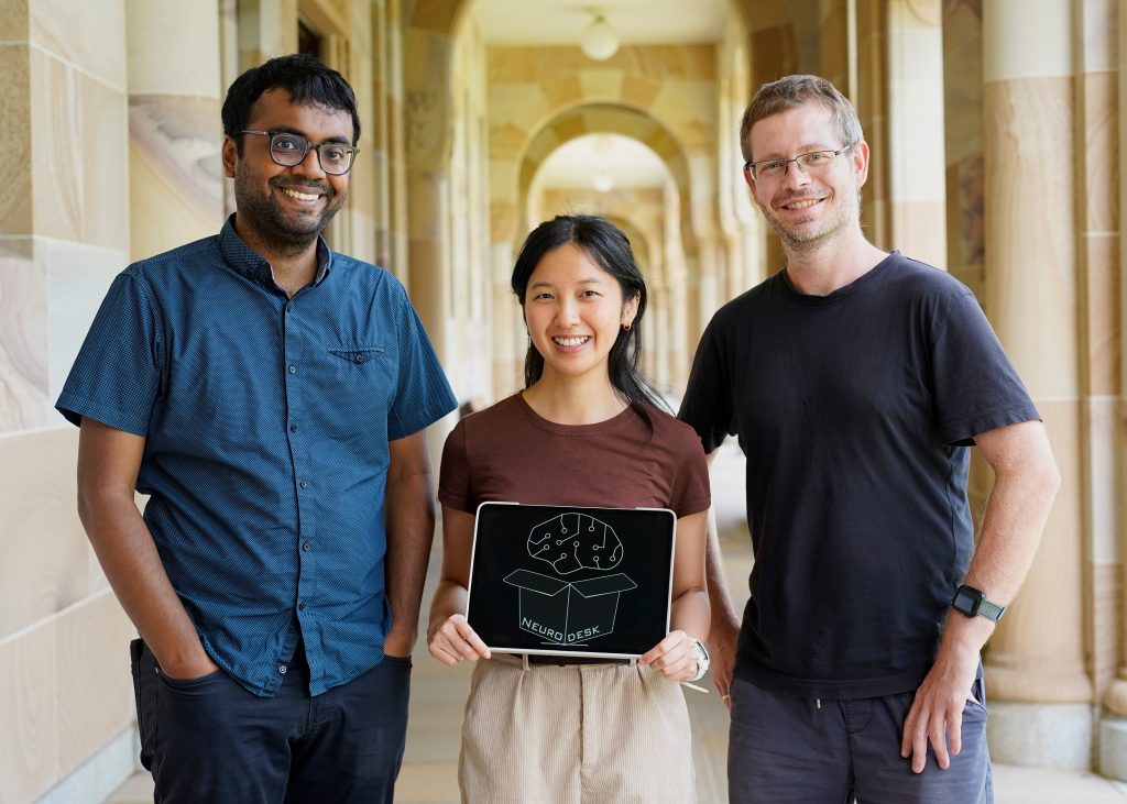
[Image: Members of the Neurodesk team, Aswin Narayanan, Thuy Dao, Dr Steffen Bollmann. Image credit: Dr Nicholas Hamilton.]

Both individual people and society stand to benefit: unravelling the brain’s mysteries helps us find cures for debilitating brain disorders, relieve people of the pain of untreated mental health issues and its societal cost, develop personalised medical treatments, inform public health policies, and understand human behaviour. 

Neurodesk is like the ultimate, seamless sci-fi control panel. Accessible through a user-friendly browser interface, it works across operating systems, pulling together different neuroimaging programs that run in all different ways, and is able to run giant research projects or small ones with its seamless access to computing power. 

Dr Bollmann sees Neurodesk’s accessibility as a leap forward in democratising neuroscience tools: “It empowers researchers worldwide to use open-source neuroimaging tools effectively and focus on their research questions, rather than grappling with technical obstacles.” 

“It is difficult to overstate how much benefit Neurodesk provides to our facility,” says Dr Tim Rosenow, NIF Facility Fellow and Neurodesk user at UWA’s Centre for Microscopy Characterisation and Analysis. “Researchers no longer need to be experienced in IT to perform their analyses, because software, dependencies, and conflicts are already handled.” 

However, the most important aim of Neurodesk’s creators was reproducibility: the process of other researchers replicating studies to check results and build a foundation of reliable scientific knowledge. The platform contains techniques that enable researchers to accurately replicate other teams’ analyses.  

Scientists can even share and publish their analyses, which can significantly reduce time and effort for collaboration and creating new knowledge. They can then tackle bigger neuroscience questions and analyse larger datasets. 

The ability to handle huge amounts of data is crucial, moving forward in brain research. Neurodesk gives researchers access to supercomputers and cloud computing to process neuroimaging data efficiently, letting them work with ‘biobanks’ of data (for instance, the UK Biobank holds imaging data from 50,000+ people). 

“By simplifying neuroimaging, fostering collaboration and empowering scientists to handle huge datasets, this breakthrough not only advances our understanding of the healthy human brain, but also sheds light on the intricacies of various brain disorders,” says Dr Bollmann, “promising improved diagnostics and treatments in the future.” 

Read the new paper published in Nature Methods here. 

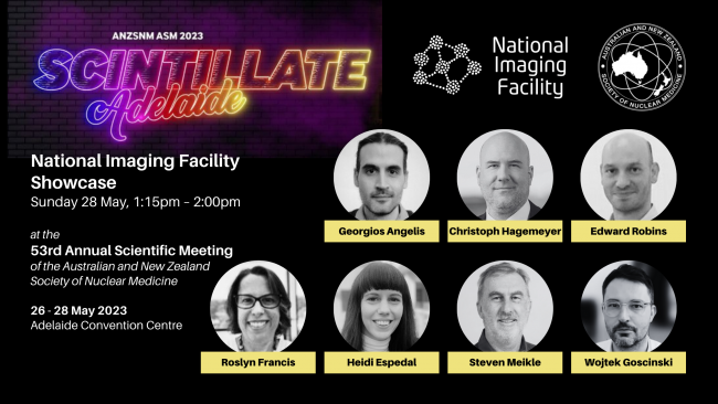
National Imaging Facility Showcase at ANZSNM 2023

National Imaging Facility will host a Showcase at ANZSNM 2023, featuring presentations from Australia’s advanced imaging network.

See the full ANZSNM Program and register here.

Sunday 28 May, 1:15pm – 2:00pm
City Room 3/4
Adelaide Convention Centre

Chair:   Prof Steven Meikle
Head, Imaging Physics Laboratory
Brain and Mind Centre, University of Sydney

Time

1:15pm

Topic

Introduction and session open

Speaker

Prof Wojtek Goscinski
Chief Executive Officer
National Imaging Facility

1:20pm

Australian National Total Body PET Facility

Dr Georgios Angelis
NIF Total Body PET Fellow
University of Sydney

1:30pm

Monash MHELTHERA Lab and cyclotron capabilities

Prof Christoph Hagemeyer
Acting Director, NIF Node Director
Monash Biomedical Imaging

1:40pm

SAHMRI Radiopharmaceutical Chemistry

Dr Edward Robins
Head, Radiopharmaceutical Research and Development
Molecular Imaging and Therapy Research Unit
SAHMRI

1:50pm

New capabilities of the Western Australia Node

A/Prof Roslyn Francis
Head, Department of Nuclear Medicine and the WA PET Service, Sir Charles Gairdner Hospital
NIF Deputy Node Director
University of Western Australia

Dr Heidi Espedal
NIF Facility Fellow
University of Western Australia

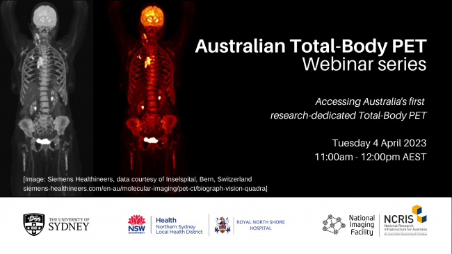
Australian Total-Body PET webinar series: Accessing Australia’s first research-dedicated Total-Body PET

Register: How to access Australia’s first research-dedicated Total-Body PET

Imaging to understand whole-body processes such as novel drug interactions during therapeutic development will soon be accessible through Australia’s first research-dedicated Total Body PET (TB-PET).

The Sydney Imaging Core Research Facility at The University of Sydney, in partnership with Northern Sydney Local Health District (NSLHD) and the National Imaging Facility (NIF), is establishing a Total Body PET facility in the Department of Nuclear Medicine at Royal North Shore Hospital.

TB-PET can be used to capture molecular processes from all organs simultaneously. The high-performance instrument can be used to study diseases that affect the entire body and build a better picture of complex processes such as ageing, metabolism, brain signalling and drug interactions.

Due to its exquisite sensitivity, it also has the potential to use much lower radiation doses compared to conventional PET scanners, making it safe to scan children, healthy volunteers, and to scan patients repeatedly to better understand disease progression and treatment effects.

TB-PET is accessible for research studies through Sydney Imaging Core Research Facility, the University of Sydney Node of the National Imaging Facility. For more information, contact Dr Georgios Angelis.

Webinar details

11:00 AM – 12:00 PM AEST
Tuesday 4 April 2023 
Register via Eventbrite here.

In this first webinar:

  • The Director of Sydney Imaging, Prof Fernando Calamante, will introduce the new facility, the operational model and the available resources to all researchers across Australia
  • Prof Steve Meikle will provide an overview of Total Body PET technology and explain how the new facility will fit within the existing clinical research imaging landscape
  • Amanda Hammond, Molecular Imaging Product Manager at Siemens Healthcare, will present an overview of the technical specifications and capabilities of the new Biograph Vision Quadra TB-PET system set to be installed.
  • The presentations will be followed by a short Q&A session.

#ImagingTheFuture Week: Enabling breakthroughs in biomedical science and technology

Chan Zuckerberg Initiative’s (CZI) Imaging the Future Week puts a spotlight on the importance of imaging science in biomedicine, and the value of the global imaging community in translating health research.

Imaging is unlocking solutions to the world’s biggest challenges across commercial, clinical and research fields and has helped innovate in bioengineering, biology, medical technology and science, pharmaceutical and non-pharmaceutical therapies.

National Imaging Facility (NIF) supports the Imaging the Future Week initiative, and the 2023 event is focused on highlighting advances in technology and the impact this has on our understanding of health and disease.

As we continue to meet the evolving needs of modern research, NIF is accelerating new technology, enabling experts to develop protocols, tools, imaging data, and the application of imaging to solve complex problems – scroll on to find out more.


Better evidence for decision-making in health

Advanced imaging methods and analysis provide critical evidence for decision-making across all aspects of health and clinical science to keep Australia healthy.


Australia’s largest investment in molecular imaging
Australia’s first open access research Total Body Positron Emission Tomography scanner is NIF’s largest investment to date, and it will deliver a transformative understanding of complex health problems. Next-generation molecular imaging and radiopharmaceuticals are revolutionising how we see biological processes, paving the way for better diagnosis and treatment of chronic, systemic adult and childhood diseases. The instrument will produce high quality data at lower doses of radiation. It can be used to capture information from all body organs simultaneously to build a better picture of complex processes such as ageing, metabolism, brain signalling, behaviour, cognition and drug interactions.

Multidisciplinary collaboration to improve epilepsy outcomes
MRI imaging technology, AI, machine learning and data analysis are helping improve the lives of 150,000 Australians with epilepsy. The Australian Epilepsy Project will combine neuroimaging with cognitive and genetic data, and integrate them using AI, to develop predictive tools that will guide diagnosis and highlight opportunities for precision treatment. Expertise from the Florey Institute of Neuroscience and Mental Health, the University of Melbourne, Monash University and Austin Health drives the project, aiming to reduce seizure frequency and the risk of injury or death.


Better health for the young and older Australians

Imaging studies that look at conditions in younger and older Australians are essential for understanding and promoting healthy development and ageing.


Understanding the development of cerebral palsy
NIF is contributing to valuable data assets, including the first collection to show the way that muscles grow in children with cerebral palsy. The MUGgLE Study is the first longitudinal study comparing muscle growth in the development of children with cerebral palsy and typically developing children. The study is a partnership between Neuroscience Research Australia, the University of NSW and the Cerebral Palsy Alliance Research Institute. Imaging is being used to study muscle tightening and shortening as it happens, with high-resolution measurements of the architecture of whole muscles, giving researchers detailed, anatomically accurate, three-dimensional reconstructions to understand disordered muscle growth. The project has included the development of imaging methods and algorithms to be able to study this, adapting the acquisition protocols as well as the imaging analysis techniques to accommodate measurement of the specific features of muscles.

Brain-computer interface restoring independence after paralysis
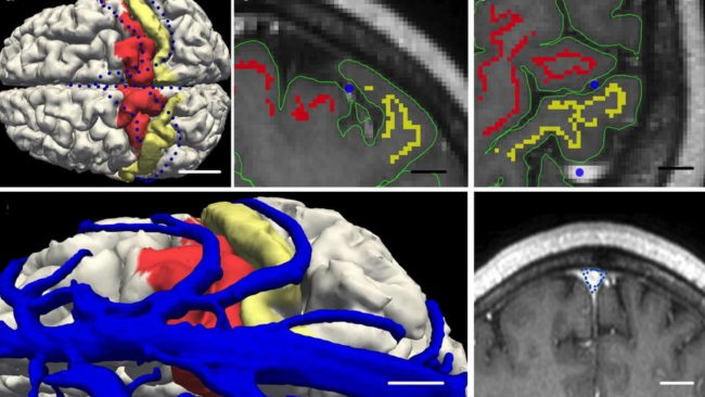
An implant the size of a paperclip is allowing people who are paralysed to operate technological devices using their thoughts without open brain surgery. NIF expertise and the 7T MRI at the University of Melbourne enabled early developments of the device which can translate brain signals from the inside of a blood vessel into commands on a computer.

The Synchron Stentrode is a world first brain-computer interface designed to restore functional independence in patients with paralysing conditions like ALS. The device was named one of TIME Magazine’s best inventions of 2021, and is currently undergoing expanded human clinical trials in preparation for submission to the FDA.


Equitable regional and rural health

Crucial to societal equity and research quality, delivering a geographically distributed network of advanced imaging to support research and personalised medicine, and taking part in medical trials, is a major national challenge.


Bringing health equity to regional and rural Australia
NIF is deploying four low-field portable MRI scanners to remote and regional sites to help researchers apply this affordable imaging technology in rural areas. The national mobile magnetic resonance (MR) network will be the first project of its kind world-wide and is a collaboration with partners including Monash University, University of Queensland, South Australian Health and Medical Research Institute (SAHMRI), the Alfred Hospital, Royal Perth Hospital, University of Western Australia and MedTech company, Hyperfine. These portable scanners will be used to understand how this fast-developing technology can help diagnose stroke, traumatic brain injury, and other conditions after testing in research laboratories at NIF nodes to build the usability of low-field MR, including developing techniques to maximise data quality and improve image processing.

Imaging mobilises ground-breaking field ventilator for deployment in the COVID-19 crisis
NIF provided critical support in preclinical testing to mobilise the now commercialised ventilator, 4DMedical ‘XV technology’ at the LARIF multipurpose fluoroscopy laboratory. A team of Australian collaborators, including biomedical company 4DMedical and University of Adelaide scientists created the ground-breaking, simple to use ‘field ventilator’ that can be locally produced at a low cost from easily acquired parts. It was developed in response to the global COVID-19 crisis, which identified potential shortages in essential medical equipment.

Announcing: Brainhack Global returns to Australia next month

Brainhack Global Australasia’s collaborative hackathon has been announced for 2022, supported by National Imaging Facility (NIF) and the Australian Research Data Commons–funded Australian Characterisation Commons at Scale (ACCS).

The Australian branch of the inclusive event will be hosted in-person for the first time at the University of Sydney from 30 Nov – 2 Dec, inviting attendance from students, post-graduates and faculty members to develop tools and collaborate on world-wide projects as part of the global hackathon.

The purpose of Brainhack is to bridge the data science and neuroscience research communities to advance the progress of brain and nervous system research, and to collaboratively build tools that foster open and reproducible practices.

Researchers from a range of disciplines and geographic locations will work together on community led projects that foster collaboration and innovation, as well as connecting the Australian neuroscientific community to international projects.

A number of successful projects have been borne out of past Australian Brainhack meetings, including Neurodesk, a flexible, scalable and browser-based data analysis environment for reproducible neuroimaging.

For more information and to register for Brainhack Australasia click here.

iSRS 2025 to be hosted in Australia

The 2025 International Symposium on Radiopharmaceutical Sciences (iSRS) will be hosted in Australia.

The National Imaging Facility supported Australia’s bid to host the 26th bi-annual global event.

The announcement was made at the 2022 event last month in Nantes, France, where it was also revealed NIF UNSW Facility Fellow Dr Giancarlo Pascali will be the upcoming Chair.

Dr Pascali said the iSRS is the most important conference in the field of basic radiochemistry developments, often debuting significant radiopharmaceutical innovations which will be introduced to clinical practice in the coming years.

“The iSRS has always been focused on the scientific grounds of our discipline and featuring examples of clinical success. It covers all the areas of radiochemistry, from targetry to imaging, preclinical imaging, radiometals, and the 18F and 11C staples,” Dr Pascali said.

The event is known for its engagement with new generations of radiochemists, with lectures mainly delivered by PhD students and early career researchers.

“In the last few years, the SRS have established the “Think Tank” team, allowing young investigators to contribute directly to the Society and the iSRS events, and even contributed to the creation of philanthropic support through the ‘Hot Atom Fund’,” he said.

“Therefore, if you are an imaging scientist interested in being among the first to witness the most promising radiopharmaceutical discoveries, or to network with the top experts and companies in the field, or simply looking to add an expert post-Doc in your lab, it is not a bad idea to join the 600+ attendees of the iSRS series!”

Dr Pascali has been a member of the event’s host institution, the Society of Radiopharmaceutical Sciences (SRS), for nearly two decades, since his first iSRS (at the time known as ISRC) attendance in Sydney 2003 as an Italian PhD student.

“I have been a member of the SRS since then, actively participating with scientific contributions, and more recently being part of the Scientific committee for the eSRS in 2021 (virtual) and the coming event in Hawaii in 2023,” he said.

As Chair for the 2025 event, Dr Pascali will coordinate and drive the conference planning and delivery alongside Co-Chair Prof Michael Kassiou,the International Scientific Program Committee and the Local Arrangements Committee.

For more information about the Society of Radiopharmaceutical Sciences, visit srsweb.org.

Meet our new Fellows

Meet the newest members of Australia’s advanced imaging network.

Their expertise is vital in applying imaging technologies, processing and interpreting imaging data, and applying imaging to solve complex problems.

We’re proud to extend our welcome to, and introduce, the newest members of NIF’s Fellows network, joining our team of experts enabling Australian imaging science to unlock solutions to major challenges.

USyd/ANSTO Facility Fellow
SAHMRI Facility Fellow
Monash Facility Fellow
Macquarie Facility Fellow

NIF Molecular Imaging and Radiochemistry Showcase to be presented at ANZSNM

National Imaging Facility enables access to imaging capabilities across the country and will present a Molecular Imaging and Radiochemistry Showcase at ANZSNM 2022, featuring presentations from a range of research leaders from Australia’s advanced imaging network.

See the full ANZSNM program here.

Register to attend ANZSNM 2022.

National Imaging Facility: Molecular Imaging and Radiochemistry Showcase
Saturday 14 May 2022, 3:15pm – 4:15pm
Session Chair: Prof Wojtek Goscinski, CEO National Imaging Facility

TimeSpeakerTopic
3:15 – 3:20Professor Wojtek Goscinski

Chief Executive Officer
National Imaging Facility

Introduction to NIF Molecular Imaging and
Radiochemistry Showcase

3:20 – 3:30Professor Steven Meikle

Head of the Imaging Physics Laboratory, Brain and Mind Research Institute, University of Sydney

Total Body PET
3:30 – 3:40Associate Professor Roslyn Francis

Head of Department of Nuclear Medicine and WA PET Service, Sir Charles Gairdner Hospital, University of Western Australia

Radiochemistry activities in Western Australia
3:40 – 3:50Professor Gary Egan

Professor and Foundation Director, Monash Biomedical Imaging

Director, ARC Centre of Excellence for Integrative Brain Function

Australian Precision Medicine Enterprise
3:50 – 4:00Prof Kristofer Thurecht

Acting Deputy Director (Research Technologies) and Group Leader – Principal Research Fellow,

Centre for Advanced Imaging, University of Queensland

Affiliate Principal Research Fellow and Group Leader,

Australian Institute for Bioengineering and Nanotechnology

Alpha therapies and activities
4:00 – 4:10Dr John Bennett

Research Infrastructure Platform Leader – Biosciences,
ANSTO

ANSTO’s new NIF Alpha Radioisotopes and
Radiopharmaceuticals Facility
Privacy Settings
Youtube
Vimeo
Google Maps