#ImagingTheFuture: The world-leading impact of the Australian biomedical imaging community

It’s Chan Zuckerberg Initiative’s #ImagingTheFuture Week, celebrating the remarkable impact of the international biomedical imaging community.

We’re privileged to partner with our NCRIS colleagues at Microscopy Australia to present some of Australia’s impactful imaging projects, supported by our national research infrastructure.

Through open access to state-of-the-art expertise, equipment, tools, data and analysis, we’re proud to empower Australian medical researchers, materials and agriculture scientists to address pressing challenges across research and industry.

The power of advanced imaging technology is driving solutions for Australia’s strategic science and research priorities, fostering innovation, supporting industry and contributing to our health and wellbeing.

Scroll down to read more about some of the impactful and innovative biomedical imaging projects we support through our Preclinical and Frontier Imaging, Advanced Human Imaging, Radiopharmaceuticals, and Imaging Data Collections and Partnerships programs, or head over to our socials and the Microscopy Australia website for more Australian imaging.

Find us on social media:

National Imaging Facility
Twitter/X | LinkedInInstagramFacebookThreads

Microscopy Australia
Twitter/XLinkedInInstagram


About Australia’s National Collaborative Research Infrastructure Strategy (NCRIS):
The Australian Government Department of Education helps maintain Australia’s reputation as an established global leader in world-class research by ensuring researchers have access to cutting edge national research infrastructure supported through the NCRIS program. More information: www.education.gov.au/ncris


Better evidence for decision-making in health

Advanced imaging methods and analysis provide critical evidence for decision-making across all aspects of health and clinical science to keep Australia healthy.
Harnessing MEG to model neural dynamics

Partner: Swinburne University of Technology

Imaging expertise:
A/Prof David White
Dr Will Woods

Instrument:
MEG Facility at Swinburne Neuroimaging.

Acknowledgements:
Miao Cao (Swinburne and Peking University), Jiayi Liu (Peking University), Wei Cui (Peking University), Xiongfei Wang (Capital Medical University Sanbo Brain Hospital), Will Woods (Swinburne), David White (Swinburne), Simon Vogrin (Swinburne), Chris Plummer (Swinburne), Changsong Zhou (Hongkong Baptist University), Jia-hong Gao (Peking University)

Image description:
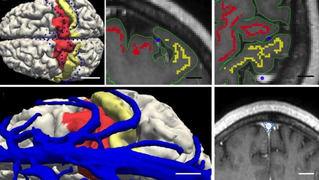
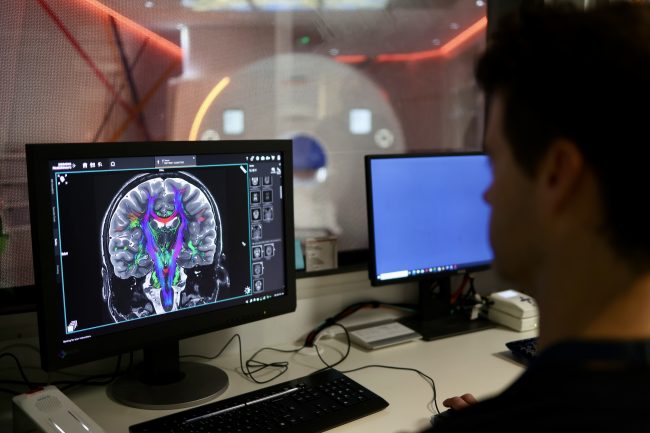
Modelling neural dynamics from non-invasive MEG recordings has the potential to provide crucial insights in the fast-evolving physiological and pathological activity that gives rise to such dynamics. Combining MEG data acquired at the Swinburne NIF node and 3-D velocity field approaches in epilepsy patients, temporospatial patterns in 3-dimensional brain space were obtained and spiral patterns (including singularities) were revealed at seizure onset region, potentially suggesting highly non-linear dynamics and locally unbalanced excitatory-inhibitory neural networks were developed and progressed at seizure onset (from onset up to 1200ms) within the localised brain region. Such developments build on existing work with MEG technology to emphasise the utility of MEG in understanding brain function in health and disease. 

Enhancing Low-Field MRI Images through Generative Deep Learning

Partner:
Monash University, The University of Queensland, Herston Imaging Research Facility, SAHMRI, The University of Western Australia

Imaging expertise:
Dr Kh Tohidul Islam

Instrument:
Utilizing paired datasets from 100 healthy participants, images were obtained at Monash Biomedical Imaging facility using Hyperfine Swoop (64mT) and Siemens Biograph mMR (3T) systems. The deep learning model was trained to generate synthetic 3T-like images from 64mT images, and their performance was compared against actual 3T images.

Acknowledgements:
This project is funded by the National Imaging Facility (NIF) and Hyperfine Inc. Special thanks are extended to the NIF at Monash Biomedical Imaging, Monash University, for their facilities and support.

View publication for more information.

Image description:
The image displays MRI scans of a 38-year-old male participant. It includes T1, T2, and FLAIR sequences, each showing four columns: 64mT, 3T, Synthetic 3T, and the difference between 3T and synthetic 3T images. The synthetic images closely resemble the 3T images, demonstrating minimal anatomical variation and highlighting the model’s effectiveness in enhancing 64mT images. This research aims to improve low-field MRI images (64mT) using a Generative Deep Learning method, making them comparable to standard high-field (3T) MRI images.


Better health for the young and older Australians 

Imaging studies that look at conditions in younger and older Australians are essential for understanding and promoting healthy development and ageing.
Early detection of biomarker changes in Huntington’s Disease using PET imaging

Partner:
SAHMRI

Imaging expertise:
Dr Muneer Ahamed
Ms Georgia Williams

Instrument:
Large animal PET scanner at SAHMRI

Acknowledgements:
Expertise and help from radiochemistry team and large animal technicians at SAHMRI and contributions from researchers at University of Antwerp, University of Cambridge and University of Warwick drives this project. The authors acknowledge CHDI Foundation, New York for providing the preclinical models.

Find out more information here.

Image description:
A representative PET image showing [18F]FDOPA uptake in neostriatum in preclinical models.

Huntington’s Disease (HD) is a devastating neurodegenerative condition that causes cognitive, movement and behavioural disturbances, which over time result in progressive disability and eventual death. SAHMRI’s NIF fellows Dr. Muneer Ahamed and Ms. Georgia Williams leading a preliminary feasibility study to validate the use of transgenic preclinical models of HD in PET imaging studies. This study is expected to monitor early-stage (disease onset) and longitudinally evaluate biomarker changes in HD following the slow progression of the HD. This will be first time PET imaging is carried out in this type of HD preclinical model, with the help of NIF’s dedicated large animal PET scanner at SAHMRI.


Growing use of imaging in agriculture and ecology

Imaging is accelerating as an important capability for agricultural and ecological sciences.
Understanding the ecology of coral reefs

Partner:
University of Western Australia

Imaging expertise:
Ms Diana Patalwala

Instrument:
MicroCT

Acknowledgements:
Mr Damian Thomson, CSIRO

Image description:
Micro computerized tomography (μCT) scans of experimental Porites sp. blocks deployed in the lagoon and on the reef slope Ningaloo Reef for 20 months (605 days). Block images show representative scans of two individual blocks (a) pre- and (b) post-deployment in green, and areas of (c) external and (d) internal erosion post-deployment in red.

Porites corals can reveal the past sea conditions by their oxygen isotopes, which reflect the temperature and rainfall of the seawater. This information is useful for studying how the climate and weather patterns have changed over time, and how physical and biological factors influence the distribution and abundance of organisms on the seafloor.

DTI tractogram of a quokka brain: Exploring how marsuipials adapt to different environments

Partner:
Biological Resource Imaging Laboratory (BRIL), UNSW

Imaging expertise:
Dr Andre Bongers
Mr Simone Zanoni

Instrument:
Bruker BioSpec Avance III 94/20 Preclinical MRI

Acknowledgements:
Jyothi Thittamranahalli KariyappaSimone ZanoniAndre BongersLydia TongKen W. S. Ashwell

View publication for more information.


Cultural contributions to materials, engineering and culture

Many varied industrial and research problems— such as chemical processes, materials science, environmental and ecosystems research, security, palaeontology and cultural preservation—are increasingly opening up to the benefits of advanced imaging technologies.
Discovery of a new nasal-emitting trident bat from early Miocene forests in northern Australia

Partner:
Biological Resource Imaging Laboratory (BRIL), UNSW

Instrument:
MILabs U-CT microCT scanner

Acknowledgements:
Suzanne J. HandMichael ArcherAnna GillespieTroy Myers

View publication for more information.

Understanding equine anatomy: Investigating intestinal muscle structure to prevent rupture

Partner:
Western Sydney University

Instrument:
Diffusion tensor image of a cross-section of equine intestine taken using the 11.7 T MRI at the Biomedical Magnetic Resonance Facility (BMRF) at WSU Campbelltown campus.

Acknowledgements:
Kate Averay1, Denis Verwilghen1, Marianne Keller3, Neil Horadagoda2, Marina Gimeno2

1Camden Equine Centre, University Veterinary Teaching Hospital Camden, Sydney School of Veterinary Science, University of Sydney, Australia

2Pathology Services, University Veterinary Teaching Hospital Camden, Sydney School of Veterinary Science, University of Sydney, Australia

3Sydney School of Veterinary Science, University of Sydney, Australia

Image description:
The research group is looking at equine jejunum rupture and whether there are any anatomical differences in the rupture region. Colour-coding shows the anisotropy of the tissue. The key is on the upper right and shows red is left-right, green is up-down and blue is in and out of the screen. You can clearly see two layers of muscle: the red-green layer wrapping around the intestine and the blue layer running longitudinally.

Victorian collaboration raises over $50m investment in critical imaging capabilities

[Image: La Trobe University – Olivia Newton-John Cancer Research Institute NIF Fellow, Dr Ingrid Burvenich with Minister Tierney]

The Victorian Government has invested $14.83m in National Imaging Facility’s (NIF) research infrastructure in Victoria, in partnership with the Victorian Biomedical Imaging Capability (VBIC), equating to a boost of just over $50m through collaborative co-investment. 

[Image: Victorian Minister for Higher Education, the Hon Gayle Tierney MP]

Victorian Minister for Higher Education, the Hon Gayle Tierney MP visited the Olivia Newton-John Cancer Research Institute on Tuesday, to highlight the impact of the collaboration and the State Government’s investment of through the Victorian Higher Education State Investment Fund (VHESIF) initiative.  

“Collaborative projects such as this demonstrate how our government is supporting higher education and industry to become international leaders in their field,” Minister Tierney said.

[Image: Victorian Minister for Higher Education, the Hon Gayle Tierney MP toured the facilities at the Olivia Newton-John Cancer Research Institute and the Austin hospital]

The funding is supporting the upgrade and expansion of imaging capabilities across NIF’s research facilities in Victoria, including The Florey, La Trobe University and the Olivia Newton-John Cancer Research Institute, Monash University, Swinburne University of Technology, and University of Melbourne in partnership with Peter MacCallum Cancer Centre and the Austin Hospital.  

Critical medical research in areas of national priority such as dementia cancer and epilepsy, as well as agriculture research will be enabled by the co-investment, which includes $26.7m from NIF through the Australian Government’s National Collaborative Research Infrastructure Strategy (NCRIS) funding scheme. 

Infrastructure funded under the collaboration includes human magnetic resonance imaging (MRI) capabilities at Swinburne University of Technology and The Florey, where state-of-the-art high-intensity focused ultrasound will support the development of new treatments for essential tremor and tremor-dominant Parkinson’s disease. 

In addition to this, the University of Melbourne upgraded their ultra-high-field 7T MRI (one of only two in Australia), and acquired a new human PET-CT. 

Preclinical capabilities including PET/MRI and PET/CT to support important drug discovery and testing have been installed at the Olivia Newton-John Cancer Research Institute/La Trobe University and Monash University. 

[Image: NIF preclinical capabilities at the Olivia Newton-John Cancer Research Institute]

A new research cyclotron at Monash (the Australian Precision Radiopharmaceutical Facility APRF) will enable the production of radioisotopes under GMP standards, and enhance Australia’s sovereign capability to produce therapeutics and diagnostics. Complementary to this, radiochemistry hotcell infrastructure upgrades at Monash, the Austin Hospital, and Peter MacCallum Cancer Centre will support the design and development of novel cancer treatments.  

[NIF radiochemistry capabilities at the Austin hospital/Olivia Newton-John Cancer Research Institute]

The funding also enables upgrades to the magnetoencephalography (MEG) at Swinburne, one of only two systems in Australia, supporting the study of brain function. 

[Image: NIF MEG at Swinburne University of Technology]

Development of specialised plant imaging capabilities at the University of Melbourne will underpin research into the effect of climate change on crops and soil, and strategies and applications for agricultural improvements to support Australia’s standing as a world leader in food and beverage production. 

NIF Chief Executive Officer, Prof Wojtek Goscinski said the co-contributed investment underpins transformational initiatives in a number of national priority areas including precision medicine, molecular imaging, drug discovery, diagnostics and plant soil imaging. 

“It’s a privilege for NIF to partner with the Victorian Government and VBIC to support Australia’s strategic science and research priorities” Prof Goscinski said. 

“These capabilities will support Australia as a world-leader in applying advanced imaging technology, resulting in better healthcare, better products, and important discoveries.”

Read the Victorian Government’s announcement here. 

National Imaging Facility Showcase at ANZSNM 2023

National Imaging Facility will host a Showcase at ANZSNM 2023, featuring presentations from Australia’s advanced imaging network.

See the full ANZSNM Program and register here.

Sunday 28 May, 1:15pm – 2:00pm
City Room 3/4
Adelaide Convention Centre

Chair:   Prof Steven Meikle
Head, Imaging Physics Laboratory
Brain and Mind Centre, University of Sydney

Time

1:15pm

Topic

Introduction and session open

Speaker

Prof Wojtek Goscinski
Chief Executive Officer
National Imaging Facility

1:20pm

Australian National Total Body PET Facility

Dr Georgios Angelis
NIF Total Body PET Fellow
University of Sydney

1:30pm

Monash MHELTHERA Lab and cyclotron capabilities

Prof Christoph Hagemeyer
Acting Director, NIF Node Director
Monash Biomedical Imaging

1:40pm

SAHMRI Radiopharmaceutical Chemistry

Dr Edward Robins
Head, Radiopharmaceutical Research and Development
Molecular Imaging and Therapy Research Unit
SAHMRI

1:50pm

New capabilities of the Western Australia Node

A/Prof Roslyn Francis
Head, Department of Nuclear Medicine and the WA PET Service, Sir Charles Gairdner Hospital
NIF Deputy Node Director
University of Western Australia

Dr Heidi Espedal
NIF Facility Fellow
University of Western Australia

International partnership launches Monash University-Helmholtz lab to solve global oncology, cardiology and infectious disease challenges

Monash University has partnered with the Helmholtz-Zentrum Dresden-Rossendorf (HZDR), a member of the Helmholtz Association of German Research Centres to establish the Monash-Helmholtz Laboratory for Radio-Immuno-Theranostics (MHELTHERA) to enable clinical translation at a launch event in Melbourne today.

The MHELTHERA Lab is a collaboration to optimise non-invasive imaging techniques and personalised therapy, drawing on diagnostic and therapeutic expertise in cancer, infectious and cardiac disease.

HZDR and Monash are developing next-generation biomedical imaging platforms and the MHELTHERA Lab will specialise in molecular imaging through nuclear techniques such as Positron Emission Tomography (PET) and Single Photon Emission Computed Tomography (SPECT), as well as optical techniques including Fluorescence Imaging (FLI).

Prof Sebastian M. Schmidt, Scientific Director of HZDR, Mr Simon McKeon AO, Chancellor of Monash University, Prof Otmar Wiestler, President of Helmholtz Association of German Research Centres and Prof Christoph Hagemeyer, National Imaging Facility Monash Biomedical Imaging Node Director opened the MHELTHERA Lab.


What are radio-immuno-theranostics?
Radio-immuno-theranostics pair diagnostic radiopharmaceuticals, applied through molecular imaging to identify tumours, with combined radiation therapy and immunotherapy to target cancer and enable clinicians to ‘see what they treat’.
Nuclear theranostics are a new and highly impactful field, revolutionising cancer therapy through a personalised approach.

The MHELTHERA Lab was officially opened at its Australian site, Monash Biomedical Imaging (MBI), a node of the National Imaging Facility (NIF) by Mr Simon McKeon AO, Chancellor of Monash University, Prof Otmar Wiestler, President of Helmholtz Association of German Research Centres, and Prof Sebastian M. Schmidt, Scientific Director of HZDR.

Mr McKeon said the partnership is an excellent example of the power of international collaborations.

“No single institution or nation has all the answers when it comes to addressing the most pressing health challenges of our time. By partnering with institutions such as Helmholtz, we can bring together the best talent, knowledge, and resources to tackle these challenges together,” Mr McKeon said.

Prof Wiestler said the Helmholtz Association is Germany’s largest research organisation, developing solutions and innovative technologies to preserve the foundations of human life.

 “By supporting MHELTHERA, we are bringing together leading international researchers to develop a highly original class of radiopharmaceuticals that will provide new treatment prospects for cancer patients in particular,” Prof Wiestler said”.

Prof Schmidt said MHELTERA will offer excellent opportunities, especially for young researchers.

“The cooperation is particularly attractive for PhD students, who will have the opportunity to improve their skills through joint research training programs of HZDR and Monash University,” Prof Schmidt said.

MHELTHERA builds upon a successful long-standing collaboration between the two organisations and is led by Prof Christoph Hagemeyer from Monash University, who is also the NIF MBI Node Director, and Prof Michael Bachmann from the HZDR Institute of Radiopharmaceutical Cancer Research. 

“The joint laboratory will leverage the research infrastructure capabilities at both sites to accelerate the development of promising technologies in the field of precision medicine,” Prof Hagemeyer said.

NIF CEO Prof Wojtek Goscinski said there are ten Helmholtz International Labs dedicated to innovative research located around the world, and it was a privilege for NIF co-funded infrastructure and staff to support the first of its kind in Australia.

“NIF has invested in world-class radiochemistry expertise and capabilities at MBI and this is an exciting opportunity for us to support a unique Helmholtz lab at the global cutting-edge of cancer research,” Prof Goscinski said.

“The application of radiopharmaceuticals and nuclear theranostics through PET imaging is revolutionising cancer treatment, and NIF is committed to improving health outcomes by supporting these innovative medical products,” Prof Goscinski said.

NIF has committed $4.95 million to a new cyclotron, enabling critical radioisotope production, and an expanded radiochemistry facility at MBI which is central to the infrastructure supporting the collaboration.

This critical infrastructure will complement the lab, providing a platform for manufacturing capability, drug innovation, treatment and advanced training.

Click here for more information about MHELTHERA.


Monash University Contact
Professor Christoph Hagemeyer
Director (Acting) of Monash Biomedical Imaging
Tel: +61 3 9905 0100
Email: Christoph.Hagemeyer@monash.edu

HZDR Contact
Professor Michael Bachmann
Director Institute of Radio­pharma­ceutical Cancer Research
Tel: +49 351 260 3170
Email: m.bachmann@hzdr.de

#ImagingTheFuture Week: Enabling breakthroughs in biomedical science and technology

Chan Zuckerberg Initiative’s (CZI) Imaging the Future Week puts a spotlight on the importance of imaging science in biomedicine, and the value of the global imaging community in translating health research.

Imaging is unlocking solutions to the world’s biggest challenges across commercial, clinical and research fields and has helped innovate in bioengineering, biology, medical technology and science, pharmaceutical and non-pharmaceutical therapies.

National Imaging Facility (NIF) supports the Imaging the Future Week initiative, and the 2023 event is focused on highlighting advances in technology and the impact this has on our understanding of health and disease.

As we continue to meet the evolving needs of modern research, NIF is accelerating new technology, enabling experts to develop protocols, tools, imaging data, and the application of imaging to solve complex problems – scroll on to find out more.


Better evidence for decision-making in health

Advanced imaging methods and analysis provide critical evidence for decision-making across all aspects of health and clinical science to keep Australia healthy.


Australia’s largest investment in molecular imaging
Australia’s first open access research Total Body Positron Emission Tomography scanner is NIF’s largest investment to date, and it will deliver a transformative understanding of complex health problems. Next-generation molecular imaging and radiopharmaceuticals are revolutionising how we see biological processes, paving the way for better diagnosis and treatment of chronic, systemic adult and childhood diseases. The instrument will produce high quality data at lower doses of radiation. It can be used to capture information from all body organs simultaneously to build a better picture of complex processes such as ageing, metabolism, brain signalling, behaviour, cognition and drug interactions.

Multidisciplinary collaboration to improve epilepsy outcomes
MRI imaging technology, AI, machine learning and data analysis are helping improve the lives of 150,000 Australians with epilepsy. The Australian Epilepsy Project will combine neuroimaging with cognitive and genetic data, and integrate them using AI, to develop predictive tools that will guide diagnosis and highlight opportunities for precision treatment. Expertise from the Florey Institute of Neuroscience and Mental Health, the University of Melbourne, Monash University and Austin Health drives the project, aiming to reduce seizure frequency and the risk of injury or death.


Better health for the young and older Australians

Imaging studies that look at conditions in younger and older Australians are essential for understanding and promoting healthy development and ageing.


Understanding the development of cerebral palsy
NIF is contributing to valuable data assets, including the first collection to show the way that muscles grow in children with cerebral palsy. The MUGgLE Study is the first longitudinal study comparing muscle growth in the development of children with cerebral palsy and typically developing children. The study is a partnership between Neuroscience Research Australia, the University of NSW and the Cerebral Palsy Alliance Research Institute. Imaging is being used to study muscle tightening and shortening as it happens, with high-resolution measurements of the architecture of whole muscles, giving researchers detailed, anatomically accurate, three-dimensional reconstructions to understand disordered muscle growth. The project has included the development of imaging methods and algorithms to be able to study this, adapting the acquisition protocols as well as the imaging analysis techniques to accommodate measurement of the specific features of muscles.

Brain-computer interface restoring independence after paralysis
An implant the size of a paperclip is allowing people who are paralysed to operate technological devices using their thoughts without open brain surgery. NIF expertise and the 7T MRI at the University of Melbourne enabled early developments of the device which can translate brain signals from the inside of a blood vessel into commands on a computer.

The Synchron Stentrode is a world first brain-computer interface designed to restore functional independence in patients with paralysing conditions like ALS. The device was named one of TIME Magazine’s best inventions of 2021, and is currently undergoing expanded human clinical trials in preparation for submission to the FDA.


Equitable regional and rural health

Crucial to societal equity and research quality, delivering a geographically distributed network of advanced imaging to support research and personalised medicine, and taking part in medical trials, is a major national challenge.


Bringing health equity to regional and rural Australia
NIF is deploying four low-field portable MRI scanners to remote and regional sites to help researchers apply this affordable imaging technology in rural areas. The national mobile magnetic resonance (MR) network will be the first project of its kind world-wide and is a collaboration with partners including Monash University, University of Queensland, South Australian Health and Medical Research Institute (SAHMRI), the Alfred Hospital, Royal Perth Hospital, University of Western Australia and MedTech company, Hyperfine. These portable scanners will be used to understand how this fast-developing technology can help diagnose stroke, traumatic brain injury, and other conditions after testing in research laboratories at NIF nodes to build the usability of low-field MR, including developing techniques to maximise data quality and improve image processing.

Imaging mobilises ground-breaking field ventilator for deployment in the COVID-19 crisis
NIF provided critical support in preclinical testing to mobilise the now commercialised ventilator, 4DMedical ‘XV technology’ at the LARIF multipurpose fluoroscopy laboratory. A team of Australian collaborators, including biomedical company 4DMedical and University of Adelaide scientists created the ground-breaking, simple to use ‘field ventilator’ that can be locally produced at a low cost from easily acquired parts. It was developed in response to the global COVID-19 crisis, which identified potential shortages in essential medical equipment.

Communities in regional Australia to benefit from world’s first mobile magnetic resonance imaging network

National Imaging Facility (NIF) is deploying four low-field portable MRI scanners to remote and regional sites to help researchers apply this affordable imaging technology in rural areas.

Australians living in regional and rural areas unduly suffer lower life expectancy and a higher burden of diseases because of poorer access to health services, including reduced screening, late detection and barriers to treatment compared with people living in metropolitan areas.

The national mobile magnetic resonance (MR) network will be the first project of its kind world-wide and is a collaboration with partners including Monash University, University of Queensland, South Australian Health and Medical Research Institute (SAHMRI), the Alfred Hospital, Royal Perth Hospital, University of Western Australia and MedTech company, Hyperfine.

The Hyperfine Swoop is the world’s first highly portable MR imaging system capable of providing neuroimaging at the point-of-care, designed to fit inside elevators and through doorways to be manoeuvred directly to a patient’s bedside, and plug into a standard electrical outlet.

These portable scanners will be used to understand how this fast-developing technology can help diagnose stroke, traumatic brain injury, and other conditions after testing in research laboratories at NIF nodes in Victoria, Queensland, South Australia and Western Australia.

The scanners will enable real time tele-reporting and either remote operation or point-of-care use with low training requirements.

NIF is uniquely positioned to support work to build the usability of low-field MR technology, including developing techniques to maximise data quality.

Researchers at these sites will scan subjects on low-field mobile MR and high-field 3T MRI instruments to build a unique database that can be used to bridge the gap in outputs.

This valuable data will be made available by NIF to researchers to develop techniques to improve image processing and better understand how low-field scans can be interpreted.

Head of the Imaging Analysis Team at Monash Biomedical Imaging and Project lead Chief Investigator, Dr Zhaolin Chen said the collaborative work across NIF Nodes was critical to the success of the project.

“This nationwide network is critically important to identify a viable pathway for point-of-care MRI technology to be used in Australia,” Dr Chen said.

“Multi-site data acquisition is already underway and AI-based solutions to expand utility in regional Australia are in development.

“The network enables our project team to share knowledge, cross-validate findings, optimise resources and plan the next steps, which ultimately provides a route from research into clinics,” Dr Chen said.

NIF Chief Executive Officer, Prof Wojtek Goscinski said there were additional long-term advantages to deploying the national mobile MR network to regional Australia.

“NIF is focused on keeping Australia at the forefront of imaging, and the national mobile MR network is an innovative application of new technology to improve accessibility,” Prof Goscinski said.

“We hope these data collections and the AI models researchers build using them will lead to better technology that will improve treatment and diagnosis for Australians.

“This work will provide the foundation for the development and application of AI in clinical practice for low-field MR scanners, with experts optimising image quality for clinical data usability with reduced noise and improved resolution.

“The national mobile MR network and NIF’s increased national human imaging reach will enable innovative health research in remote populations, improve low-field MR technology, and over the long run will help increase access to better healthcare, professional training and socio-economic equity,” Prof Goscinski said.

ISMRM and ISMRT ANZ Chapters’ Annual Meetings shine a light on national imaging expertise and infrastructure

[Image: Presentation award winners at ISMRM ANZ, Honours student, Arunan Srirengan, Dr Ed Green, Dr Gwen Schroyen and Dr Myrte Strik. Photo credit: Dr Adam Clemente]

The Australian and New Zealand Chapters of the International Society for Magnetic Resonance in Medicine (ISMRM) and the International Society for Magnetic Resonance Radiographers and Technologists (ISMRT) held their Annual Meetings in Sydney last month, highlighting the work of leading national researchers and clinicians, including members of the NIF network.

NIF enables coordinated open access to magnetic resonance expertise and infrastructure to support leading national researchers and clinicians, and proudly supported the events.

ISMRM ANZ Joint Chapter Annual Meeting 9-10 Nov

ISMRM ANZ hosted sessions on revolutionising MRI technology, advances in neuroimaging, and clinical applications of advanced MRI, in addition to keynote speakers neurologist and leader in stroke medicine, Prof Mark Parsons and Director of the Institute of Medical Physics at the University of Sydney, Prof Annette Haworth.

Dr Zhaolin Chen was a key speaker in the Revolutionising MRI technology session, presenting the NIF Point-of-Care project, a collaboration between NIF, Australian hospitals, and US medical device manufacturer, Hyperfine, to build the usability of low-field MRI and bring critical imaging to remote Australia and deploy imaging in challenging clinical environments such as COVID wards.

[Image: Dr Zhaolin Chen presenting the NIF Point-of-Care Magnetic Resonance project at ISMRM ANZ]

A number of other NIF users spoke at ISMRM ANZ, including:

  • Rebecca Glarin from the University of Melbourne, presenting findings from her PhD on ‘Optimising functional brainstem imaging of sympathetic drive with ultra-high field MRI’.
  • Dr Shahrzad Moinian from the University of Queensland Centre for Advanced Imaging, presenting ‘In vivo microstructural border delineation between areas of the human cerebral cortex using magnetic resonance fingerprinting (MRF) residuals’.
  • Honours student Arunan Srirengan presenting ‘Early identification of cerebral small vessel disease in obstructive sleep apnoea patients using magnetic resonance spectroscopy: a pilot study’, featuring data obtained on the NIF 3T MRI at NeuRA. This session was awarded second prize in the oral presentation awards.
  • Dr Myrte Strik from the University of Melbourne, presenting ‘Altered network topology in patients with visual snow syndrome: a resting-state 7 Tesla MRI study’, winning the award for best Early Career Researcher Data Blitz presentation.

[Image: Dr Shahrzad Moinian from the University of Queensland Centre for Advanced Imaging. Photo credit: Dr Adam Clemente]

Congratulations to University of Melbourne NIF Fellow, Prof Brad Moffatt as ANZ ISMRM Chapter President on the success of the 2022 meeting hosted at UNSW.

ISMRT ANZ Joint Chapter Annual Meeting 12-13 Nov

The ISMRT ANZ 2022 joint meeting program theme was MRI: Past, Present and Future, and featured a range of internationally renowned speakers demonstrating future technologies and cutting-edge imaging techniques.

Keynote presenters included Medical physicist and human brain imaging academic researcher Dr Samantha Holdsworth, Chief of the Quantitative Medical Imaging Laboratory, USA National Institute of Biomedical Imaging and Bioengineering, Dr Carlo Pierpaoli, and founding member of the Society of Cardiovascular Magnetic Resonance and Principal Investigator for the Cardiac Atlas Project, Prof Alistair Young.

NIF Senior Manager and Senior Research Scientist – National Magnetic Resonance Capability, Dr Shawna Farquharson was a key speaker at the Diffusion Weighted Imaging (DWI) Forum, presenting on ‘DWI: Principles and practical applications’.

[Image: Dr Shawna Farquharson, National Imaging Facility]

NIF users showcased at ISMRT ANZ included:

  • Prof Lynne Bilston from NeuRA, presenting ‘Brain Elastography’.
  • Sarah Daniel from the University of Queensland Centre for Advanced Imaging, presenting ‘Image quality enhancement using deep learning for in vivo human kidney MRI’.

[Image: Ms Sarah Daniel from the University of Queensland Centre for Advanced Imaging]

Congratulations to all presenters at ISMRM and ISMRT ANZ.

Victorian imaging network meets to map out innovative future

[Pictured: VBIC Annual Network Meeting guest speakers Prof Lindy Fitzgerald and Prof Amy Brodtmann] 

Innovation, industry partnerships and commercialisation will be among the topics discussed at a meeting bringing together NIF’s capabilities from around Victoria on November 24. 

The annual Victorian Biomedical Imaging Capability (VBIC) network meeting will attract researchers, clinicians and industry partners to Monash Biomedical Imaging in Melbourne. 

Participants will represent NIF nodes at the University of Melbourne, Monash, Swinburne, the Florey, Olivia Newton John Cancer Research Institute and La Trobe University. 

Neurologist Prof Amy Brodtmann will draw on her interests in imaging, stroke and dementia to present research findings from the Cognitive Health Initiative at Monash and Alfred Health  

Guest speaker Prof Melinda Fitzgerald from Curtin University and the Perron Institute will present on a national initiative she heads as CEO, called Connectivity, the Mission for Traumatic Brain Injury.  The initiative includes use of MRI and clinical biomarkers in a national trial to improve the diagnosis and prognosis of traumatic brain injury. 

Sessions at the meeting will also cover the work of imaging experts in research programs employing ultra-high field MRI, CT and PET; as well as presentations from experts in industry partnering and commercialisation. 

NIF Chief Executive Officer Prof Wojtek Goscinski said the meeting would provide a platform for early career researchers and emerging leaders, a showcase for new-generation imaging, and opportunities for important in-person networking. 

It would also enable discussion on the NIF Imaging Roadmap, including supporting innovation and ensuring Australia’s international comparative advantage, Prof Goscinski said. 

The roadmap will add to the substantial impact and jobs that VBIC and NIF have already delivered, with a recent report estimating more than $350 million in economic activity for Victoria. 

VBIC nodes have grown to employ more than 150 FTE imaging staff, and partner with more than 90 organisations, including Austin Health, CSIRO, Melbourne Health, Mental Health Research Institute and the Peter MacCallum Cancer Centre. 

Capital investments have reached $37 million, providing access to human and preclinical MRI and PET-CT, preclinical DEXA scanners and confocal endomicroscopy, as well as magnetoencephalography and nuclear scintigraphy. 

A massive $235 million in major grants has been secured, enabling new research projects in cancer, infection and inflammation, brain function, epilepsy, dementia and even long-term aspirin use. 

Click here for more information about the VBIC network meeting and full programme.

NIF to demonstrate impact of coordinated data and AI at RANZCR ASM 2022

National Imaging Facility (NIF) will host a session at the Royal Australian and New Zealand College of Radiologists (RANZCR) 72nd Annual Scientific Meeting (ASM) this month.

The NIF Showcase will highlight critical expertise and human imaging projects from across Australia’s advanced imaging network, including regional MRI and life-changing imaging for Australians living with epilepsy.


Point-of-care imaging leveraging AI to grow healthcare equity in regional Australia

Head of Diagnostic and Interventional Radiology research at Alfred Hospital, Prof Meng Law will present on new technology for point-of-care imaging and regional MRI. Prof Law is an expert on neuroimaging and AI, and his presentation will focus on federated deep learning for signal-to-noise ratio imaging and motion correction, using NIF’s low-field magnetic resonance network.

NIF is deploying four low-field MRI scanners to remote and regional sites to help researchers apply this affordable imaging technology in rural areas. These scanners will be used to understand how this fast-developing technology can be used to diagnose stroke, traumatic brain injury, and other conditions after testing in research laboratories at NIF nodes.


Imaging networks and datasets to support life-changing platform for more than 150,000 Australians living with epilepsy

Clinical Director of The Florey Institute of Neuroscience and Mental Health, Prof Graeme Jackson will present on the Australian Epilepsy Project (AEP), reducing diagnosis uncertainty and fast-tracking optimal treatment by combining advanced imaging, genetics, cognition, and artificial intelligence (AI).

Prof Jackson is the Chief Investigator on the AEP Platform, which will drive major advances in decision support tools for epilepsy, and NIF’s national human MR network is set to enable scanning across Australia.

The data collected by the AEP will provide a rich resource for addressing many other traditional science and mechanistic questions in epilepsy to progress epilepsy research worldwide.


NIF CEO Prof Wojtek Goscinski said the invitation to showcase NIF at the RANZCR ASM was an opportunity to highlight the transformation of imaging through AI and big data, and to underline the unique capabilities that NIF provides.

“We’re privileged to have world-class speakers Prof Meng Law and Prof Graeme Jackson presenting on two projects that are supported by data collections and the AI models around them, which will lead to better treatment and diagnosis for Australians,” Prof Goscinski said.

The impact of imaging in radiology is only increasing, with experts now able to extract quantifiable information from ever larger data collections by applying machine learning methods such as deep learning and convolutional neural networks.

Big data and AI have a transformative effect on radiology, enhancing patient outcomes by distinguishing irregularities and patterns in data collections, and enabling diagnosis with speed and accuracy.

“NIF is focused on keeping Australia at the forefront of imaging technology and imaging data analytics, and is exploring a range of activities to increase uptake of machine learning in imaging, including data infrastructure and imaging quality,” he said.

The NIF Showcase session will also see a panel of experts discuss opportunities for collaboration between NIF and RANZCR for the benefit of medical research.

View the NIF Showcase agenda below:

RANZCR ASM NIF Showcase: Friday 28 October, 08:30-10:00

TIMETOPICSPEAKER
8:30IntroductionA/Prof Sanjay Jeganathan
RANZCR President
8:35Introduction to National Imaging FacilityProf Wojtek Goscinski
NIF Chief Executive Officer
8:45Point of Care Imaging and Regional MRI 
NIF Low Field MR Network
Federated Deep Learning for SNR, Motion Correction
Prof Meng Law
Professor and Director of Radiology, Alfred Health
Director of iBRAIN
Monash University
9:05The Australian Epilepsy Project
MR guided focused ultrasound
Prof Graeme Jackson
Chief Investigator, Australian Epilepsy Project
Clinical Director, The Florey Institute of Neuroscience and Mental Health
9:25Panel discussion
RANZCR and NIF: Opportunities for collaboration for the benefit of Australian healthcare
Chair: Prof Paul Parizel
NIF UWA Node Director
Chair, UWA Medical School
David Hartley Chair in Radiology, UWA Medical School

Prof Wojtek Goscinski

Prof Meng Law

Prof Graeme Jackson

A/Prof Christen Barras
Radiologist
Co-Convenor RANZCR ASM 2022

Dr Lauren Oakden Rayner
Director, Research
Royal Adelaide Hospital Medical Imaging

The RANZCR ASM will take place at the Adelaide Convention Centre on 27–30 October 2022.

Under the theme of Reflect, Revive, Reimagine, the 72nd RANZCR ASM will be the largest meeting to date, with an innovative scientific program of over 250 presentations across 70+ sessions.

The four-day conference has lined up leading international and local radiologists to share best practices and highlight emerging medical advancements.

Inaugural NIF Scientific Symposium kicks off #NationalScienceWeek

Leading researchers, clinicians and industry attended the inaugural National Imaging Facility (NIF) Scientific Symposium on 12 August.

The event kicked off National Science Week for NIF, highlighting the critical role of collaboration in translating research challenges to benefit industry and keep Australians healthy, with the theme ‘National partnerships for innovation and impact’.

NIF CEO Prof Wojtek Goscinski said the Symposium was an excellent opportunity to highlight ground-breaking work from Australia’s world-class imaging community.

“It was a privilege to host experts from across Australia, including keynote speakers Prof Graeme Jackson, Prof Louise Emmet and Prof Gemma Figtree, whose work is at the leading edge of imaging globally,” Prof Goscinski said.

“I’d also like to extend my thanks to the presenters who delivered an excellent Technology Showcase session, and Health and Medical Translational Challenges session.

“A particular highlight was hearing from our industry partners, including Telix Pharmaceuticals, Clarity Pharmaceuticals, Cochlear and Nyrada, who discussed the way they engage with national imaging research infrastructure.

“NIF is privileged to have a strong network of world-leading expertise at our fingertips and it was an honour to bring some of these people together to present their work and share ideas at the 2022 Symposium,” he said.

Keynote presentations of the Symposium included:

  • ‘The Australian Epilepsy Project’, Prof Graeme Jackson
  • From mouse to Medicare: the PSMA story in Australia’, Prof Louise Emmett
  • Coronary artery imaging to inform the next Frontier of heart attack prevention’, Prof Gemma Figtree

The Technology Showcase session highlighted NIF’s latest capabilities, including tools for processing and interpreting data, and applications of imaging to solve complex problems, including:

  • ‘Ultra-high field magnetic resonance imaging’, Prof Leigh Johnston and Prof Markus Barth
  • ‘Bringing imaging to rural Australia with a national network of low field mobile MR scanners’, Dr Zhaolin Chen
  • ‘Australian Imaging Service: The national platform for trusted data management and analysis’, Dr Ryan Sullivan
  • ‘Magnetic Particle Imaging’, Dr Andre Bongers
  • An insight into MicroCT imaging: recent advances, applications and impact on research and innovation’, Ms Diana Patalwala
  • Preclinical Research: The Crucial Step in Medical Advancements’, Dr Chris Christou

The Health and Medical Translation Challenges session provided an opportunity for attendees to hear from clinicians and researchers about their journey to making translational impact, including:

  • Neuroimaging in clinical trials: Perspectives of a clinician-researcher’, A/Prof Sylvia Gustin
  • The Australasian Radiopharmaceutical Trials network (ARTnet)’, A/Prof Ros Francis

The Industry Discussion Panel opened up conversation on how imaging accelerates and underpins innovation and future opportunities, with speakers:

  • Dr David Cade, Chief Executive Officer, Telix Pharmaceuticals Asia Pacific
  • Dr Matt Harris, Chief Scientific Officer, Clarity Pharmaceuticals
  • Dr Zachary Smith, Director, Algorithms and Applications, Cochlear
  • Dr Jasneet Parmar, Neuroscience Researcher, Nyrada Inc
Privacy Settings
Youtube
Vimeo
Google Maps