Members of the NIF network recognised internationally as in-person conferences return
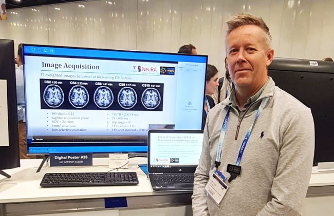
[Pictured: UNSW-NeuRA Facility Fellow, Dr Michael Green presented a study titled “Effect of Compressed SENSE on Freesurfer parcellation precision” which was a collaboration between NeuRA researchers, Philips Australia and New Zealand, and UNSW.]
In-person events have returned – and over the last few months, leading edge experts from the NIF network have attended, presented, and taken the opportunity to collaborate at conferences like ANZSNM and ISMRM.
We’re proud to acknowledge the members of the NIF network who have presented their globally significant work to the greater imaging communities.
We congratulate University of Sydney-ANSTO Node Co-Director, Prof Fernando Calamante as President of ISMRM on the success of the 2022 31st Annual Meeting hosted in London, UK in May.
We also recognise the incredible achievement of Dr Shawna Farquharson as recipient of the ISMRT 2022 Distinguished Service Award at the same event.
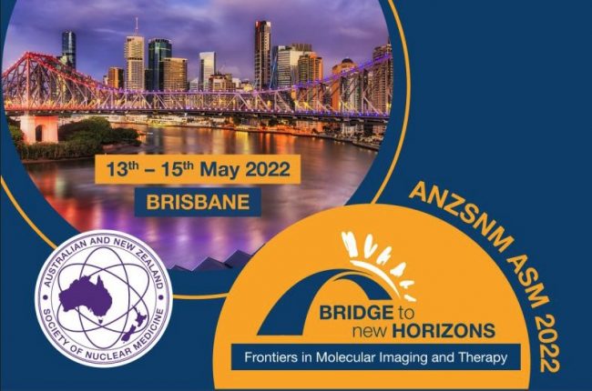
Back in Australia, NIF kicked off events with a Molecular imaging and Radiopharmaceuticals Capability Showcase at ANZSNM. We were honoured to invite world-class speakers from within our network, Prof Steven Meikle, A/Prof Roslyn Francis, Prof Gary Egan, Prof Kristofer Thurecht and Dr John Bennett to present during the NIF session.
We look forward to seeing more of our network at upcoming events – stay tuned for the NIF Scientific Symposium next month in Sydney. Save the date for Friday 12 August.
Here are some more highlights from the NIF network attending events so far this year:
QLD Node Director ISMRM
| Why did you attend? Many reasons: present group results; moderator of sessions; member of study groups and initiatives
What was the highlight of the event for you? Catching up with fellow researchers
What would you say to someone considering attending next meeting? Best check the hybrid setup, i.e. what is available in person and what is available online |
NeuRA Facility Fellow ISMRM | Why did you attend? Primarily it was a great way to re-connect with colleagues and share ideas in an old-fashioned, non-Zoom type of way. I presented a study titled “Effect of Compressed SENSE on Freesurfer parcellation precision” which was a collaboration between NeuRA researchers, Philips Australia and New Zealand, and UNSW. The study assessed the reliability of an MRI acceleration techniques designed to speed up the time it takes to acquire images. We wanted to provide a guideline for MR researchers wanting to reduce scan time while acquiring high quality data.
What was the highlight of the event for you? The face-to-face aspect of a conference was a real highlight. It was a nice compliment and surprise to see Philips also present data from our study to a global audience as validation for their acceleration techniques employed on their MRI machines. I also received some interesting feedback regarding the study analysis which I may implement before publishing the manuscript.
What would you say to someone considering attending next meeting? Study the conference schedule well before attending then pick and choose which seminars you’d like to attend. Then talk to as many people as possible. In person! |
UWA Facility Fellow ANZSNM | Why did you attend? This is a good meeting to attend to connect with the other radiochemists in Australia. Due to COVID I had not had a chance to do this in a long while. I was also very keen to see the Q-TRaCE labs at Royal Brisbane, as we have a good working relationship between them and us at Sir Charles Gairdner Hospital. I was able to let people know I had moved across to the NIF Node at UWA and was able to speak about our new lab and facilities being built now in Perth during my talk on the Saturday What was the highlight of the event for you? While ANZSNM was a great chance to hear some great talks and connect with a lot of people, it was also exciting to tour the labs at Q-TRaCE and the Centre for Advanced Imaging at UQ, where we also had our national Cyclotron User Group meeting. What would you say to someone considering attending next meeting? There are just not that many radiochemists in Australia, and the ANZSNM (along with the EPSM) is a great opportunity to see meet each other in person and see how the radiopharmaceuticals we make are being used to image and treat disease around the country.
|
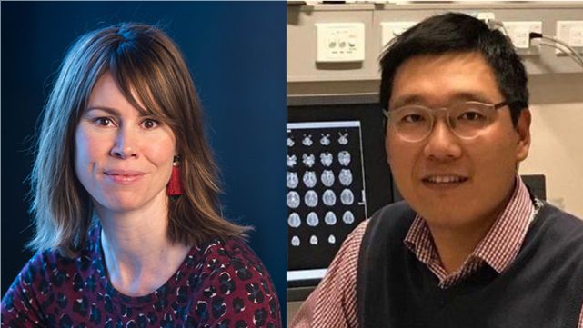
UWA Facility Fellow ISMRM
| Why did you attend? I presented a project shared between my current role as NIF fellow and my previous job in London.
What was the highlight of the event for you? My highlight was discussing potential new collaborations within Australia and internationally.
What would you say to someone considering attending next meeting? I think this is also a key reason to go to these conferences – to help explore new collaborations to benefit our imaging centres and community. |
Monash Informatics Fellow ISMRM (Virtual) | Why did you attend? My abstract was accepted as an online power pitch presentation in the ISMRM 2022 conference. And I virtually co-chaired one of the gather.town sessions in the theme of imaging processing and analysis. What was the highlight of the event for you? The main highlight was the talk provided by one of the famous AI researchers, Yann LeCun, and his topic was ‘Future AI research in medical imaging‘. The key take-home message is the shifting from supervised to self-supervised learning framework in general AI and medical imaging research. |