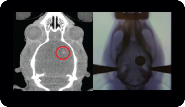
Development of Microbeam Radiation Therapy

Only two in ten people diagnosed with brain cancer will survive for at least five years, a figure that has barely improved in the last 30 years. Due to the blood-brain barrier and the risk of damaging normal brain tissue, brain cancer treatments are complex and multidisciplinary. Despite advances in surgical techniques, radiotherapy and chemotherapeutics, brain tumours remain challenging to treat. Hence, image-guided microbeam radiation therapy (MRT) with nanoparticle enhancement is being investigated for the treatment of brain cancer in animal models.

Read More

National Preclinical PET QA

The NIF Molecular Imaging & Radiochemistry (MIR) Theme is a group of NIF Fellows, Directors, and users of NIF facilities that focus on state-of-the-art radiochemistry and molecular imaging applications using PET, SPECT, and MRI.

Integrating preclinical PET systems into a national resource requires the development of defined QA programs to monitor and integrate the data from individual systems. Hence, the MIR Theme initiated a national quality assurance (QA) program for the NIF preclinical PET instruments.

Read More

Hyperpolarized 129Xe MR imaging of lung

Magnetic Resonance Imaging (MRI) has a range of applications in medical diagnosis, and more than 25,000 scanners are estimated to be in use worldwide. However, lung imaging suffers from some technical challenges, limiting its application in pulmonary disease diagnosis and treatment.


Due to low proton density, movement and high susceptibility difference between air and tissue, conventional proton MRI struggles to image lung tissue and function. These can be partially overcome by the introduction of contrast agents into the lung. Hyperpolarised gases are promising contrast agents for imaging lung structure and function. The two most common gasses are helium (3He) and xenon (129Xe) isotopes. Helium isotopes are challenging and expensive to obtain, and do not provide significant functional readouts. Xenon can be challenging to work with, but promises novel physiological measurements not previously feasible. This resulting technique, termed hyperpolarized 129Xe MR imaging, has revolutionised the field of functional lung imaging.


Five years ago, an Australian Research Council (ARC) grant was awarded to researchers at Monash Biomedical Imaging (MBI) and ANSTO to design and build a machine that could reliably produce hyperpolarized xenon. In this project, Dr Wai Tung Lee and NIF Facility Fellow Dr Gang Zheng have teamed up to develop a new multimodal technology capable of high-quality investigations of the human lungs. While Dr Lee, of ANSTO, was responsible for the construction of the polarizer and production of polarized 129Xe for MR imaging, Dr Zheng, of Monash University, designed the MR experiments and adapted imaging protocols.

Figure 1. Schematic diagram of the polarizer system. Note: Some details omitted for clarity.

Hyperpolarized 129Xe (HP-Xe) gas was generated in a custom designed and constructed SEOP system at Monash Biomedical Imaging (Figure 1 and Figure 2). The system consists of a gas-polarizing unit and a gas injection and recycle unit. The core component of the gas-polarizing unit is an optical pumping cell (OPC) where the gas is polarized. The OPC is placed in an oven to regulate the amount of Rb vapour by maintaining a constant temperature between 60ºC-90ºC. The optical pumping process uses two 240W narrow-bandwidth lasers tuned to the Rb D1 transition wavelength of 794.7nm. Additional optics condition the laser light to circular polarization and shape the laser profile to fully illuminate the OPC. HP-Xe is produced in batches rather than using a continuous-flow process.

Figure 2. The spin-exchange-optical pumping system. A. Gas polarizing unit in Room 1; B. Gas injection and recycling unit in Room 2.

MR experiments were performed on a clinical 3T whole-body system (Skyra, Siemens Medical Solutions, Erlangen, Germany) with a broadband RF amplifier. HP-129Xe MRI was enabled using a bird-cage transmit/receive chest coil (RAPID Biomedical GmbH, Wuerzburg, Germany). A 2D-GRE sequence for X-nuclei MRI was used to image HP-Xe gas in lung. Proton channel images were acquired for the localization of the lung. The resonance frequency of 129Xe was set to 34.09 MHz on the 3T scanner. The human subject first quickly flushed his lung with pure nitrogen, and then immediately inhaled HP-Xe in the Tedlar bag. After inhalation of HP-Xe, the subject held their breath during the scan. The total imaging time was less than 10s.


Dr Zheng outlines the results to date, “We have successfully imaged gas-phase HP-129Xe in a range of phantoms, such as the Tedlar bag, syringe and the glass cell (Figure 3). We repeated the gas-phase imaging in both ex-vivo (Figure 4) and in-vivo lamb lungs (Figure 5). Imaging results supported that our polarizer can provide sufficient polarization for lung imaging. In 2019, we successfully performed a human experiment and a gas signal from the lungs was clearly identified (Figure 6). In addition, all studied showed a clear Rf peak of gaseous and dissolved xenon which should allow us to develop methods to assess lung function. We plan to study human pulmonary disease in the near future and hope to find additional collaborators to help translate this modality into clinical use.”

Gallery Notice : Images have either not been selected or couldn't be found

Construction of the hyperpolarizer is complete; it can routinely produce HP-129Xe for research use. The technique has successfully imaged HP-129Xe in phantoms, ex-vivo and in-vivo lamb lungs and a human lung. If you are considering lung research, please see more on the Monash website, or get in touch with Dr Gang Zheng to discuss your project needs.


Publications:

Zheng G, Lee WT, Tong X, et al., A SEOP Filling Station at the Monash Biomedical Imaging Centre. Conference on polarization in Noble gases (PiNG), 2017.


Zheng G, Lee WT, Tong X, et al., Phase imaging of hyperpolarized 129Xe gas in a human lung. ISMRM 2020, submitted on 6 Nov 2019.


Acknowledgements:

National Imaging Facility, ARC (Grant LE130100035); NHMRC (Grant APP606944); CASS Foundation; Monash Biomedical Imaging, ANSTO


This story was contributed by Dr Gang Zheng of the Monash University NIF Node.


National Network of Trusted Data Repositories

During 2017 the National Imaging Facility (NIF) nodes at the University of Western Australia (UWA), University of Queensland (UQ), University of New South Wales (UNSW) and Monash University collaborated on a national project to enhance the quality, durability and reliability of data generated by NIF through the Trusted Data Repository project.

●        Quality pertains to a NIF user’s expectation that an animal, plant or material can be scanned and from that data reliable outcomes/characterisations can be obtained (e.g. signal, volume, morphology) over time and across NIF sites.

●        Durability refers to guaranteed long-term availability of the data.

●        Reliability means that the data is useful for future researchers, i.e. stored in one or more open data formats and with sufficient evidential metadata.

The Project, Delivering durable, reliable, high-quality image data, was jointly funded by the Australian National Data Service (ANDS) and Research Data Services (RDS). It was motivated both by NIF’s desire to enhance the quality of the data associated with the use of its facilities, and the desire of ANDS/RDS to facilitate the establishment of Trusted Data Repositories that enable access to data for at least 10 years and includes metadata that documents both the quality of the data and its provenance.

A trusted data repository service is essential for sharing data and ensures that project data created and used by researchers is “managed, curated, and archived in such a way to preserve the initial investment in collecting them” and that the data “remain useful and meaningful into the future” (https://www.coretrustseal.org).

The scope of the Project was limited to MRI data with the understanding that the developed requirements and trusted data repository services could be adapted to, or serve as a basis for other instruments/modalities.

The key outcomes from the Project include:

  1. The NIF agreed process for acquiring trusted data (NAP) – Lists the requirements that must be satisfied to obtain high-quality data, i.e. NIF-certified data, suitable for ingestion in a NIF trusted data repository service. They cover provisioning of a unique instrument identifier, instrument registration with Research Data Australia (https://researchdata.ands.org.au), quality control (QC), quality assurance measures, requisite metadata (including cross-reference to the QC data),  the process by which data is moved from the instrument to the digital repository service and the format(s) of the data.
  2. The NIF requirements for a trusted data repository service – Provides a platform-agnostic checklist of requirements that a basic NIF trusted data repository service should satisfy, including: identification of data by a unique Project identifier, ingestion of data from NIF-compliant instruments, authentication via the Australian Access Federation (https://aaf.edu.au), interoperability and easy deployment across NIF nodes.
  3. Implementations of trusted data repository services for two exemplars:
    1. Preclinical MRI data (with mouse brain data as an example) acquired across three NIF nodes—UNSW, UQ and UWA—using a Bruker BioSpec 9.4T MRI. The services have been implemented using the open source MyTardis/ImageTrove (https://www.mytardis.org) platform.
    2. Clinical ataxia MRI data acquired using a Siemens Skyra 3T MRI scanner in support of a Monash-proposed International Ataxia Imaging Repository (IAIR). The service has been Implemented using the open source XNAT (https://www.xnat.org) platform.

Software developed to support the implementation of the repository services includes: Docker (https://www.docker.com) Compose scripts to permit easy deployment at differents sites, client-side scripts for uploading NIF-certified data to ImageTrove/MyTardis and an XNAT plugin for uploading non-DICOM files.

  1. Assessments of the resulting trusted data repository services against a relevant international metric, the CoreTrustSeal (https://www.coretrustseal.org) Core Trustworthy Data Repositories Requirements.

For NIF users and the broader imaging research community the benefits and impact of this Project include:

  • Reliable and durable access to data
  • Improved reliability of research outputs and the provenance associated with it
  • Making NIF data more FAIR (Findable, Accessible, Interoperable, Reusable – https://www.ands.org.au/working-with-data/the-fair-data-principles)
  • Easier linkages between publications and data
  • Stronger research partnerships

For research institutions they include:

  • Enhanced reputation management
  • A means by which to comply with the Australian Code for the Responsible Conduct of Research
  • Enhanced ability to engage in multi-centre imaging research projects

For NIF they include

  • Improved data quality
  • Improved international reputation
  • The ability to run multi-centre trials

The transition plan post-funding includes: maintenance of existing services for 10 years; the integration of additional instruments; creation of a project web portal; planned new national and international service deployments; refinements and improvements; and CoreTrustSeal certification.

Project documents have been archived in the NIF Customer Relationship Management (CRM) system (accessible by NIF staff). Project software is hosted on GitHub and is freely available for download here: https://github.com/NIF-au/TDR. For further information please contact either the national Project Manager or NIF.

Project Manager and UWA lead: Andrew Mehnert (NIF Informatics Fellow, Centre for Microscopy, Characterisation and Analysis).
NIF lead – Graham Galloway (Chief Executive Officer, NIF)
UQ lead – Andrew Janke (NIF Informatics Fellow, Centre for Advanced Imaging)
UNSW lead – Marco Gruwel (Senior Research Associate, Mark Wainwright Analytical Centre)
Monash lead – Wojtek Goscinski (Associate Director, Monash eResearch Centre)

New diagnostic strategies to determine cardiovascular risk

Despite significant advances in diagnostic and therapeutic technologies, cardiovascular disease (CVD) remains the global leading cause of death, accounting for 17.3 million deaths per year, and is expected to grow to more than 23.6 million by 2030. Currently, the prevention of MI and stroke is limited due to the lack of sensitive imaging methods. Those available usually involve invasive procedures such as coronary angiograms, which are potentially associated with complications, including death caused by MI or bleeding. Hence, there is a great need for new diagnostic strategies to determine whether the individual patient is at risk of MI or stroke, which then would allow for effective and early preventative treatment and improved clinical outcome.

This project is a multicentre collaboration led by the University of Queensland (UQ), Australian Institute for Bioengineering and Nanotechnology (AIBN), including the Queensland nodes of the National Imaging Facility and Australian National Fabrication Facility, Monash University, Baker IDI Heart and Diabetes Institute and the SooChow University. Together this project developed novel molecular imaging nanoparticles to enhance for MRI detection of activated platelets which is associated with unstable vulnerable atherosclerotic plaques.


A complete description of the project, including the particles and imaging methods, is available via the a publication in Biomaterials journal.


Bringing extinct Australia fauna back to life

What do long extinct pig-footed bandicoots, anatomical education and 3D printing have in common? Justin Adams and Paul McMenamin of the Centre for Human Anatomy Education at the Monash Department of Anatomy and Developmental Biology are using imaging data generated by the Monash NIF node fellows and the radiography team at Monash Biomedical Imaging. Their work developing highly detailed tissue models of human anatomy and long extinct Australian fauna is bring the dead back to life.

Neurosurgical training using simulators is now becoming more commonplace. High resolution CT and MRI data of cadaver hands, and biomechanical data available on the different tissues of the hand are being utilized to maximize anatomical and biomechanical accuracy in the development of a hand surgery simulator. These types of 3D datasets and rendering methods are being used to help develop the next generation of surgical assist robots by developing methods for generating precise tissue maps from clinical imaging data. Justin, Paul and his team including PhD student Raf Ratinam use the small bore Inveon CT and extended structural scans on the Skyra 3T MRI to generate high resolution images of iodine preserved human tissue specimens.

The CT and MR images are then reconstructed and “stitched” together to generate the tissue maps, enable further segmentation, 3D rendering and visualisation highlighting to allow students to visualise the tissue morphology during surgical training and anatomy classes. The datasets are then 3D printed using multiple materials to mimic tissue densities and generate highly realistic physical models of hand anatomy for surgical practice and training.

The researchers have also used the techniques with patient clinical CT scans to 3D print fractured bones to produce 3D models that allow surgeons to visualise the fracture and locate fastening points that speeds up the actual surgery. Patients could even collect a souvenir of their bone fracture or break, which would make an interesting trophy in the pool room cupboard at home!


Collaborators:

Justin Adams

Paul McMenamin

Raf Ratinam

Michael de Veer

Tara Sepehrizadeh

Gang Zheng

National network of trusted data repositories establish standard for the future

Imaging equipment such as MRI, PET and CT scanners are capable of producing vast amounts of valuable research data. In order to maximise research outcomes, data must be stored securely, have its quality verified, and should be accessible to the wider research community.

Informatics fellows from around Australia have combined their expertise to build a series of Trusted Data Repositories (TDR’s) to provide researchers with a secure location to store, share and curate their data.

This national project, Delivering durable, reliable, high-quality image data, jointly funded by the Australian National Data Service (ANDS) and Research Data Services (RDS), guarantees the storage of data for at least 10 years for use in future research.

Led by the National Imaging Facility (NIF), the project brought together researchers and informatics specialists from UQ’s Centre for Advanced Imaging (CAI), Monash Biomedical Imaging (MBI), Monash eResearch Centre, the University of Western Australia, RCC (Research Computing Centre, UQ) and the University of NSW. Together, the team has established best practices for TDR’s to store imaging data nationally, through the NIF network.

To read the full article, please click on the following link:

https://cai.centre.uq.edu.au/article/2017/12/national-network-trusted-data-repositories-establish-standard-future

Privacy Settings
Youtube
Vimeo
Google Maps