ICYMI, Horridus, the world’s most complete and finely preserved Triceratops was unveiled to the public (for the first time in 67 million years) last month at Melbourne Museum, as part of their new exhibition, Triceratops: Fate of the Dinosaurs.
Advanced Imaging experts from Monash Biomedical Imaging (MBI) and the National Imaging Facility (NIF) worked with Melbourne Museum to create a digital record of Horridus and further examine how the dinosaur would have walked the earth back-in-the-day for the exhibition’s immersive digital experience.
Longstanding research collaborations exist between the Melbourne Museum palaeontology team and the Biomedicine Discovery Institute and School of Biological Sciences at Monash University, with MBI facilitating imaging of many important fossil specimens for collaborative scientific study, teaching and outreach.
Imaging is critical to a broad variety of research problems including environmental and ecosystems research, palaeontology and preservation. The National Imaging Facility (NIF) makes cutting edge imaging capabilities accessible to Australian researchers and companies, and NIF’s world-class network of Fellows provides expertise in processing and interpreting imaging data and applying imaging to solve complex challenges.
Why make a digital record of Horridus?
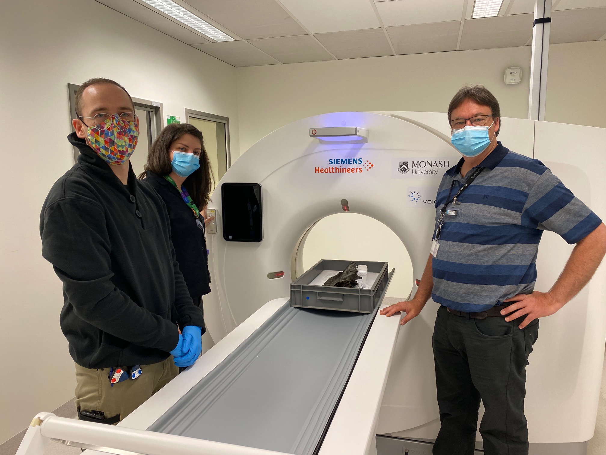
As the most complete real dinosaur fossil in any Australasian Museum, Horridus was scanned in MBI’s large bore Siemens CT scanner before it was assembled for display, to enable further study of the Triceratops by the global scientific community.
The imaging of rare and high-value specimens such as Horridus allows the preservation of information from fragile objects, in addition to the non-destructive exploration of the interior of specimens.
Monash University PhD student Hazel Richards conducted the scans and created 3D models of the Triceratops bones for the exhibition as part of her role as research assistant on the project.
“When we combine the internal images produced by these CT scans with the external surface scan images, we can create a complete 3D model of the Triceratops bones that allows us to research a range of exciting biological and evolutionary questions,” Ms Richards said.
“The team at MBI are always enthused and accommodating when we come to them with proposals for scanning weird and often unwieldy objects like these Triceratops fossils.”
“With their support and expertise, we have been able to maximise the scientific potential of these remarkable pieces of natural history,” Ms Richards said.

Applying imaging expertise to solve challenges
NIF Facility Fellow, and MBI’s Head of Pre-Clinical Imaging, Dr Michael de Veer worked with Ms Richards to provide training and operational guidance on the optimal use of the instrument to generate the data.
“Fossilised bone is very dense, so our scanning challenge was to manipulate the CT settings so that the X-rays would penetrate the bones, allowing visualisation of internal structures such as the dinosaur’s brain case,” Dr de Veer said.
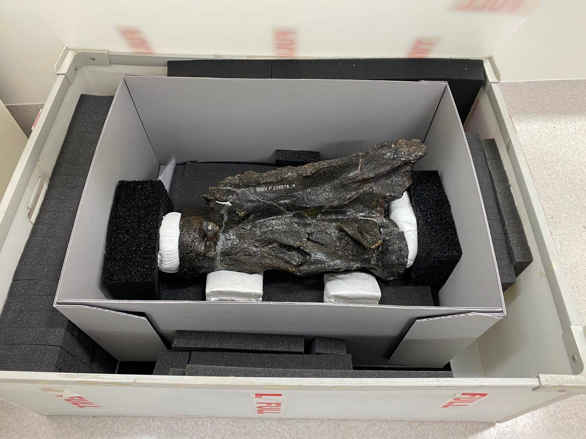
“Different parts of the dinosaur fossil were scanned over a number of visits, and the bones were transported in special crates to reduce the possibility of damage.
“As the CT scanner can penetrate plastic and foam, we were able to keep the bones in their protective stillages during scanning, a capability that made Museums Victoria palaeontologist Tim Ziegler very happy” Dr de Veer said.
As Collection Manager Vertebrate Palaeontology, Tim Ziegler manages the preservation of Victoria’s fossil collections of backboned animals, plants, and microfossils, including dinosaurs.
“Fossils are surprisingly fragile once they are uncovered and brought out of the ground,” Mr Ziegler said.
“We take any opportunity to improve the safety of specimens under research. As part of Victoria’s State Collection, this skeleton will be kept and preserved in perpetuity, and will offer scientific potential for years to come.”
What can a CT tell us about life 67 million years ago?
Visitors to the Triceratops exhibition will see the CT data captured first-hand – both virtually –through animated projections from the scans, and physically – as life-size touchable resin casts made from the 3D printed models.
This information can be used to tell deeper stories about Triceratops, including its evolution, behaviour and how it sensed its Cretaceous world.
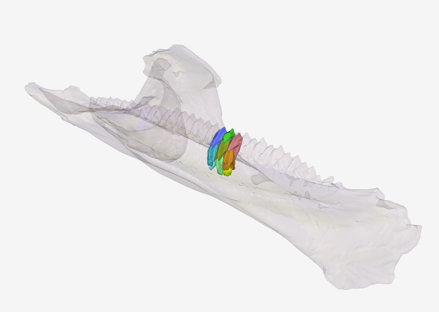
The data of the upper and lower jaws reveal Horridus had more than 800 teeth. These CT scans can contribute to investigations of feeding biomechanics and diet in Triceratops and other highly specialised herbivorous dinosaurs.
Scans of the dinosaur’s well-preserved braincase provided 3D models of the internal cranial cavity, allowing the team to examine the size and shape of regions of the brain and inner ear.
These provide important data for research reconstructing what sorts of sounds Triceratops was adapted to hear, and the relative importance of vision, smell and hearing in the daily lives of these long-extinct beasts.
Say hi to Horridus
You can visit Horridus in the flesh (er.. bones?) at Melbourne Museum’s exhibition, Triceratops: Fate of the Dinosaurs.
For more information, contact Dr Michael de Veer, Head of Pre-Clinical Imaging and Monash Biomedical Imaging NIF Facility Fellow.
Images courtesy of Museums Victoria and Monash Biomedical Imaging.