#ImagingTheFuture: The world-leading impact of the Australian biomedical imaging community

It’s Chan Zuckerberg Initiative’s #ImagingTheFuture Week, celebrating the remarkable impact of the international biomedical imaging community.

We’re privileged to partner with our NCRIS colleagues at Microscopy Australia to present some of Australia’s impactful imaging projects, supported by our national research infrastructure.

Through open access to state-of-the-art expertise, equipment, tools, data and analysis, we’re proud to empower Australian medical researchers, materials and agriculture scientists to address pressing challenges across research and industry.

The power of advanced imaging technology is driving solutions for Australia’s strategic science and research priorities, fostering innovation, supporting industry and contributing to our health and wellbeing.

Scroll down to read more about some of the impactful and innovative biomedical imaging projects we support through our Preclinical and Frontier Imaging, Advanced Human Imaging, Radiopharmaceuticals, and Imaging Data Collections and Partnerships programs, or head over to our socials and the Microscopy Australia website for more Australian imaging.

Find us on social media:

National Imaging Facility
Twitter/X | LinkedInInstagramFacebookThreads

Microscopy Australia
Twitter/XLinkedInInstagram


About Australia’s National Collaborative Research Infrastructure Strategy (NCRIS):
The Australian Government Department of Education helps maintain Australia’s reputation as an established global leader in world-class research by ensuring researchers have access to cutting edge national research infrastructure supported through the NCRIS program. More information: www.education.gov.au/ncris


Better evidence for decision-making in health

Advanced imaging methods and analysis provide critical evidence for decision-making across all aspects of health and clinical science to keep Australia healthy.
Harnessing MEG to model neural dynamics

Partner: Swinburne University of Technology

Imaging expertise:
A/Prof David White
Dr Will Woods

Instrument:
MEG Facility at Swinburne Neuroimaging.

Acknowledgements:
Miao Cao (Swinburne and Peking University), Jiayi Liu (Peking University), Wei Cui (Peking University), Xiongfei Wang (Capital Medical University Sanbo Brain Hospital), Will Woods (Swinburne), David White (Swinburne), Simon Vogrin (Swinburne), Chris Plummer (Swinburne), Changsong Zhou (Hongkong Baptist University), Jia-hong Gao (Peking University)

Image description:
Modelling neural dynamics from non-invasive MEG recordings has the potential to provide crucial insights in the fast-evolving physiological and pathological activity that gives rise to such dynamics. Combining MEG data acquired at the Swinburne NIF node and 3-D velocity field approaches in epilepsy patients, temporospatial patterns in 3-dimensional brain space were obtained and spiral patterns (including singularities) were revealed at seizure onset region, potentially suggesting highly non-linear dynamics and locally unbalanced excitatory-inhibitory neural networks were developed and progressed at seizure onset (from onset up to 1200ms) within the localised brain region. Such developments build on existing work with MEG technology to emphasise the utility of MEG in understanding brain function in health and disease. 

Enhancing Low-Field MRI Images through Generative Deep Learning

Partner:
Monash University, The University of Queensland, Herston Imaging Research Facility, SAHMRI, The University of Western Australia

Imaging expertise:
Dr Kh Tohidul Islam

Instrument:
Utilizing paired datasets from 100 healthy participants, images were obtained at Monash Biomedical Imaging facility using Hyperfine Swoop (64mT) and Siemens Biograph mMR (3T) systems. The deep learning model was trained to generate synthetic 3T-like images from 64mT images, and their performance was compared against actual 3T images.

Acknowledgements:
This project is funded by the National Imaging Facility (NIF) and Hyperfine Inc. Special thanks are extended to the NIF at Monash Biomedical Imaging, Monash University, for their facilities and support.

View publication for more information.

Image description:
The image displays MRI scans of a 38-year-old male participant. It includes T1, T2, and FLAIR sequences, each showing four columns: 64mT, 3T, Synthetic 3T, and the difference between 3T and synthetic 3T images. The synthetic images closely resemble the 3T images, demonstrating minimal anatomical variation and highlighting the model’s effectiveness in enhancing 64mT images. This research aims to improve low-field MRI images (64mT) using a Generative Deep Learning method, making them comparable to standard high-field (3T) MRI images.


Better health for the young and older Australians 

Imaging studies that look at conditions in younger and older Australians are essential for understanding and promoting healthy development and ageing.
Early detection of biomarker changes in Huntington’s Disease using PET imaging

Partner:
SAHMRI

Imaging expertise:
Dr Muneer Ahamed
Ms Georgia Williams

Instrument:
Large animal PET scanner at SAHMRI

Acknowledgements:
Expertise and help from radiochemistry team and large animal technicians at SAHMRI and contributions from researchers at University of Antwerp, University of Cambridge and University of Warwick drives this project. The authors acknowledge CHDI Foundation, New York for providing the preclinical models.

Find out more information here.

Image description:
A representative PET image showing [18F]FDOPA uptake in neostriatum in preclinical models.

Huntington’s Disease (HD) is a devastating neurodegenerative condition that causes cognitive, movement and behavioural disturbances, which over time result in progressive disability and eventual death. SAHMRI’s NIF fellows Dr. Muneer Ahamed and Ms. Georgia Williams leading a preliminary feasibility study to validate the use of transgenic preclinical models of HD in PET imaging studies. This study is expected to monitor early-stage (disease onset) and longitudinally evaluate biomarker changes in HD following the slow progression of the HD. This will be first time PET imaging is carried out in this type of HD preclinical model, with the help of NIF’s dedicated large animal PET scanner at SAHMRI.


Growing use of imaging in agriculture and ecology

Imaging is accelerating as an important capability for agricultural and ecological sciences.
Understanding the ecology of coral reefs

Partner:
University of Western Australia

Imaging expertise:
Ms Diana Patalwala

Instrument:
MicroCT

Acknowledgements:
Mr Damian Thomson, CSIRO

Image description:
Micro computerized tomography (μCT) scans of experimental Porites sp. blocks deployed in the lagoon and on the reef slope Ningaloo Reef for 20 months (605 days). Block images show representative scans of two individual blocks (a) pre- and (b) post-deployment in green, and areas of (c) external and (d) internal erosion post-deployment in red.

Porites corals can reveal the past sea conditions by their oxygen isotopes, which reflect the temperature and rainfall of the seawater. This information is useful for studying how the climate and weather patterns have changed over time, and how physical and biological factors influence the distribution and abundance of organisms on the seafloor.

DTI tractogram of a quokka brain: Exploring how marsuipials adapt to different environments

Partner:
Biological Resource Imaging Laboratory (BRIL), UNSW

Imaging expertise:
Dr Andre Bongers
Mr Simone Zanoni

Instrument:
Bruker BioSpec Avance III 94/20 Preclinical MRI

Acknowledgements:
Jyothi Thittamranahalli KariyappaSimone ZanoniAndre BongersLydia TongKen W. S. Ashwell

View publication for more information.


Cultural contributions to materials, engineering and culture

Many varied industrial and research problems— such as chemical processes, materials science, environmental and ecosystems research, security, palaeontology and cultural preservation—are increasingly opening up to the benefits of advanced imaging technologies.
Discovery of a new nasal-emitting trident bat from early Miocene forests in northern Australia

Partner:
Biological Resource Imaging Laboratory (BRIL), UNSW

Instrument:
MILabs U-CT microCT scanner

Acknowledgements:
Suzanne J. HandMichael ArcherAnna GillespieTroy Myers

View publication for more information.

Understanding equine anatomy: Investigating intestinal muscle structure to prevent rupture

Partner:
Western Sydney University

Instrument:
Diffusion tensor image of a cross-section of equine intestine taken using the 11.7 T MRI at the Biomedical Magnetic Resonance Facility (BMRF) at WSU Campbelltown campus.

Acknowledgements:
Kate Averay1, Denis Verwilghen1, Marianne Keller3, Neil Horadagoda2, Marina Gimeno2

1Camden Equine Centre, University Veterinary Teaching Hospital Camden, Sydney School of Veterinary Science, University of Sydney, Australia

2Pathology Services, University Veterinary Teaching Hospital Camden, Sydney School of Veterinary Science, University of Sydney, Australia

3Sydney School of Veterinary Science, University of Sydney, Australia

Image description:
The research group is looking at equine jejunum rupture and whether there are any anatomical differences in the rupture region. Colour-coding shows the anisotropy of the tissue. The key is on the upper right and shows red is left-right, green is up-down and blue is in and out of the screen. You can clearly see two layers of muscle: the red-green layer wrapping around the intestine and the blue layer running longitudinally.


Communities in regional Australia to benefit from world’s first mobile magnetic resonance imaging network

National Imaging Facility (NIF) is deploying four low-field portable MRI scanners to remote and regional sites to help researchers apply this affordable imaging technology in rural areas.

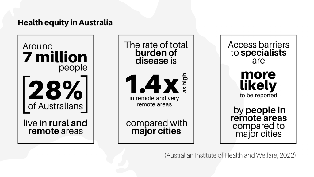
Australians living in regional and rural areas unduly suffer lower life expectancy and a higher burden of diseases because of poorer access to health services, including reduced screening, late detection and barriers to treatment compared with people living in metropolitan areas.

The national mobile magnetic resonance (MR) network will be the first project of its kind world-wide and is a collaboration with partners including Monash University, University of Queensland, South Australian Health and Medical Research Institute (SAHMRI), the Alfred Hospital, Royal Perth Hospital, University of Western Australia and MedTech company, Hyperfine.

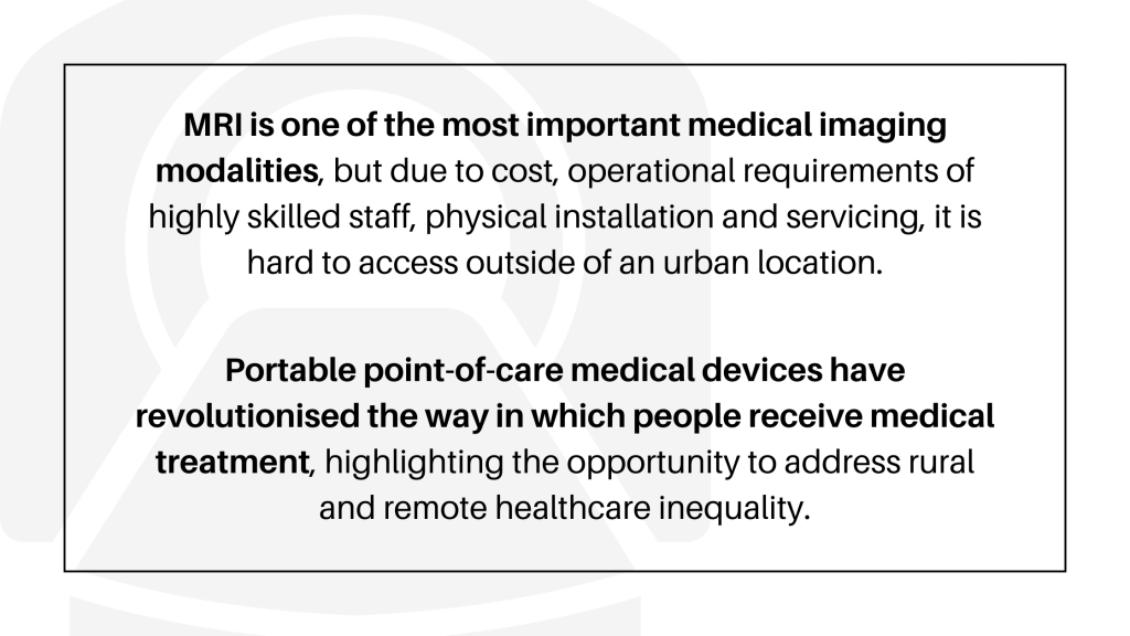
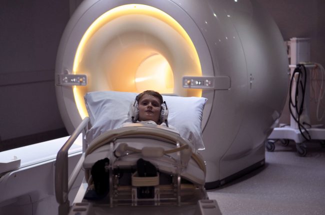
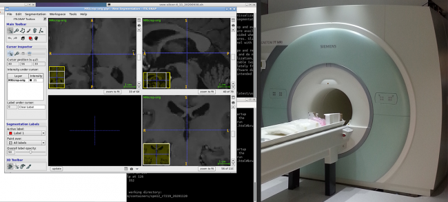
The Hyperfine Swoop is the world’s first highly portable MR imaging system capable of providing neuroimaging at the point-of-care, designed to fit inside elevators and through doorways to be manoeuvred directly to a patient’s bedside, and plug into a standard electrical outlet.

These portable scanners will be used to understand how this fast-developing technology can help diagnose stroke, traumatic brain injury, and other conditions after testing in research laboratories at NIF nodes in Victoria, Queensland, South Australia and Western Australia.

The scanners will enable real time tele-reporting and either remote operation or point-of-care use with low training requirements.

NIF is uniquely positioned to support work to build the usability of low-field MR technology, including developing techniques to maximise data quality.

Researchers at these sites will scan subjects on low-field mobile MR and high-field 3T MRI instruments to build a unique database that can be used to bridge the gap in outputs.

This valuable data will be made available by NIF to researchers to develop techniques to improve image processing and better understand how low-field scans can be interpreted.

Head of the Imaging Analysis Team at Monash Biomedical Imaging and Project lead Chief Investigator, Dr Zhaolin Chen said the collaborative work across NIF Nodes was critical to the success of the project.

“This nationwide network is critically important to identify a viable pathway for point-of-care MRI technology to be used in Australia,” Dr Chen said.

“Multi-site data acquisition is already underway and AI-based solutions to expand utility in regional Australia are in development.

“The network enables our project team to share knowledge, cross-validate findings, optimise resources and plan the next steps, which ultimately provides a route from research into clinics,” Dr Chen said.

NIF Chief Executive Officer, Prof Wojtek Goscinski said there were additional long-term advantages to deploying the national mobile MR network to regional Australia.

“NIF is focused on keeping Australia at the forefront of imaging, and the national mobile MR network is an innovative application of new technology to improve accessibility,” Prof Goscinski said.

“We hope these data collections and the AI models researchers build using them will lead to better technology that will improve treatment and diagnosis for Australians.

“This work will provide the foundation for the development and application of AI in clinical practice for low-field MR scanners, with experts optimising image quality for clinical data usability with reduced noise and improved resolution.

“The national mobile MR network and NIF’s increased national human imaging reach will enable innovative health research in remote populations, improve low-field MR technology, and over the long run will help increase access to better healthcare, professional training and socio-economic equity,” Prof Goscinski said.

ISMRM and ISMRT ANZ Chapters’ Annual Meetings shine a light on national imaging expertise and infrastructure

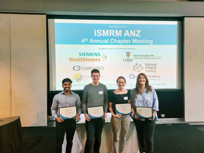
[Image: Presentation award winners at ISMRM ANZ, Honours student, Arunan Srirengan, Dr Ed Green, Dr Gwen Schroyen and Dr Myrte Strik. Photo credit: Dr Adam Clemente]

The Australian and New Zealand Chapters of the International Society for Magnetic Resonance in Medicine (ISMRM) and the International Society for Magnetic Resonance Radiographers and Technologists (ISMRT) held their Annual Meetings in Sydney last month, highlighting the work of leading national researchers and clinicians, including members of the NIF network.

NIF enables coordinated open access to magnetic resonance expertise and infrastructure to support leading national researchers and clinicians, and proudly supported the events.

ISMRM ANZ Joint Chapter Annual Meeting 9-10 Nov

ISMRM ANZ hosted sessions on revolutionising MRI technology, advances in neuroimaging, and clinical applications of advanced MRI, in addition to keynote speakers neurologist and leader in stroke medicine, Prof Mark Parsons and Director of the Institute of Medical Physics at the University of Sydney, Prof Annette Haworth.

Dr Zhaolin Chen was a key speaker in the Revolutionising MRI technology session, presenting the NIF Point-of-Care project, a collaboration between NIF, Australian hospitals, and US medical device manufacturer, Hyperfine, to build the usability of low-field MRI and bring critical imaging to remote Australia and deploy imaging in challenging clinical environments such as COVID wards.

[Image: Dr Zhaolin Chen presenting the NIF Point-of-Care Magnetic Resonance project at ISMRM ANZ]

A number of other NIF users spoke at ISMRM ANZ, including:

  • Rebecca Glarin from the University of Melbourne, presenting findings from her PhD on ‘Optimising functional brainstem imaging of sympathetic drive with ultra-high field MRI’.
  • Dr Shahrzad Moinian from the University of Queensland Centre for Advanced Imaging, presenting ‘In vivo microstructural border delineation between areas of the human cerebral cortex using magnetic resonance fingerprinting (MRF) residuals’.
  • Honours student Arunan Srirengan presenting ‘Early identification of cerebral small vessel disease in obstructive sleep apnoea patients using magnetic resonance spectroscopy: a pilot study’, featuring data obtained on the NIF 3T MRI at NeuRA. This session was awarded second prize in the oral presentation awards.
  • Dr Myrte Strik from the University of Melbourne, presenting ‘Altered network topology in patients with visual snow syndrome: a resting-state 7 Tesla MRI study’, winning the award for best Early Career Researcher Data Blitz presentation.

[Image: Dr Shahrzad Moinian from the University of Queensland Centre for Advanced Imaging. Photo credit: Dr Adam Clemente]

Congratulations to University of Melbourne NIF Fellow, Prof Brad Moffatt as ANZ ISMRM Chapter President on the success of the 2022 meeting hosted at UNSW.

ISMRT ANZ Joint Chapter Annual Meeting 12-13 Nov

The ISMRT ANZ 2022 joint meeting program theme was MRI: Past, Present and Future, and featured a range of internationally renowned speakers demonstrating future technologies and cutting-edge imaging techniques.

Keynote presenters included Medical physicist and human brain imaging academic researcher Dr Samantha Holdsworth, Chief of the Quantitative Medical Imaging Laboratory, USA National Institute of Biomedical Imaging and Bioengineering, Dr Carlo Pierpaoli, and founding member of the Society of Cardiovascular Magnetic Resonance and Principal Investigator for the Cardiac Atlas Project, Prof Alistair Young.

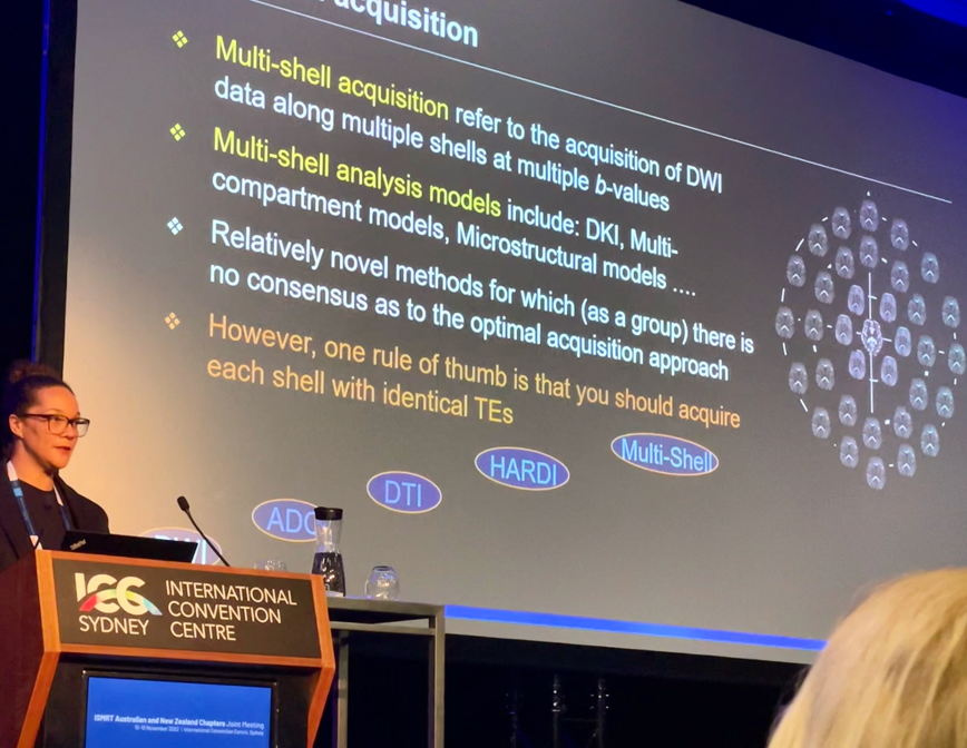
NIF Senior Manager and Senior Research Scientist – National Magnetic Resonance Capability, Dr Shawna Farquharson was a key speaker at the Diffusion Weighted Imaging (DWI) Forum, presenting on ‘DWI: Principles and practical applications’.

[Image: Dr Shawna Farquharson, National Imaging Facility]

NIF users showcased at ISMRT ANZ included:

  • Prof Lynne Bilston from NeuRA, presenting ‘Brain Elastography’.
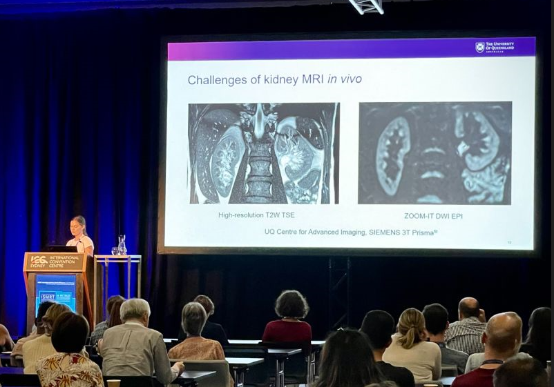
  • Sarah Daniel from the University of Queensland Centre for Advanced Imaging, presenting ‘Image quality enhancement using deep learning for in vivo human kidney MRI’.

[Image: Ms Sarah Daniel from the University of Queensland Centre for Advanced Imaging]

Congratulations to all presenters at ISMRM and ISMRT ANZ.

World’s first longitudinal muscle study grows understanding of cerebral palsy development

NIF infrastructure is enabling the Muscle Growth in the Lower Extremity (MUGgLE) Study, the first longitudinal study comparing muscle growth in children with cerebral palsy and typically developing children.

The project is a collaboration between Neuroscience Research Australia (NeuRA), the University of NSW (UNSW) and the Cerebral Palsy Alliance Research Institute.

The National Health and Medical Research Council-funded study is using magnetic resonance imaging (MRI) to compare muscle growth between typically developing children and children with cerebral palsy, using high-resolution measurements of the architecture of whole muscles.

Researcher Dr Bart Bolsterlee said the longitudinal study will see the lower legs of over 300 children scanned, between the ages of 0-3 months and 5-14 years.

“They will be scanned three times, with one-and-a-half years in between scans. We analyse the images to look at the individual muscles and how they change in size and structure over time,” Dr Bolsterlee said.

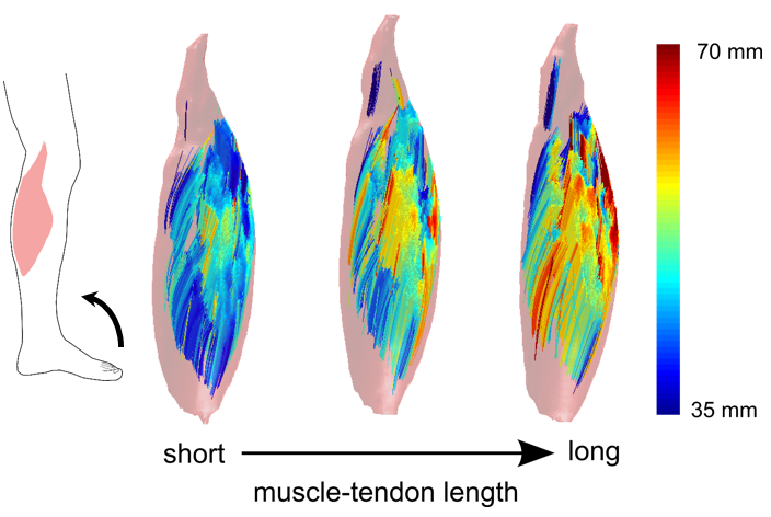
“The key measures we are getting out of this study are not just the volume of muscles, but also the orientations and lengths of their muscle fibres, which is a key determinant of the function of a muscle.

“We also look at the fat content which is a compositional feature of muscles that is quite different between diseased muscles and healthy muscles.

The impacts of this research have real implications for children growing up in Australia, with one-in-seven hundred babies born with cerebral palsy.

“This is very much a fundamental research study – we don’t have any direct clinical outcomes that we are assessing – but what we do know about children with cerebral palsy, the leading cause of childhood physical disability in the western world, is that outcomes can be pretty poor,” Dr Bolsterlee said.

“One-in-three children with cerebral palsy cannot walk independently, and we know this has got something to do with disordered muscle growth.

“It’s obvious from cross-sectional studies that there are quite some differences between the muscles of children with cerebral palsy and their typically developing peers, but nobody has actually studied this longitudinally, so we don’t know when these changes occur.

“We believe that information is necessary to develop new treatments.”

Currently there is no cure for cerebral palsy, and often children undergo severe interventions including complex surgical procedures with drugs to improve daily functioning. These interventions can change muscle growth, but how that affects musculoskeletal function is poorly understood.

Dr Bolsterlee is part of the team developing imaging methods and algorithms to be able to study this, and they are now generating the first data to give a comprehensive picture of how muscles develop typically – and how they develop in children with cerebral palsy.

“Many of the tools that are out there were developed for the brain – I’d say 99% of diffusion imaging software is used to reconstruct the neuronal architecture of the brain. We had to adapt the acquisition protocols as well as the imaging analysis techniques to accommodate measurement of the specific features of muscles we are interested in,” Dr Bolsterlee said.

In addition to configuring the imaging software to analyse data for the muscles, Dr Bolsterlee said there was a lot to consider when optimising scanning protocols to get the best images possible, while scanning children within a limited time.

“I’ve been working at NeuRA for the better part of eight years on this – and it’s really nice to see the first proof of principle demonstrations being taken to large-scale research – and hopefully to clinical practice as well.

“We’ve developed algorithms that several groups around the world are now using,” Dr Bolsterlee said.

This research into muscle imaging has grown the understanding of the architecture of muscles globally.

“Most anatomical knowledge comes from textbooks that are based on dissections of cadaver legs, and these are usually from older people who’ve donated their bodies to science.

“We have a rough understanding of the fibre structure within muscles and how they sit between muscles, but it’s been very difficult to get any information from living human muscles.

“Muscle is one of the most adaptable human tissues in the human body – when you exercise, they get bigger and when you’re lying in bed for too long, they get smaller very quickly.

“So, it’s very important if you want to understand how muscles respond to various stimuli, to have in-vivo imaging methods – or methods that can be applied to living humans,” Dr Bolsterlee said.

Previously, researchers were limited to ultrasound in living patients, which was 2D and only able to capture muscles superficial to the skin because the ultrasonic waves have limited penetration depth.

The MRI diffusion imaging technique allows researchers to look at whole human muscles in 3D, which has led to discoveries in the complex fibre structure of muscles and how it changes when they contract, lengthen or are diseased.

For more information, listen to our podcast with Dr Bolsterlee and NIF Fellow, Dr Michael Green from NeuRA: The MUGgLE Study: Imaging to understand how muscles grow.

Neuro Imaging to examine high rates of dementia in older Aboriginal Australians

Early life stress (ELS) has been linked to abnormalities in brain structure and function and may contribute to increased risk of cognitive decline and dementia later in life. ELS has also been associated with the high prevalence of dementia observed in older Aboriginal Australians.

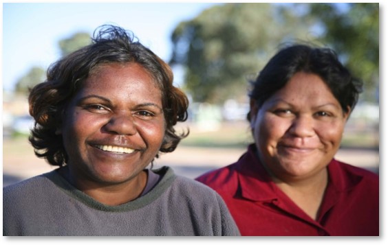
A study at NIF’s UNSW Node, NeuRA Imaging is engaging the Australian Aboriginal community to investigate structural and pathological brain changes that underlie in high rates of dementia and cognitive decline in older Aboriginal Australians.

This will be the first study that investigates neuroimaging in cognitive impairment in older Aboriginal Australians and will inform dementia prevention, diagnosis and policy. It will also contribute to the wider literature on vascular risk in the pathogenesis of Alzheimer’s disease and associated biomedical and social risk factors.

After extensive community engagement with partnering Aboriginal communities including La Perouse, NSW, the initial consultation stage of NeuRA’s Koori Growing Old Well Study indicated that neuroimaging should be included in future dementia studies (Lavrencic et al., 2020, Int Psychogeriatr). Led by NeuRA’s, researchers including Dr Kylie Radford, Professor Tony Broe AM and Dr Louise Lavrencic, the Koori Growing Old Well Study included a community planning survey, pilot MRI study and guidance from an Aboriginal and Torres Strait Islander Steering Committee.

“NIF’s capabilities are allowing this study to investigate underlying brain changes and pathology in ageing and dementia in partnership with Aboriginal communities. The study will give greater detail and is using sophisticated and novel MRI techniques. By having the facility in-house at NeuRA it also means we can ensure a culturally safe and welcoming environment for our participants. With a rapidly ageing population and high rates of dementia, we hope that this ground breaking study will shed light on important ways to promote healthy brain ageing with Aboriginal and Torres Strait Islander peoples,” said Dr Kylie Radford, Senior Research Scientist and Group Leader, Neuroscience Research Australia.

The neuroimaging sub-study is a prospective, cross-sectional non-interventional study where participants will first complete a comprehensive interview and diagnostic assessment as part of the Koori Growing Old Well study. Consenting participants (200) aged 55+ will undergo MR scans with an expected study completion by 2023.

The outcome analyses will include identifying associations between cognitive impairment and hippocampal atrophy/volume and vascular indices on MR. Vascular pathology will be examined for cases of possible or probable Alzheimer’s disease compared to a cognitively intact control group. Correlations between MR measures and early life stress, adult risk and protective factors, cognitive function, and clinically diagnosed cognitive impairment will be investigated.

Better detection and treatment of dementia

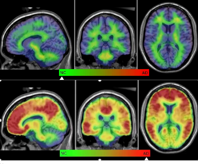
Biogen’s Aducanumab (Aduhelm) is the first disease modifying therapy for AD approved by the The United States Food and Drug Administration (FDA). NIF’s positron emission tomography (PET) imaging facilities at the University of Melbourne, HIRF and the Hunter Medical Research Institute (HMRI) supported the Australian trial recruitment of the Biogen Phase 3 trial by screening potentially suitable participants with amyloid PET scans in collaboration with the Australian Imaging Biomarkers and Lifestyle Study of Ageing at the Florey Institute of Neuroscience and Mental Health and Austin Health.

To prescribe the treatment for prodromal and early clinical AD it will be necessary to use imaging both PET and magnetic resonance imaging (MRI) and cerebrospinal fluid (CSF)/blood biomarkers to ensure that subjects being treated have AD and that side effects from Aduhelm are properly managed.

Alzheimer’s disease (AD) is the most common type of dementia making up 70% of all dementia. There are about 300,000 Australians currently living with the disease, with the average disease duration of 10 years equating to 30,000 new cases each year. These numbers are predicted to triple by 2050.

The degeneration within the brain begins two to three decades before overt symptoms, highlighting early detection is critical. NIF’s University of Melbourne Node and the Herston Imaging Research Facility (HIRF) have been involved in several dementia trials, studying different aspects of the disease including early biomarker detection, combining state of the art multimodality imaging, genetics and neuropsychology. NIF is working to assess novel radiotracers as a diagnostic tool for early detection of AD and the development of a national network of radiotracers for dementia screening in collaboration with QTRaCE and the Australia Dementia Network (ADNeT). This research aids the success of new preventative medicines, both through repurposing an existing drug and novel drug development and improves classification of AD subtypes, which impact treatment profiles.

NIF has played a major role in helping Australia to maintain its leadership in imaging applied to the dementias, particularly in AD. This has occurred at multiple sites around the country since 2012. The advent of disease modifying therapies for AD will cause an increase in demand for services and it will be crucial for new innovative and cost effective methods of service delivery in our increasingly ageing population.


Changes in the brain related to parenthood

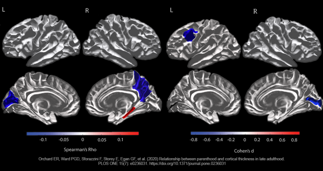
Parents can tell you that having kids changes your life. Recent studies by the Cognitive Neuroimaging Team at Monash Biomedical Imaging have shown just how deep those changes go by analysing brain structure in older adults that have had children. Parenthood was shown to cause life-long changes in both mothers and fathers, with a follow-up study suggesting that motherhood can protect the ageing maternal brain.

Read More

fMRI Short Course at UQ

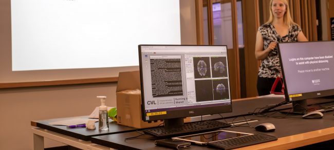
From Friday 20th – Sunday 22nd November 2020, a broad audience of PhD students, postdocs, associate professors, a radiographer and a clinician attended the University of Queensland (UQ) Centre for Advanced Imaging (CAI) functional Magnetic Resonance Imaging (fMRI) short course.

Read More
Privacy Settings
Youtube
Vimeo
Google Maps