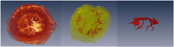
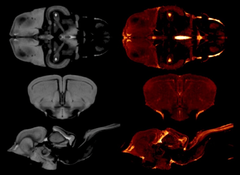
Creating a Lizard Brain Atlas

Until recently, reptilian evolutionary studies lacked an important resource – a lizard brain atlas. As the subject of numerous ecological and behavioural studies, the Australian tawny dragon (Agamidae: Ctenophorus decresii) was an ideal candidate for creating a high-resolution MRI atlas of a representative scaled reptile (squamata). Such data is not only a resource for studies of the genus but also informs environmental decision making through an improved understanding of animal adaptation and evolution.

Read More

CT as part of the Forensic Science SA toolkit

A post-mortem examination, or autopsy, is a forensic technique for learning about the conditions of a person’s health at the end of life. These are typically carried out as part of a coronial inquiry to establish the cause of death. A post-mortem may also provide information about undiagnosed medical problems of relevance to family members and the research community.

Read More

Understanding Feto-Placental Vasculature

Proper vascular development of the human placenta is crucial for meeting the metabolic needs of the developing fetus during pregnancy. Maternal environmental stressors such as malnutrition disrupt the elaboration of the feto-placental vasculature that, in turn, impacts on placental function and results in reduced fetal growth. The ramifications of this are not only on short-term foetal health but also on long-term health outcomes. Indeed, distortion in placental shape and size strongly associate with later adult health outcomes such as cardiovascular disease, obesity and cancer.

Read More

Detecting atherothrombosis using targeted MRI

Cardiovascular disease (CVD) is responsible for more than a quarter of all deaths in Australia and remains the global leading cause of death accounting for 17.9 million deaths per year. Of all CVDs, stroke and coronary artery disease account for the majority of deaths. A common underlying cause in these conditions in atherosclerosis, characterised by the build-up of abnormal deposits inside the arteries. Atherosclerotic plaques can rupture and cause thrombosis, or blood clots, resulting in stroke and myocardial infarction. Diagnostic strategies for the detection of thrombi are currently invasive and may not be sensitive to early biomarkers such as localised coagulation and inflammation.

A/Prof Ta, of Griffith University, has teamed up with researchers across Australia and internationally to develop a new form of MRI contrast agent. These ultra-small dual positive and negative contrast iron oxide nanoparticles (DCIONs) provide both T1-positive and T2-negative contrast effects, overcoming the limitations of single modality contrast agents. This duality is particularly important for imaging intravascular thromboses, as current single-contrast nanoparticles results in a black dye against a black artery. Further, the DCIONs are monodisperse, water-soluble, and biocompatible, of critical importance to biomedical applications.

In-vivo MRI of carotid artery thrombus (green arrows) detection after injection with a conjugated DCION. Image from Ta et al 2017.

Using non-invasive MRI at the NIF QLD Node, the application of a DCION conjugated to an enzyme found in activated platelets demonstrated accurate and sensitive detection of intravascular thrombosis. Work is continuing to further optimise the early detection of thrombi, expected to allow for earlier and more effective preventative treatments and improved clinical outcomes for patients at risk of stroke and myocardial infarction.

A/Prof Ta is enthusiastic about the future applications of DCIONs beyond thrombosis diagnoses, stating that “these nanoparticles have the potential to replace traditional gadolinium-based contrast agents due to their stronger T1 contrast effect. Existing alternatives cannot do what these nanoparticles can.”

If you have any concerns about heart disease or atherosclerosis, please talk to your GP and check this website.

Stent-electrode array for cortical neural activity

Minimally invasive endovascular stent-electrode array for high-fidelity, chronic recordings of cortical neural activity

This news has been contributed by Assoc. Prof. Bradford Moffat of the Melbourne Brain Centre Imaging Unit, Department of Radiology and Medicine, The University of Melbourne, Parkville.

National Imaging Facility Fellow, Assoc. Prof. Bradford Moffat collaborated with Dr. Tom Oxley’s group at the University of Melbourne for this high profile publication[1] that appeared in the journal “Nature Biotechnology”.

Read More

MRtrix3

MRtrix3: Advanced tools for the analysis of diffusion MRI data

Diffusion-weighted MRI (dMRI) is a commonly-used medical imaging modality for the investigation of tissue microstructure, exploiting the local hindrance and restriction of water diffusion as indirect probes. The neuroimaging research community utilises this technology extensively for the study of brain white matter in particular, reconstructing structural connectivity pathways and analysing estimated tissue properties.

Read More

MRI investigations of placental structure and function

Preeclampsia is a medical condition affecting up to 3% of pregnant women in Australia. Characterised by high blood pressure and protein in the urine, it is a leading cause of morbidity and mortality in both mothers and infants. Furthermore, preeclampsia has been linked to long-term health consequences for both mother and child.

3-D reconstructions of the placenta from MRI images. (left) Foetal surface view of the placenta. (middle) Maternal surface view of the placenta with an overlay showing maternal vasculature. (right) Side view showing maternal vasculature alone

Hampering early diagnosis, prevention, and treatment efforts is a lack of understanding of preeclampsia pathophysiology.  Currently, the cause of this condition is unknown. Prof Annemarie Hennessy and a team of researchers at Western Sydney University are utilising the WSU NIF Node, in collaboration with NIF Fellow Dr Timothy Stait-Gardner, to learn more about this serious condition.

In this project, high-resolution magnetic resonance imaging is being used to examine placental changes in vivo in mouse models of preeclampsia. In addition to the in vivo studies, high-resolution scans of fixed mouse placentas, normal and abnormal, have been used to create a placental atlas. The creation of a placental atlas and a number of publications have provided important information on mouse models of preeclampsia, including its characterisation and how to differentiate between different models of preeclampsia from T2 maps of the mouse placentas. These works have provided some of the basis for investigations of new treatments of preeclampsia.

Publications:

This story was contributed by the Western Sydney University NIF Node. For further information, please contact Dr Timothy Stait-Gardner.

Privacy Settings
Youtube
Vimeo
Google Maps