Unwrapping the mystery of ancient Egyptian mummies

Reviving an ancient Egyptian Mummy sounds like something out of a science fiction movie, but researchers at the University of Melbourne have done the next best thing. In a multidisciplinary project with the Faculty of Medicine, Dentistry and Health Sciences headed by Dr Varsha Pilbrow, the head of Meritamun – a young Egyptian woman who lived more than 2000 years ago – has been imaged using CT and reconstructed using 3D-printing technology.


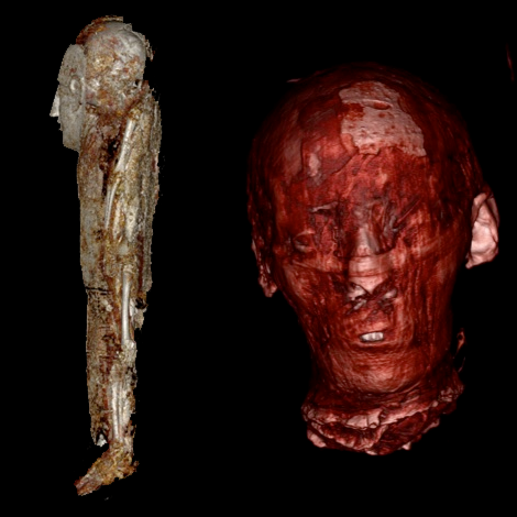
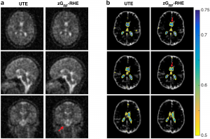
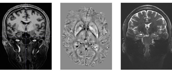
On the left, CT reconstructions of a baby mummy (still being studied). On the right, the CT reconstruction of the head of the mummy named Meritamun from the University of Melbourne’s collection within the School of Biomedical Sciences


Without disturbing the rare specimen and adhering to the controls and procedures of the Human Tissue Act 1982, Meritamun was imaged using the Siemens Human PET/CT in the University of Melbourne National Imaging Facility (NIF) Node. Digital and volumetric displays of tomographic data were acquired and reconstructed for 3D printing to create a skull replica.


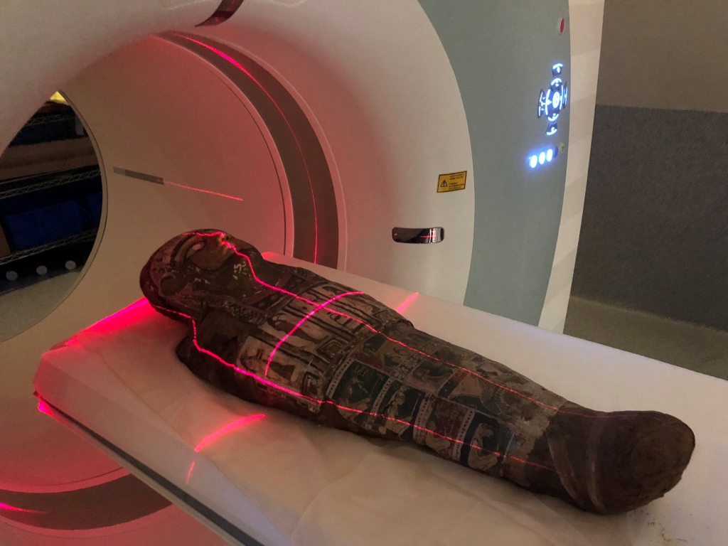
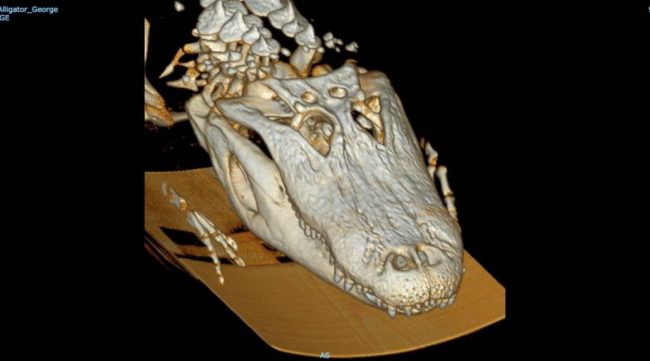
The scanning of a mummy with no adverse affects


Biomedical science Masters Student Stacey Gorski has used the CT data to diagnose Meritanum with anaemia, and hypothesizes a cause of death due to parasitic infection.  “The fact that she lived to adulthood suggests that she was infected later in life,” says Gorski. She and supervisor Dr Pilbrow are continuing the investigation, hoping to learn more about the life and death of the ancient Egyptian using forensic pathology.


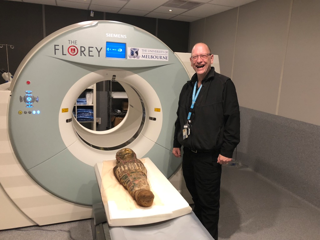
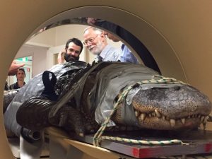
NIF Facility Fellow Mr Rob Williams facilitating access and providing expertise to the Human PET/CT scanner


In addition to learning more about the pathology and environments of population groups of 2000 years ago, the capability to replicate body parts and organs from CT imaging of specimens offers an opportunity for students to interact with old rare samples without damage to the original.


This story was contributed by the University of Melbourne. Acknowledgements go to Varsha Pilbrow, Julietta Capodistrias, Nina Sellars, Quentin Fogg, Michelle Gough, Gavan Mitchell, Peter Mayal, and Natalie Langowski.

For more information, please contact Rob Williams.

NIF Annual Meeting 2019


The NIF Annual Meeting was held June 18 – 20th 2019. At the green St Lucia campus of the University of Queensland, Fellows, Directors, and Board Members from across the country gathered in the Centre for Advanced Imaging (CAI).

The Fellows’ Program kicked off with a FAIR data principles and Characterisation Virtual Laboratory (CVL) workshop. Here, Fellows were reminded about FAIR data principles and had assistance in opening CVL accounts.


A seminar room in workshop configuration filled with people focussed on laptops and a presenter at a lectern

If you’re interested in CVL, why not sign up for the CVL Champions Program? Applications close July 31st! https://characterisation-virtual-laboratory.github.io/CVL_Community/champions/


four images of people seated around various tables, eating and drinking

The first day was wrapped up by an informal BBQ in the fresh Brisbane air


On day 2, we enjoyed the Fellows Mini-Symposium boasting the theme of ‘collaboration’. These talks showcased the cutting-edge projects and facilities that 11 of our NIF Facility and Informatics Fellows have been working on. An open discussion followed lunch, spawning ideas to collaborate on a new atlasing project, enhance the utility of the CVL Program, and the identity of NIF as a go-to imaging brand.


People standing in groups chatting while holding plates of food

Morning and afternoon tea was always stimulating and delicious!


The three NIF Thematic Groups met in the afternoon of Day 2 to discuss their latest challenges, share expertise, and develop action plans for their National Initiatives. Each of the Themes is working towards a National Initiative relevant to their user base, improving research quality and availability across the country. These fascinating discussions and more continued on into the evening for our final dinner together.


Graham standing before a projection of a Powerpoint slide on the right with NIF Fellows and Associates seated seminar style on the left


Day 3 opened early with an address by the NIF CEO, Graham Galloway, congratulating the three NIF Professional Development Grant winners, Dr Karine Mardon, Dr Tom Close, and Ms Diana Patalwala. Their awards have taken them to laboratories and events around the globe! We also welcomed Dr Rob Smith, Dr Tonima Ali, and Dr Paula Martinez Villegas to the ranks. At the same time, we bid a fond farewell to Dr Kirk Feindel, and wish him every success in the next stage of his career. NIF has enjoyed great successes over the past year, including an additional $53m investment via the NCRIS program. With the transition from a representative Board to an independent governing Board only a few months ago, NIF can expect exciting challenges ahead. Watch this space to see how we plan to continue engaging and collaborating across Australia!


NIF Fellows seated in groups focusing on a brainstorming activity


Next, we all discussed ways of sharing our outcomes and facilitating reporting, wrapped up with some fun group activities to ignite creativity and collaborative communication!


An emoticon face with a question mark

What is informatics, and how can NIF help you with your data needs? Get in touch today! Email us at NIF Central or contact your local NIF Fellow: https://anif.org.au/contact/


The meeting was finished off with an open discussion focussed on informatics, data curation, and repository systems. Two great ideas came from this discussion; persistent identifiers for instruments, allowing researchers to cite the instrument in publications, and an online knowledge repository (such as a wiki) for sharing workflows and processes. We look forward to supporting these initiatives!


The NIF Group standing on the steps of CAI


It was sad to say goodbye to all the NIF Fellows, Node Directors, Board Members and Associates at the end of our three-day meeting, but we are reassured by the plan to meet again in April or May 2020 in Sydney! Until then, we will continue to share our stories and build on the initiatives that we’re so excited about.


NIF pays its respects to the traditional custodians of the land upon which we met. We acknowledge both the Jagera people and the Turrbul people and their Elders, past, present, and emerging, for they hold the hopes, dreams, traditions and cultures of Aboriginal Australia.

Messages inside Porites: Are corals exposed to repeated heatwaves coping?

Researchers at the Australian Research Council Centre of Excellence for Coral Reef Studies, the University of Western Australia, Curtin University, Aix-Marseille University in France, and the US National Oceanic and Atmospheric Administration wanted to know the history of coral bleaching events on the Great Barrier Reef and how corals are responding to climate change.


Hard long-lived corals, such as Porites, are the backbone of reef ecosystems such as the Great Barrier Reef. Such reef-building corals are sensitive to light levels and temperature. Reef-building corals are already reaching their limits with every heatwave. With ocean temperatures rising, can they hope to survive more frequent extreme temperature events?


Underwater photograph with two scubba divers insertingg a longg metal rod into a large coral bed

Dr. Thomas DeCarlo drilling a 2+ meter core from a massive coral on the Great Barrier Reef


The team of researchers collected Porite core samples across the northern Great Barrier Reef, the Coral Sea, and New Caledonia, bring them to the WA NIF Node at the University of Western Australia, where they were scanned on the Bruker Skyscan 1176 in vivo micro-CT. 3-dimensional image stacks of density variations revealed ‘bands’ within the coral skeletons, corresponding to age. Also seen were high/low density ‘stress bands’, corresponding to environmental stressors such as exceptionally high water temperature.


CT images showing the bands inside coral cores from 1815 - 2017

μCT scans (dark/light shading = low/high density) reveal high-density stress bands and partial mortality scars preserved within the skeletons of long-lived Porites corals.


Some of the oldest (and longest) core samples had over 200 annual bands, meaning they were a living coral that has been growing for two centuries. By comparing stress bands to age, change-induced bleaching episodes were mapped, providing a timeline of coral bleaching events. Three striking observations followed: First, the researchers found the first piece of evidence that coral bleaching has occurred prior to the 1980s. Second, a significant increase in the frequency of stress bands was seen over time, consistent with the effects of global warming sparking more frequent coral bleaching events. Finally, recent (within the past few years) acclimatization was seen, whereby corals became less sensitive to heat stress follow repeated exposure to marine heatwaves. These results, published in early 2019, offer hope that reef-building corals surviving heatwaves may be able to adapt for future heatwaves.


This story was contributed by the University of Western Australia. For enquiries, please contact Ms Diana Patalwala.


Radiographers visit LARIF

In conjunction with the Australian Society of Medical Imaging and Radiation Therapy (ASMIRT) conference, the International Association of Forensic Radiographers (IAFR) organised a site visit to the South Australian NIF Node, Large Animal Research and Imaging Facility (LARIF), on the 31st of March 2019 to view the facilities used for post-mortem imaging (CT & MRI). Presentations included the practical aspects of post-mortem imaging using CT & MRI by Mr Raj Perumal and using CT & MRI in forensic practice by A/Prof Neil Langlois.

The participants were keen to understand the CT & MRI protocols used by Forensic Science South Australia (FSSA). LARIF provides a unique opportunity for post-mortem MRI to assist forensic investigations and there was a lot of interest from the participants to learn about the technical aspects of forensic MRI imaging.

A group photo in front of NIF and IAFR banners

The day before, at the ASMIRT conference, presentations were given about imaging opportunities and translation research at SAHMRI including a site tour of the rodent imaging facility at SAHMRI North Terrace by Dr. Marianne Keller.


This story was contributed by LARIF. For more information, please contact Mr Raj Perumal

High-Resolution MRI for Exposing Cancer Spread in the Brain

Sunny Queensland has a high rate of skin cancer, with melanoma as the second most common cancer in Queenslanders. In the area of the head and neck, this can lead to invasion of facial nerves and spread via the base of the skull to reach the brain stem. The extent of the progression, the so-called perineural spread, defines the therapeutic approach, informing the extent of required resection, and the complexity and duration of the surgical procedure. Imaging the extent of nerve involvement is critical to guiding treatment decisions and MRI neurography is the accepted imaging modality of choice.

Standard MRI may underestimate disease extent, but a current clinical trial explores the improved resolution and sensitivity using the 7 Tesla ultra-high field MRI scanner at the Centre for Advance Imaging at UQ. This improves visualisation of cranial nerves (see Figure and insert with zoomed nerve fibres) and potentially improves the definition of the perineural spread. The clinical trial is led by Dr. Sommerville from the Royal Brisbane and Women’s Hospital, QLD, and involves a Brisbane-based multidisciplinary team of head and neck surgeons, radiologists and scientists in the UHF MR Research team (Head: Dr. Barth, NIF Fellows Dr. Bollmann and Dr. Ali) at the UQ Centre for Advanced Imaging. The first patient scan has been performed, with 20 patients more to follow after a successful funding application sponsored by the 2018 round of the RBWH Diamond Care Grants.


MRI images showing a slice of brain with perineural cancer spread. The images highlight a region which is blurry in 3T and well defined in 7T.


If you would like to know more about skin cancer, please check the Cancer Council website and speak to your GP.


This story was contributed by the University of Queensland. For further information, contact Dr Tonima Ali.

Closing the acquisition time gap – a new sequence scheme for optimised Sodium MRI

Magnetic Resonance (MR) opens a window for the non-invasive investigation of MR-observable nuclei, i.e. nuclei with a non-zero spin. Common nuclei in organic substances, like Carbon-12 and Oxygen-16; however, possess a zero-spin consequently rendering Hydrogen-1 (commonly referred as ‘proton‘ due to the single proton in the nucleus) the first and most frequently imaged nuclei.

Despite the much lower signal regime, also the potential of imaging ‘other nuclei’, so-called x-Nuclei MRI was approached in the early years of MRI. In 1985, only a few years after the first proton-based images were published, Sodium-23 was the second nucleus in the human body that was imaged non-invasively using MRI by Hilal and coworkers [1]. In spite of these early beginnings, x-Nuclei imaging in general and Sodium MRI, in particular, did not see the same high-paced progression as proton-based MRI techniques. This downturn was mostly caused by significant SNR limitations resulting from the low in vivo concentration and complicated NMR-signal characteristics of Sodium nuclei.

While SNR issues are more and more overcome through hardware improvements, particularly the recent advent of high field systems, the challenging signal characteristics continue to encourage the development of dedicated acquisition methods. Sodium possesses a 3/2-spin and as such exhibits a fast bi-exponential signal decay. Consequently, Sodium MRI sequences are commonly performed in 3D with k-space sampled via centre-out trajectory schemes resulting in inherent drawbacks for sampling efficiency and SNR. While previous research in Sodium MRI method development focused on the optimisation of sampling trajectories, we present a new acquisition method that tackles Sodium image quality improvements via a sequence timing optimisation.

Recently, we introduced zero-gradient excitation ramped hybrid encoding (zGRF-RHE) Sodium MRI, a timing optimisation that utilises the sequence dead-time delay (time-gap between transmit and receive mode) for gradient pre-ramping. This concept improves encoding time across k-space and in so doing alleviates signal decay effects during the acquisition. It utilises a hybrid sequence encoding approach where the central k-space gap, resulting from gradient activity before receiver onset, is filled with Cartesian single point acquisitions. A sequence diagram of zGRF-RHE is displayed in Figure 1.


Figure 1: Radial zero‐gradient‐excitation ramped hybrid encoding (zGRF‐RHE) sequence diagram (A) and 2D illustration of hybrid k‐space encoding scheme (B). Gradient‐free excitation is followed by an immediate gradient ramping, data sampling commences after dead‐time. Central k-space is measured separately as single points with the same gradient pre‐ramping condition. TRO, readout duration; td, dead‐time. Figure taken from [2].

Hybrid imaging techniques, like PETRA or its generalised concept RHE, are frequently used in short-T2 proton imaging. Their application to low SNR problems like Sodium was, however, hampered by the employed gradient modulation during signal excitation. In that regard, we establish zero-gradient excitation for an artefact-free excitation profile maximizing image quality. The gradient-free excitation supports high flip angle acquisition, an essential requirement for low SNR imaging like Sodium, but also provides the opportunity to employ advanced RF pulse shapes without introducing a gradient-based excitation bias. Additionally, it should be highlighted that the zGRF-RHE sequence approach essentially describes a timing concept that is not just independent of the RF-excitation but also is not restricted to a particular readout-scheme.

An investigation of 3D-radial zGRF-RHE with standard 3D-radial ultra-short echo time (UTE) imaging can be found in our recent publication [2]. Compared to simulations, phantoms and in-vivo human brain acquisitions confirmed that our proposed sequence timing concept improves on image quality independent of the readout bandwidth and shows reduced image blurring and higher SNR. Figure 2 displays results from an in vivo human brain experiment highlighting improved image sharpness.


Figure 2: (A) Cross sections through matched slices in in vivo experiments at TRO of 2 ms UTE and zGRF‐RHE. zGRF‐RHE shows sharper details around the brain stem (red arrow). (B) Three consecutive transverse slices of CSF masks with contour lines of normalized UTE and zGRF‐RHE at TRO of 2 ms. zGRF‐RHE provides better delineation between lateral ventricles (red arrow). Figure taken from [2].

In summary, this work introduces an enhanced sequence timing concept with particular applicability to challenging low SNR-problems. Ultimately, this approach is expected to be of use for a wider range of x-Nuclei applications with spin > ½.

For further information, the interested reader is pointed to our full publication which can be accessed at https://onlinelibrary.wiley.com/doi/abs/10.1002/mrm.27484.

References
[1] Hilal, S. K., Maudsley, A. A., Ra, J., Simon, H. E., Roschmann, P., Wittekoek, S., Cho, Z., and Mun, S. (1985). In vivo NMR imaging of sodium-23 in the human head. Journal of Computer Assisted Tomography, 9(1):1–7.
[2] Blunck Y, Moffat BA, Kolbe SC, Ordidge RJ, Cleary JO, Johnston LA. Zero-Gradient-Excitation Ramped Hybrid Encoding (zGRF-RHE) Sodium MRI. Magnetic Resonance in Medicine 2018. https://doi.org/10.1002/mrm.27484.


This story was contributed by Yasmin Blunck of the Melbourne Brain Centre Imaging Unit and Department of Biomedical Engineering, University of Melbourne, Parkville

The First Australian Multi-Centre Study of Dementia using Ultra-High Field MRI

Australia is at the forefront of dementia research with world leading studies such as the Australian Imaging and Lifestyle study of Ageing (AIBL) led by a consortium of Australia’s leading Dementia centres, and the recently started Prospective Study of Ageing (PISA) led by the QIMR Berghofer.

The installation of the first human ultra-high field MRI scanner in the southern hemisphere at the Centre for Advance Imaging, the Qld node of the National Imaging Facility, in 2014 opened up a new era of imaging research. The Siemens 7T whole-body MRI scanner brought Australia to the forefront of ultra-high field research enabling examination of the human brain with an unprecedented level of detail.

Subsequently, a second 7T scanner was installed at the Melbourne Brain Centre providing a unique opportunity for a national multi-centre collaboration in ultra-high field MRI and the capability to explore new imaging biomarkers for diagnosis of neurodegenerative disease. A major project is underway, led by the Brisbane-based CSIRO eHealth Research Centre, co-funded by the CRC for Mental Health and in collaboration with the QIMR Berghofer, University of Melbourne and Florey Institute for Neuroscience with the broad aim of characterising new bioimaging biomarkers of neurodegeneration in the aging population. A suite of MRI methods is being applied at both sites on large cohorts of healthy aging subjects and patients diagnosed with fronto-temporal dementia. The scanning part of the project has been successfully completed with superb image quality obtained using state of the art sequences. A significant effort is now underway to analyse this valuable data which may contain a wealth of diagnostic information not otherwise available.


This story was contributed by The University of Queensland

Feature image: (Left) 3D MP2RAGE 0.9mm isotropic showing exceptional tissue contrast, (centre) example of a Quantitative Susceptibility Mapping (QSM), a mechanism for useful chemical identification and quantification of specific biomarkers, and (right) T2W TSE using coronal accquisition for hippocampus subfield examination. 

Wildlife Matters


A restrained alligator inside a CT scanner with veterinarians and researchers looking on

Dr Tim Kuchel and zoo veterinarian Dr David McLelland talking about George’s exam

The Large Animal Research and Imaging Facility had an unusual visitor. George the Alligator from the Adelaide Zoo came by for a temporal mandibular joint and dental check-up, courtesy of our 16 slice Phillips CT scanner.

A CT scan showing 3D structure of an alligator head

A CT scan of George, revealing bone structure

It had been recognised over the past two years that George did not open his mouth wide or quickly, and instead took food gently and played with it before swallowing it slowly in a most unlikely Alligator way. The differential diagnosis most likely was temporomandibular joint (TMJ) pathology which may have been able to be diagnosed/described by a CT scan. George was physically restrained, and his mouth secured (the muscles to open the mouth are quite weak, fortunately) and his 180kg body carried by 6 staff and placed into the NIF funded 16 slice CT scanner. Then the Alligator was scanned prone in a 16-slice Philips CT scanner using a 3D protocol (KV/mA: 120/350; field of view: 350 mm; matrix: 256×256; thickness/overlap: 1 mm/0.5 mm). The CT protocol was designed to give good high-resolution data, optimal contrast and good signal-to-noise ratios. Unfortunately, whilst good quality images were taken of the TMJ and adjoining tissue, no obvious abnormality was detected. Normally treatment with a non-steroidal anti-inflammatory drug (NSAIDs) would be employed to help with a diagnosis and potential efficacious treatment, it has been decided that the downside of the use of NSAIDs in a slow metabolising reptile was not outweighed by the clinical difficulty the ‘jaw opening problem’. George is not losing weight, is an old Alligator, so careful observation of his wellbeing will be undertaken over the next 6 months to ensure that his welfare is not adversely affected.

Two alligators resting on the ground

George, on the left, next to his girlfriend, after coming home from LARIF

This story was contributed by LARIF and the Adelaide Zoo

Imaging shows Alzheimer’s decline

Researchers at the Florey have invented a breakthrough imaging technique to describe in micro-detail the brain degeneration occurring in people with early Alzheimer’s and the full-blown disease.

Using the Siemens 3T Trio scanner at the Florey node of the National Imaging Facility (NIF), researchers have identified the precise locations of brain degeneration in a cohort of living Alzheimer’s patients. The work is important as it sheds new light on the underlying cognitive degeneration in Alzheimer’s, helping us focus our efforts to slow the decline.

To develop the technique, the team analysed brain scans from 177 Australians as part of the Australian Imaging, Biomarkers and Lifestyle study, who were either healthy, had an early form of Alzheimer’s or had the full-blown disease.

The brain pathways identified by the team have all been implicated in Alzheimer’s disease previously; those known to be crucial for memory formation, emotion and reasoning.

Alzheimer’s disease is usually thought to be caused by abnormal production and buildup of a peptide called amyloid beta.

Professor Alan Connelly, who led the study, said, “Interestingly, the mildly affected patients with low amyloid had more fibre degeneration in particular brain regions than those with high amyloid levels. This suggests that firstly, specific degeneration of certain brain areas will not necessarily be useful in predicting which mildly impaired individuals will progress to Alzheimer’s disease, and secondly that degeneration of this pathway is related to cognitive impairment, regardless of the buildup of the amyloid peptide.

“This is an important advance for a field still struggling to come to grips with what exactly causes Alzheimer’s. Our study shows we still have a way to go in interrogating the natural history of this insidious disease,” Alan says.

Lead author Remika Mito says, “This study was conducted by comparing the averages of each group of patients against each other, in order to give us the most statistically, and biologically, relevant results. In the future, we want to be able to compare an individual patient against a normal, healthy standard, to see how far along the disease trajectory they are. Or we could compare back to their previous scans to determine what effect a new medication is having as part of a clinical trial for example.”

Remika recently explained her results in an online abstract for Brain. If you would like to more about the details of the study, head over to Youtube to view Remika explain her work.


This story was contributed by the Florey Institute of Neuroscience and Mental Health. 

National Network of Trusted Data Repositories

During 2017 the National Imaging Facility (NIF) nodes at the University of Western Australia (UWA), University of Queensland (UQ), University of New South Wales (UNSW) and Monash University collaborated on a national project to enhance the quality, durability and reliability of data generated by NIF through the Trusted Data Repository project.

●        Quality pertains to a NIF user’s expectation that an animal, plant or material can be scanned and from that data reliable outcomes/characterisations can be obtained (e.g. signal, volume, morphology) over time and across NIF sites.

●        Durability refers to guaranteed long-term availability of the data.

●        Reliability means that the data is useful for future researchers, i.e. stored in one or more open data formats and with sufficient evidential metadata.

The Project, Delivering durable, reliable, high-quality image data, was jointly funded by the Australian National Data Service (ANDS) and Research Data Services (RDS). It was motivated both by NIF’s desire to enhance the quality of the data associated with the use of its facilities, and the desire of ANDS/RDS to facilitate the establishment of Trusted Data Repositories that enable access to data for at least 10 years and includes metadata that documents both the quality of the data and its provenance.

A trusted data repository service is essential for sharing data and ensures that project data created and used by researchers is “managed, curated, and archived in such a way to preserve the initial investment in collecting them” and that the data “remain useful and meaningful into the future” (https://www.coretrustseal.org).

The scope of the Project was limited to MRI data with the understanding that the developed requirements and trusted data repository services could be adapted to, or serve as a basis for other instruments/modalities.

The key outcomes from the Project include:

  1. The NIF agreed process for acquiring trusted data (NAP) – Lists the requirements that must be satisfied to obtain high-quality data, i.e. NIF-certified data, suitable for ingestion in a NIF trusted data repository service. They cover provisioning of a unique instrument identifier, instrument registration with Research Data Australia (https://researchdata.ands.org.au), quality control (QC), quality assurance measures, requisite metadata (including cross-reference to the QC data),  the process by which data is moved from the instrument to the digital repository service and the format(s) of the data.
  2. The NIF requirements for a trusted data repository service – Provides a platform-agnostic checklist of requirements that a basic NIF trusted data repository service should satisfy, including: identification of data by a unique Project identifier, ingestion of data from NIF-compliant instruments, authentication via the Australian Access Federation (https://aaf.edu.au), interoperability and easy deployment across NIF nodes.
  3. Implementations of trusted data repository services for two exemplars:
    1. Preclinical MRI data (with mouse brain data as an example) acquired across three NIF nodes—UNSW, UQ and UWA—using a Bruker BioSpec 9.4T MRI. The services have been implemented using the open source MyTardis/ImageTrove (https://www.mytardis.org) platform.
    2. Clinical ataxia MRI data acquired using a Siemens Skyra 3T MRI scanner in support of a Monash-proposed International Ataxia Imaging Repository (IAIR). The service has been Implemented using the open source XNAT (https://www.xnat.org) platform.

Software developed to support the implementation of the repository services includes: Docker (https://www.docker.com) Compose scripts to permit easy deployment at differents sites, client-side scripts for uploading NIF-certified data to ImageTrove/MyTardis and an XNAT plugin for uploading non-DICOM files.

  1. Assessments of the resulting trusted data repository services against a relevant international metric, the CoreTrustSeal (https://www.coretrustseal.org) Core Trustworthy Data Repositories Requirements.

For NIF users and the broader imaging research community the benefits and impact of this Project include:

  • Reliable and durable access to data
  • Improved reliability of research outputs and the provenance associated with it
  • Making NIF data more FAIR (Findable, Accessible, Interoperable, Reusable – https://www.ands.org.au/working-with-data/the-fair-data-principles)
  • Easier linkages between publications and data
  • Stronger research partnerships

For research institutions they include:

  • Enhanced reputation management
  • A means by which to comply with the Australian Code for the Responsible Conduct of Research
  • Enhanced ability to engage in multi-centre imaging research projects

For NIF they include

  • Improved data quality
  • Improved international reputation
  • The ability to run multi-centre trials

The transition plan post-funding includes: maintenance of existing services for 10 years; the integration of additional instruments; creation of a project web portal; planned new national and international service deployments; refinements and improvements; and CoreTrustSeal certification.

Project documents have been archived in the NIF Customer Relationship Management (CRM) system (accessible by NIF staff). Project software is hosted on GitHub and is freely available for download here: https://github.com/NIF-au/TDR. For further information please contact either the national Project Manager or NIF.

Project Manager and UWA lead: Andrew Mehnert (NIF Informatics Fellow, Centre for Microscopy, Characterisation and Analysis).
NIF lead – Graham Galloway (Chief Executive Officer, NIF)
UQ lead – Andrew Janke (NIF Informatics Fellow, Centre for Advanced Imaging)
UNSW lead – Marco Gruwel (Senior Research Associate, Mark Wainwright Analytical Centre)
Monash lead – Wojtek Goscinski (Associate Director, Monash eResearch Centre)
Privacy Settings
Youtube
Vimeo
Google Maps