Magnetic Lymphatic Mapping in Pigs

Working Towards a New Gold Standard for Cancer Care


The lymphatic system is one of the key mechanisms for metastatic spread, whereby cancer cells that have disseminated from a primary tumour are taken up into lymphatic vessels and transported to other locations in the body, beginning with the lymph nodes. Conventionally, surgeons map the lymphatic flow from a tumour site into the first draining nodes – known as sentinel lymph nodes – using a Tc99m radiocolloid. Drainage pathways of tracers may be imaged before surgery with gamma cameras (lymphoscintigraphy), and individual nodes detected during surgery with handheld gamma probes. These sentinel nodes are the ‘canary in the coal mine’ and can offer valuable information to help stage the disease and determine the most appropriate treatment options, so it is ideal for removing just these nodes while leaving uninvolved nodes intact.

While this technique is successfully and routinely applied to breast cancer and melanoma, accuracy is limited in cancers where the nodes are in close proximity to the primary tumour or other nodes, such as cancers of the head and neck. Without an accurate method to identify sentinel nodes in oral cancer, extensive dissection of all nodes in the neck region is routinely performed to ensure any potential draining nodes are harvested, yet approximately 75 % of patients undergoing neck dissection are exposed to the complications and morbidity of this invasive procedure without clinical benefit.

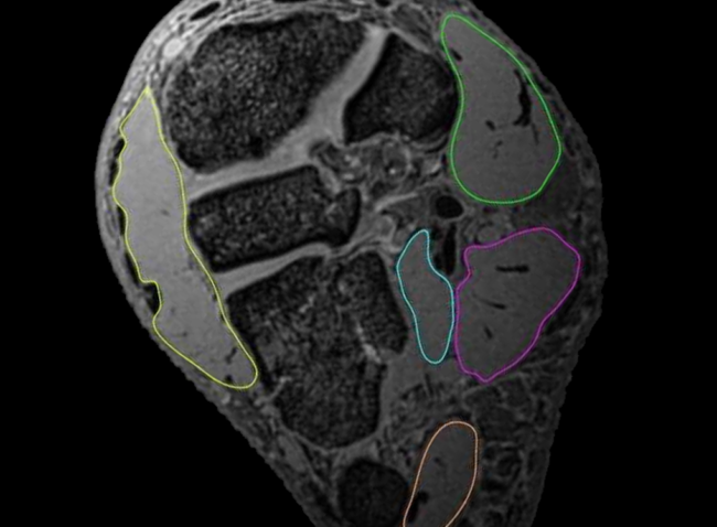
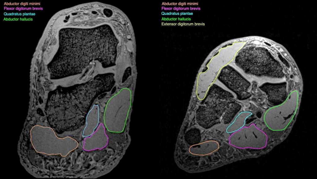
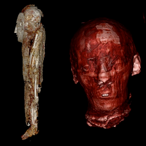
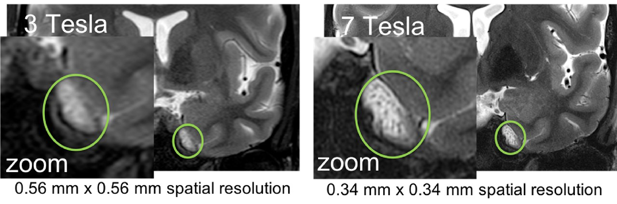
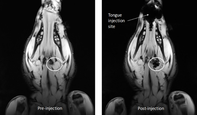
Dr Aidan Cousins and Dr Giri Krishnan have been working as part of a collaborative project between the University of South Australia and the University of Adelaide to demonstrate a novel, high-resolution technique for lymphatic mapping using magnetic nanoparticle-based tracers. Studies conducted at the South Australian Health and Medical Research Institute (SAHMRI) involve the injection of the magnetic tracer to the oral cavity of female Large White pigs, which are then scanned with a 3.0T Siemens MRI located at the large animal research and imaging facility (LARIF) NIF Node. Post-injection scans give Dr Cousins and Dr Krishnan detailed anatomical information of the drainage patterns of the tracer, which is then used to plan the surgery. During surgery, a handheld magnetometer probe developed by Dr Cousins is used, along with MRI data and visual identification, to pinpoint the draining nodes of interest from other, uninvolved nodes.

Two men looking at a screen in front of an MRI

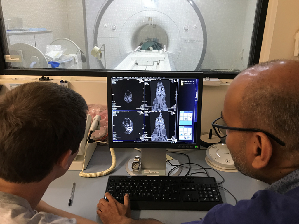
Dr Cousins with Raj Perumal (LARIF) examining an MRI scan of a pig following injection of the magnetic tracer

The results of this experiment showed the magnetic alternative to be adept for mapping lymphatic drainage in complex environments. Triangulating the location draining nodes with high precision before surgery was made possible by the high-resolution soft-tissue detail afforded by the 3.0T MRI. During surgery, the handheld probe was able to identify all draining nodes by way of detecting their magnetic ‘signature’. This final confirmation of draining nodes is analogous to the use of handheld gamma probes but has the distinct advantage of pin-point resolution, meaning nodes can be in very close (touching) proximity to each other and it is still possible to differentiate between the individual nodes’ magnetic signals. This spatial resolution is currently unmatched by any other probe technology commercially available and is key to the application of sentinel node mapping in complex cancers.

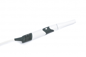
Photo of the high-resolution magnetometer probe developed by Dr Cousins


Results from this study are being used to design a world-first clinical trial applying magnetic tracers and a high-resolution probe to human patients with cancer of the oral cavity.


This story was contributed by SAHMRI. For further inquiries, please contact Mr Raj Perumal

Exploration of the deep foot muscles at ultra-high field

The arches of the human foot are unique structures that are important for functions like walking and running. The deep intrinsic muscles of the foot, such as the adductor halluces and interossei, are thought to play key roles in arch control; yet little is known about how they are controlled during functional tasks. The traditional measurement techniques can only provide information regarding muscle size, which is inadequate to evaluate the force-generating capacity of the muscles, assess the process of force generation by the muscles, and understand the involvement of the neural drive sent by the body to the muscles to regulate force production.


Using the 7T human MRI at the Centre for Advanced Imaging, a research study is currently investigating the muscle architecture of the adductor halluces and interossei. This research aims to quantify the force-generating capacities of these deep-foot muscles by measuring their MRI muscle volumes, estimate their force production by shear wave Elastography and measure their neural drive by using Electromyography.

black and white MRI images showing a cross-section of the foot with deep foot muscles outlined in colour

Deep foot muscles outlined in two cross-sections of a structural MRI scan obtained at ultra-high field: abductor halluces (green), abductor digiti minimi (pink), flexor digitorium brevis (magenta), quadrature plantae (cyan) and extensor digitorium brevis (yellow). With thanks to Dr Natalie Collins, University of Queensland


MRI images are obtained from male and female volunteers with no lower limb pain or injury. To date, structural MR images of deep foot muscles have been obtained using T1 VIBE 3D Transverse Oblique sequence and MRI muscle volumes have been measured. In future, diffusion tensor imaging will be performed to measure apparent diffusion coefficient and fractional anisotropy of the deep foot muscles.


This story was contributed by the University of Queensland, with acknowedgements to Dr Natalie Collins of the School of Health and Rehabilitation Sciences, Health and Behavioural Sciences, University of Queensland.

For further information, please contact Dr Tonima Ali.

NIF User Satisfaction Survey 2019 – Results

Thank you to everyone who participated in the National Imaging Facility User Satisfaction Survey! These results will be used to inform NIF direction and strategy through reports to the NIF Governing Board and the Department of Education and Training, ensuring we continue to meet the needs of the Australian research community.

pie chart showing the breakdown of current role/level of respondents

We received a total of 149 responses from users affiliated with over 25 Universities, institutes, and research organisations. Students were slightly underrepresented compared to other academic roles; could it be that they’re unaware that the facilities they access are NIF? If you’re a student accessing NIF Facilities, tell us what you think!

Bubble chart showing the breakdown of respondents research fields


The fields that NIF users identified with was heavily focused on biomedical and health-related research, with neuroscience topping the list at 37% of responses. It came as no surprise, then, to see that more than half our users identify with the Human Imaging theme.

Pie chart showing 54% of users identify with the human imaging theme

NIF has excellent instrumentation for investigating the brain, including two impressive ultra-high-field 7T MRIs, a network of 3T MRIs, human PET/CT and MEG capabilities. It’s worth noting that we have a great array of imaging capabilities that are used in areas as diverse as palaeontology, veterinary science, and agriculture, contributing to non-health research. So, don’t be shy if you’re not researching the brain or the human body. Ask us how we can help image your samples today!

pie chart showing 43% of respondents had utilised other NCRIS facilities, with a gauge chart breakdown of those facilities

More than half of the respondents indicated they have never utilised another NCRIS facility! NCRIS is the National Collaborative Research Infrastructure Strategy, enabling the world-class instrumentation available across Australia for the research community to access.  We can see that NIF users commonly access the computation and data infrastructure capabilities, supporting imaging data storage, analysis, management, and curation. NIF Fellows have a great deal of expertise in this area, let us know if you would like help with data practices or to help point you to the right NCRIS capability for your research.

bar chart showing that most respondents rated their satisfaction as 4 or 5 (highly satisfactory)

We are so pleased to see that the majority of our users are satisfied with the level of support they receive when accessing NIF facilities! We can see an area for improvement surrounding the data analysis and management. We hear you, and we are planning to bring on more Informatics Fellows to support NIF users across the country in this area. The odd result here is ‘communications with NIF Fellows’, especially given the high levels of satisfaction with other areas of support! I wonder how we could get this rating to 5 for more users? We are always improving the user experience; to walk the walk, we need your feedback!

Tell us about your experiences:

Did you miss your chance to give feedback in this survey? You can email us, chat with your local NIF Facility Fellow, or you can follow this link to submit your ideas to a rolling survey. We won’t be using these results for reporting, but can still accept anonymous feedback this way!

Once again, a huge thank you to all users participating in this survey.


Diffusion Tensor Imaging of the lower leg: Learnings for muscle contracture and cerebral palsy

Diffusion tensor imaging (DTI) is a magnetic resonance imaging (MRI) technique that exploits the movement of water molecules to reveal microscopic details about tissue architecture. DTI is commonly used in brain imaging studies, used to track neural tracts through the brain. The technique is also ideal for investigating the 3D architecture of muscles, as DTI can be used to obtain detailed, quantitative measurements of the anatomy of complex skeletal muscles in living humans. Prof Robert Herbert’s group at NeuRA utilised the 3T MRI located in the UNSW Node of NIF to take a first look at the compartmentalised soleus muscle to provide reference values for further modelling.

CT and DTI slices through leg muscle with regions highlighting the front back side and rear of the leg followed by 3D reconstructions of fibre muscles coloured to indicate the same regions as shown in the slices above

Reconstruction of the architecture of the human soleus muscle using MRI and DTI, taken from ref., showing (A) the MRI slice (midway between ankle and knee) and (B) the corresponding DTI slice taken on a healthy child participant, with (C – F) showing the 3D reconstruction of the surface of all muscle compartments based on the outlines on the anatomical scan.


The human soleus muscle is particularly difficult to study using conventional techniques, such as ultrasound, due to the depth of the anterior and proximal compartments and difficulty in accurate orientation. Hence, DTI is an ideal method to quantify the macroscopic arrangement of muscle fibres of the soleus and help develop comprehensive, quantitative atlases of human muscle architecture.

Prof Herbert’s team have recently used the method to investigate the leg muscles of children with cerebral palsy. Measurements of the medial gastrocnemius muscles were obtained from structural MRI and DTI scans of 20 children with unilateral spastic CP and 20 typically developing children. The study showed that children with unilateral spastic cerebral palsy had reduced range and muscle volume in the calf on the more affected side compared to typically developing children.

The calf plays a vital role in standing and walking, and the differences detected here provide insight into the pathophysiology of muscle contractures and functional impairments in children with cerebral palsy. This knowledge is essential for orthopaedic surgeons and physiotherapists supporting affected children in learning to walk independently.


For further information, please contact NIF Fellow Dr Michael Green.

This story was contributed by NeuRA.

Unwrapping the mystery of ancient Egyptian mummies

Reviving an ancient Egyptian Mummy sounds like something out of a science fiction movie, but researchers at the University of Melbourne have done the next best thing. In a multidisciplinary project with the Faculty of Medicine, Dentistry and Health Sciences headed by Dr Varsha Pilbrow, the head of Meritamun – a young Egyptian woman who lived more than 2000 years ago – has been imaged using CT and reconstructed using 3D-printing technology.


On the left, CT reconstructions of a baby mummy (still being studied). On the right, the CT reconstruction of the head of the mummy named Meritamun from the University of Melbourne’s collection within the School of Biomedical Sciences


Without disturbing the rare specimen and adhering to the controls and procedures of the Human Tissue Act 1982, Meritamun was imaged using the Siemens Human PET/CT in the University of Melbourne National Imaging Facility (NIF) Node. Digital and volumetric displays of tomographic data were acquired and reconstructed for 3D printing to create a skull replica.


The scanning of a mummy with no adverse affects


Biomedical science Masters Student Stacey Gorski has used the CT data to diagnose Meritanum with anaemia, and hypothesizes a cause of death due to parasitic infection.  “The fact that she lived to adulthood suggests that she was infected later in life,” says Gorski. She and supervisor Dr Pilbrow are continuing the investigation, hoping to learn more about the life and death of the ancient Egyptian using forensic pathology.


NIF Facility Fellow Mr Rob Williams facilitating access and providing expertise to the Human PET/CT scanner


In addition to learning more about the pathology and environments of population groups of 2000 years ago, the capability to replicate body parts and organs from CT imaging of specimens offers an opportunity for students to interact with old rare samples without damage to the original.


This story was contributed by the University of Melbourne. Acknowledgements go to Varsha Pilbrow, Julietta Capodistrias, Nina Sellars, Quentin Fogg, Michelle Gough, Gavan Mitchell, Peter Mayal, and Natalie Langowski.

For more information, please contact Rob Williams.

NIF Annual Meeting 2019


The NIF Annual Meeting was held June 18 – 20th 2019. At the green St Lucia campus of the University of Queensland, Fellows, Directors, and Board Members from across the country gathered in the Centre for Advanced Imaging (CAI).

The Fellows’ Program kicked off with a FAIR data principles and Characterisation Virtual Laboratory (CVL) workshop. Here, Fellows were reminded about FAIR data principles and had assistance in opening CVL accounts.


A seminar room in workshop configuration filled with people focussed on laptops and a presenter at a lectern

If you’re interested in CVL, why not sign up for the CVL Champions Program? Applications close July 31st! https://characterisation-virtual-laboratory.github.io/CVL_Community/champions/


four images of people seated around various tables, eating and drinking

The first day was wrapped up by an informal BBQ in the fresh Brisbane air


On day 2, we enjoyed the Fellows Mini-Symposium boasting the theme of ‘collaboration’. These talks showcased the cutting-edge projects and facilities that 11 of our NIF Facility and Informatics Fellows have been working on. An open discussion followed lunch, spawning ideas to collaborate on a new atlasing project, enhance the utility of the CVL Program, and the identity of NIF as a go-to imaging brand.


People standing in groups chatting while holding plates of food

Morning and afternoon tea was always stimulating and delicious!


The three NIF Thematic Groups met in the afternoon of Day 2 to discuss their latest challenges, share expertise, and develop action plans for their National Initiatives. Each of the Themes is working towards a National Initiative relevant to their user base, improving research quality and availability across the country. These fascinating discussions and more continued on into the evening for our final dinner together.


Graham standing before a projection of a Powerpoint slide on the right with NIF Fellows and Associates seated seminar style on the left


Day 3 opened early with an address by the NIF CEO, Graham Galloway, congratulating the three NIF Professional Development Grant winners, Dr Karine Mardon, Dr Tom Close, and Ms Diana Patalwala. Their awards have taken them to laboratories and events around the globe! We also welcomed Dr Rob Smith, Dr Tonima Ali, and Dr Paula Martinez Villegas to the ranks. At the same time, we bid a fond farewell to Dr Kirk Feindel, and wish him every success in the next stage of his career. NIF has enjoyed great successes over the past year, including an additional $53m investment via the NCRIS program. With the transition from a representative Board to an independent governing Board only a few months ago, NIF can expect exciting challenges ahead. Watch this space to see how we plan to continue engaging and collaborating across Australia!


NIF Fellows seated in groups focusing on a brainstorming activity


Next, we all discussed ways of sharing our outcomes and facilitating reporting, wrapped up with some fun group activities to ignite creativity and collaborative communication!


An emoticon face with a question mark

What is informatics, and how can NIF help you with your data needs? Get in touch today! Email us at NIF Central or contact your local NIF Fellow: https://anif.org.au/contact/


The meeting was finished off with an open discussion focussed on informatics, data curation, and repository systems. Two great ideas came from this discussion; persistent identifiers for instruments, allowing researchers to cite the instrument in publications, and an online knowledge repository (such as a wiki) for sharing workflows and processes. We look forward to supporting these initiatives!


The NIF Group standing on the steps of CAI


It was sad to say goodbye to all the NIF Fellows, Node Directors, Board Members and Associates at the end of our three-day meeting, but we are reassured by the plan to meet again in April or May 2020 in Sydney! Until then, we will continue to share our stories and build on the initiatives that we’re so excited about.


NIF pays its respects to the traditional custodians of the land upon which we met. We acknowledge both the Jagera people and the Turrbul people and their Elders, past, present, and emerging, for they hold the hopes, dreams, traditions and cultures of Aboriginal Australia.

Messages inside Porites: Are corals exposed to repeated heatwaves coping?

Researchers at the Australian Research Council Centre of Excellence for Coral Reef Studies, the University of Western Australia, Curtin University, Aix-Marseille University in France, and the US National Oceanic and Atmospheric Administration wanted to know the history of coral bleaching events on the Great Barrier Reef and how corals are responding to climate change.


Hard long-lived corals, such as Porites, are the backbone of reef ecosystems such as the Great Barrier Reef. Such reef-building corals are sensitive to light levels and temperature. Reef-building corals are already reaching their limits with every heatwave. With ocean temperatures rising, can they hope to survive more frequent extreme temperature events?


Underwater photograph with two scubba divers insertingg a longg metal rod into a large coral bed

Dr. Thomas DeCarlo drilling a 2+ meter core from a massive coral on the Great Barrier Reef


The team of researchers collected Porite core samples across the northern Great Barrier Reef, the Coral Sea, and New Caledonia, bring them to the WA NIF Node at the University of Western Australia, where they were scanned on the Bruker Skyscan 1176 in vivo micro-CT. 3-dimensional image stacks of density variations revealed ‘bands’ within the coral skeletons, corresponding to age. Also seen were high/low density ‘stress bands’, corresponding to environmental stressors such as exceptionally high water temperature.


CT images showing the bands inside coral cores from 1815 - 2017

μCT scans (dark/light shading = low/high density) reveal high-density stress bands and partial mortality scars preserved within the skeletons of long-lived Porites corals.


Some of the oldest (and longest) core samples had over 200 annual bands, meaning they were a living coral that has been growing for two centuries. By comparing stress bands to age, change-induced bleaching episodes were mapped, providing a timeline of coral bleaching events. Three striking observations followed: First, the researchers found the first piece of evidence that coral bleaching has occurred prior to the 1980s. Second, a significant increase in the frequency of stress bands was seen over time, consistent with the effects of global warming sparking more frequent coral bleaching events. Finally, recent (within the past few years) acclimatization was seen, whereby corals became less sensitive to heat stress follow repeated exposure to marine heatwaves. These results, published in early 2019, offer hope that reef-building corals surviving heatwaves may be able to adapt for future heatwaves.


This story was contributed by the University of Western Australia. For enquiries, please contact Ms Diana Patalwala.


Radiographers visit LARIF

In conjunction with the Australian Society of Medical Imaging and Radiation Therapy (ASMIRT) conference, the International Association of Forensic Radiographers (IAFR) organised a site visit to the South Australian NIF Node, Large Animal Research and Imaging Facility (LARIF), on the 31st of March 2019 to view the facilities used for post-mortem imaging (CT & MRI). Presentations included the practical aspects of post-mortem imaging using CT & MRI by Mr Raj Perumal and using CT & MRI in forensic practice by A/Prof Neil Langlois.

The participants were keen to understand the CT & MRI protocols used by Forensic Science South Australia (FSSA). LARIF provides a unique opportunity for post-mortem MRI to assist forensic investigations and there was a lot of interest from the participants to learn about the technical aspects of forensic MRI imaging.

A group photo in front of NIF and IAFR banners

The day before, at the ASMIRT conference, presentations were given about imaging opportunities and translation research at SAHMRI including a site tour of the rodent imaging facility at SAHMRI North Terrace by Dr. Marianne Keller.


This story was contributed by LARIF. For more information, please contact Mr Raj Perumal

High-Resolution MRI for Exposing Cancer Spread in the Brain

Sunny Queensland has a high rate of skin cancer, with melanoma as the second most common cancer in Queenslanders. In the area of the head and neck, this can lead to invasion of facial nerves and spread via the base of the skull to reach the brain stem. The extent of the progression, the so-called perineural spread, defines the therapeutic approach, informing the extent of required resection, and the complexity and duration of the surgical procedure. Imaging the extent of nerve involvement is critical to guiding treatment decisions and MRI neurography is the accepted imaging modality of choice.

Standard MRI may underestimate disease extent, but a current clinical trial explores the improved resolution and sensitivity using the 7 Tesla ultra-high field MRI scanner at the Centre for Advance Imaging at UQ. This improves visualisation of cranial nerves (see Figure and insert with zoomed nerve fibres) and potentially improves the definition of the perineural spread. The clinical trial is led by Dr. Sommerville from the Royal Brisbane and Women’s Hospital, QLD, and involves a Brisbane-based multidisciplinary team of head and neck surgeons, radiologists and scientists in the UHF MR Research team (Head: Dr. Barth, NIF Fellows Dr. Bollmann and Dr. Ali) at the UQ Centre for Advanced Imaging. The first patient scan has been performed, with 20 patients more to follow after a successful funding application sponsored by the 2018 round of the RBWH Diamond Care Grants.


MRI images showing a slice of brain with perineural cancer spread. The images highlight a region which is blurry in 3T and well defined in 7T.


If you would like to know more about skin cancer, please check the Cancer Council website and speak to your GP.


This story was contributed by the University of Queensland. For further information, contact Dr Tonima Ali.

Closing the acquisition time gap – a new sequence scheme for optimised Sodium MRI

Magnetic Resonance (MR) opens a window for the non-invasive investigation of MR-observable nuclei, i.e. nuclei with a non-zero spin. Common nuclei in organic substances, like Carbon-12 and Oxygen-16; however, possess a zero-spin consequently rendering Hydrogen-1 (commonly referred as ‘proton‘ due to the single proton in the nucleus) the first and most frequently imaged nuclei.

Despite the much lower signal regime, also the potential of imaging ‘other nuclei’, so-called x-Nuclei MRI was approached in the early years of MRI. In 1985, only a few years after the first proton-based images were published, Sodium-23 was the second nucleus in the human body that was imaged non-invasively using MRI by Hilal and coworkers [1]. In spite of these early beginnings, x-Nuclei imaging in general and Sodium MRI, in particular, did not see the same high-paced progression as proton-based MRI techniques. This downturn was mostly caused by significant SNR limitations resulting from the low in vivo concentration and complicated NMR-signal characteristics of Sodium nuclei.

While SNR issues are more and more overcome through hardware improvements, particularly the recent advent of high field systems, the challenging signal characteristics continue to encourage the development of dedicated acquisition methods. Sodium possesses a 3/2-spin and as such exhibits a fast bi-exponential signal decay. Consequently, Sodium MRI sequences are commonly performed in 3D with k-space sampled via centre-out trajectory schemes resulting in inherent drawbacks for sampling efficiency and SNR. While previous research in Sodium MRI method development focused on the optimisation of sampling trajectories, we present a new acquisition method that tackles Sodium image quality improvements via a sequence timing optimisation.

Recently, we introduced zero-gradient excitation ramped hybrid encoding (zGRF-RHE) Sodium MRI, a timing optimisation that utilises the sequence dead-time delay (time-gap between transmit and receive mode) for gradient pre-ramping. This concept improves encoding time across k-space and in so doing alleviates signal decay effects during the acquisition. It utilises a hybrid sequence encoding approach where the central k-space gap, resulting from gradient activity before receiver onset, is filled with Cartesian single point acquisitions. A sequence diagram of zGRF-RHE is displayed in Figure 1.


Figure 1: Radial zero‐gradient‐excitation ramped hybrid encoding (zGRF‐RHE) sequence diagram (A) and 2D illustration of hybrid k‐space encoding scheme (B). Gradient‐free excitation is followed by an immediate gradient ramping, data sampling commences after dead‐time. Central k-space is measured separately as single points with the same gradient pre‐ramping condition. TRO, readout duration; td, dead‐time. Figure taken from [2].

Hybrid imaging techniques, like PETRA or its generalised concept RHE, are frequently used in short-T2 proton imaging. Their application to low SNR problems like Sodium was, however, hampered by the employed gradient modulation during signal excitation. In that regard, we establish zero-gradient excitation for an artefact-free excitation profile maximizing image quality. The gradient-free excitation supports high flip angle acquisition, an essential requirement for low SNR imaging like Sodium, but also provides the opportunity to employ advanced RF pulse shapes without introducing a gradient-based excitation bias. Additionally, it should be highlighted that the zGRF-RHE sequence approach essentially describes a timing concept that is not just independent of the RF-excitation but also is not restricted to a particular readout-scheme.

An investigation of 3D-radial zGRF-RHE with standard 3D-radial ultra-short echo time (UTE) imaging can be found in our recent publication [2]. Compared to simulations, phantoms and in-vivo human brain acquisitions confirmed that our proposed sequence timing concept improves on image quality independent of the readout bandwidth and shows reduced image blurring and higher SNR. Figure 2 displays results from an in vivo human brain experiment highlighting improved image sharpness.


Figure 2: (A) Cross sections through matched slices in in vivo experiments at TRO of 2 ms UTE and zGRF‐RHE. zGRF‐RHE shows sharper details around the brain stem (red arrow). (B) Three consecutive transverse slices of CSF masks with contour lines of normalized UTE and zGRF‐RHE at TRO of 2 ms. zGRF‐RHE provides better delineation between lateral ventricles (red arrow). Figure taken from [2].

In summary, this work introduces an enhanced sequence timing concept with particular applicability to challenging low SNR-problems. Ultimately, this approach is expected to be of use for a wider range of x-Nuclei applications with spin > ½.

For further information, the interested reader is pointed to our full publication which can be accessed at https://onlinelibrary.wiley.com/doi/abs/10.1002/mrm.27484.

References
[1] Hilal, S. K., Maudsley, A. A., Ra, J., Simon, H. E., Roschmann, P., Wittekoek, S., Cho, Z., and Mun, S. (1985). In vivo NMR imaging of sodium-23 in the human head. Journal of Computer Assisted Tomography, 9(1):1–7.
[2] Blunck Y, Moffat BA, Kolbe SC, Ordidge RJ, Cleary JO, Johnston LA. Zero-Gradient-Excitation Ramped Hybrid Encoding (zGRF-RHE) Sodium MRI. Magnetic Resonance in Medicine 2018. https://doi.org/10.1002/mrm.27484.


This story was contributed by Yasmin Blunck of the Melbourne Brain Centre Imaging Unit and Department of Biomedical Engineering, University of Melbourne, Parkville

Privacy Settings
Youtube
Vimeo
Google Maps