Local Connectivity Networks Disrupted by Sports-Related Concussion

From https://journals.sagepub.com/doi/10.1177/2059700219861200
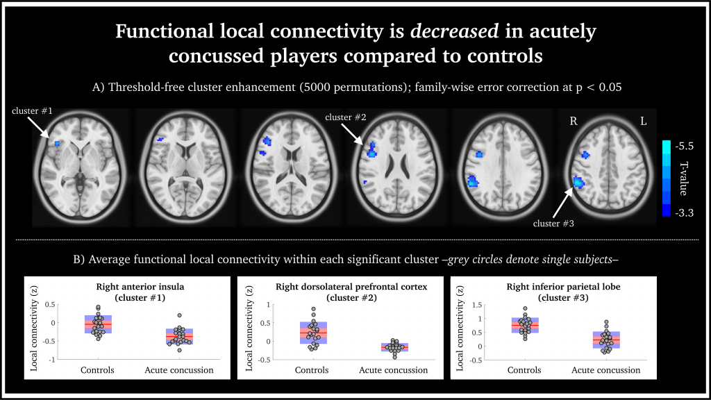
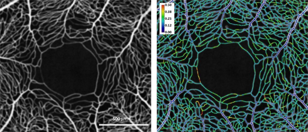
Functional local connectivity is decreased in acutely concussed players compared to controls. (a) Statistically significant brain regions at family-wise error rate p < 0.05; 5000 permutations using threshold-free cluster enhancement. Cool-scaled color bar denotes the magnitude of voxel t-values. (b) Individual-level z-normalized fMRI local connectivity values where each grey circle denotes an individual subject; red line is the group mean; red shaded area is the 95th percentile of the mean value; and blue shaded area is one standard deviation from the mean.

Head injuries, including concussion, are taken very seriously in sporting professions. To date, making an accurate diagnosis of acute concussion has been made difficult by the lack of a reliable direct biomarker for injury and recovery. This diagnostic gap can lead to unknown recovery periods and potentially long-term impacts for athletes.


Researchers at the Florey Institute of Neuroscience and Mental Health set out to understand functional brain changes in professional players in the Australian Football League who had been diagnosed with acute sport-related concussion.


The world-first study, published in the Journal of Concussion, utilised NIF infrastructure, the 3T Trio and Skyra MRI scanners, and NIF expertise, Facility Fellows Shawna Farquharson and David Abbott.

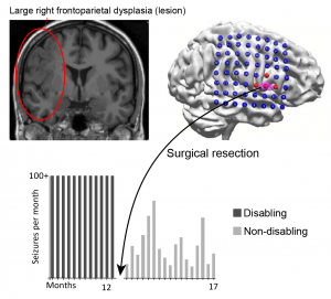
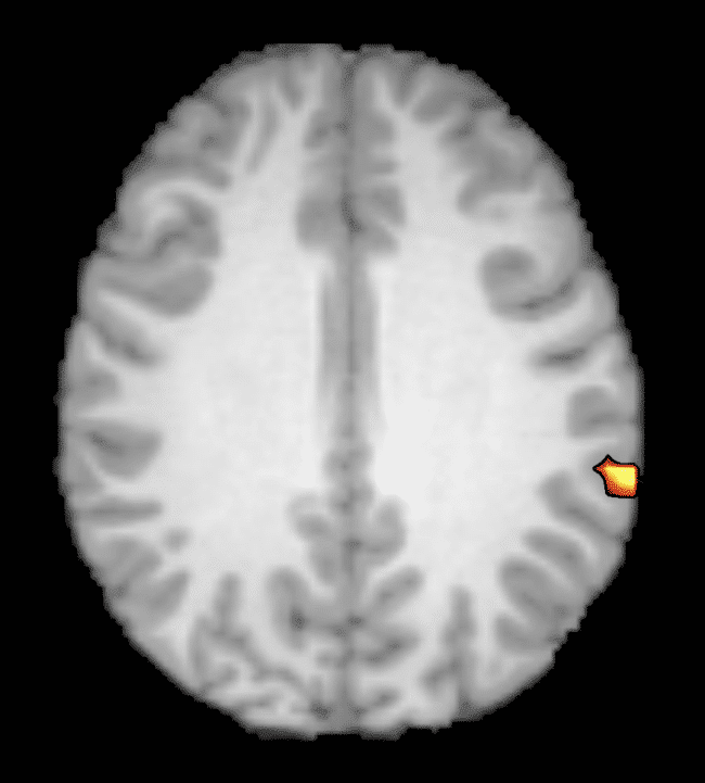
 Functional MRI (fMRI) was undertaken to assess functional connectivity alongside anatomical imaging. Although no anatomical damage was observed, the authors described a decreased intrinsic fMRI connectivity within the right frontoparietal regions in acutely concussed footballers. In other words, all 20 concussed athletes showed reduced activity in parts of the brain responsible for executive function, working memory and switching tasks.


“By looking at how the different parts of the brain talk to each other, we can see how these three brain networks are affected, and these changes may help explain the symptoms we see in concussed players.” – Dr Mangor Pedersen, study co-author, from the Florey Institute of Neuroscience and Mental Health.


One interesting finding in this study is that concussion appears to affect particular networks in the brain. These findings are in agreement with some of the typical clinical features of concussion; however, they are based on trends seen as a group. In future, the authors intend to investigate individual brain networks and develop guidelines for personalised treatment and recovery.


This story was contributed by the Florey Institute of Neuroscience and Mental Health NIF Node. For more information, please contact Shawna Farquharson or David Abbott.


More information about concussion is available here, or by speaking to your GP.

Hyperpolarized 129Xe MR imaging of lung

Magnetic Resonance Imaging (MRI) has a range of applications in medical diagnosis, and more than 25,000 scanners are estimated to be in use worldwide. However, lung imaging suffers from some technical challenges, limiting its application in pulmonary disease diagnosis and treatment.


Due to low proton density, movement and high susceptibility difference between air and tissue, conventional proton MRI struggles to image lung tissue and function. These can be partially overcome by the introduction of contrast agents into the lung. Hyperpolarised gases are promising contrast agents for imaging lung structure and function. The two most common gasses are helium (3He) and xenon (129Xe) isotopes. Helium isotopes are challenging and expensive to obtain, and do not provide significant functional readouts. Xenon can be challenging to work with, but promises novel physiological measurements not previously feasible. This resulting technique, termed hyperpolarized 129Xe MR imaging, has revolutionised the field of functional lung imaging.


Five years ago, an Australian Research Council (ARC) grant was awarded to researchers at Monash Biomedical Imaging (MBI) and ANSTO to design and build a machine that could reliably produce hyperpolarized xenon. In this project, Dr Wai Tung Lee and NIF Facility Fellow Dr Gang Zheng have teamed up to develop a new multimodal technology capable of high-quality investigations of the human lungs. While Dr Lee, of ANSTO, was responsible for the construction of the polarizer and production of polarized 129Xe for MR imaging, Dr Zheng, of Monash University, designed the MR experiments and adapted imaging protocols.

Figure 1. Schematic diagram of the polarizer system. Note: Some details omitted for clarity.

Hyperpolarized 129Xe (HP-Xe) gas was generated in a custom designed and constructed SEOP system at Monash Biomedical Imaging (Figure 1 and Figure 2). The system consists of a gas-polarizing unit and a gas injection and recycle unit. The core component of the gas-polarizing unit is an optical pumping cell (OPC) where the gas is polarized. The OPC is placed in an oven to regulate the amount of Rb vapour by maintaining a constant temperature between 60ºC-90ºC. The optical pumping process uses two 240W narrow-bandwidth lasers tuned to the Rb D1 transition wavelength of 794.7nm. Additional optics condition the laser light to circular polarization and shape the laser profile to fully illuminate the OPC. HP-Xe is produced in batches rather than using a continuous-flow process.

Figure 2. The spin-exchange-optical pumping system. A. Gas polarizing unit in Room 1; B. Gas injection and recycling unit in Room 2.

MR experiments were performed on a clinical 3T whole-body system (Skyra, Siemens Medical Solutions, Erlangen, Germany) with a broadband RF amplifier. HP-129Xe MRI was enabled using a bird-cage transmit/receive chest coil (RAPID Biomedical GmbH, Wuerzburg, Germany). A 2D-GRE sequence for X-nuclei MRI was used to image HP-Xe gas in lung. Proton channel images were acquired for the localization of the lung. The resonance frequency of 129Xe was set to 34.09 MHz on the 3T scanner. The human subject first quickly flushed his lung with pure nitrogen, and then immediately inhaled HP-Xe in the Tedlar bag. After inhalation of HP-Xe, the subject held their breath during the scan. The total imaging time was less than 10s.


Dr Zheng outlines the results to date, “We have successfully imaged gas-phase HP-129Xe in a range of phantoms, such as the Tedlar bag, syringe and the glass cell (Figure 3). We repeated the gas-phase imaging in both ex-vivo (Figure 4) and in-vivo lamb lungs (Figure 5). Imaging results supported that our polarizer can provide sufficient polarization for lung imaging. In 2019, we successfully performed a human experiment and a gas signal from the lungs was clearly identified (Figure 6). In addition, all studied showed a clear Rf peak of gaseous and dissolved xenon which should allow us to develop methods to assess lung function. We plan to study human pulmonary disease in the near future and hope to find additional collaborators to help translate this modality into clinical use.”

Gallery Notice : Images have either not been selected or couldn't be found

Construction of the hyperpolarizer is complete; it can routinely produce HP-129Xe for research use. The technique has successfully imaged HP-129Xe in phantoms, ex-vivo and in-vivo lamb lungs and a human lung. If you are considering lung research, please see more on the Monash website, or get in touch with Dr Gang Zheng to discuss your project needs.


Publications:

Zheng G, Lee WT, Tong X, et al., A SEOP Filling Station at the Monash Biomedical Imaging Centre. Conference on polarization in Noble gases (PiNG), 2017.


Zheng G, Lee WT, Tong X, et al., Phase imaging of hyperpolarized 129Xe gas in a human lung. ISMRM 2020, submitted on 6 Nov 2019.


Acknowledgements:

National Imaging Facility, ARC (Grant LE130100035); NHMRC (Grant APP606944); CASS Foundation; Monash Biomedical Imaging, ANSTO


This story was contributed by Dr Gang Zheng of the Monash University NIF Node.


Impact of surgical lymph node removal

The impact of surgical lymph node removal on metastatic disease and the response to immunotherapy

Surgical resection of cancer remains the frontline therapy for millions of cancer patients every year, but disease recurrence after surgery is common with a relapse rate of around 45% for lung cancer. Relapse rates are expected to decline, with new immunotherapies producing extraordinary successes in several solid cancers. Immunotherapy administered after surgery could potentially ‘mop up’ small persisting cancer deposits that lead to disease recurrences. However, uninvolved (tumour-free) draining lymph nodes are the primary ‘factory’ for generating anti-cancer T cell responses; hence, should they be removed, subsequent immunotherapy may be negatively impacted. The aim of this project is to determine in murine models if the response of metastatic disease to immunotherapy is reduced following tumour lymph node resection.

Dr Vanessa Fear of the School of Biomedical Sciences, at The University of Western Australia, is investigating if the response of metastatic disease to immunotherapy is reduced following tumour draining lymph node resection. To do this, the Tumour Immunology Group is using an AB1 Model of metastatic disease. Tumour progression is visualised using IVIS imaging and histology following resection to track the effectiveness of treatment regimes. Ultimately, the team will seek to determine the impact of lymph node removal at the time of tumour resection to subsequent immunotherapeutic outcomes.

Fig 1: IVIS imaging from AB1-HA tumour model. Mice received AB1-HA_LUC i.v. and lung tumour development monitored on the InVivo Imaging System (IVIS, Lumina II imager). At the imaged timepoints mice received intrapertioneal injections of luciferin (150µg/g) and tumour burden was measured on the IVIS in photons/sec (p/s). A, tumour progression day 14 to day 19.

The research project involves collaboration with the Centre for Microscopy, Characterisation and Analysis, the West Australian Node for the National Imaging Facility to image, visualise and characterise the development of lung metastatic disease using the IVIS Lumina II in vivo bioluminescence imager with the help of Living Image Software (Caliper Life Sciences).

Fig 2: IVIS imaging and histology from AB1-HA tumour model.Comparison of IVIS reading with lung H&E staining showing AB1-HA tumour from the same mouse. Tumour volume determined using FIJI software.

The team have completed preliminary studies determining a 55% metastatic disease onset after surgical resection of the primary tumour. Current investigations in tumour resection and lymph node resection indicate temporal changes in onset of metastatic disease compared to mice with intact lymph nodes.

Further investigations into the impact of lymph node resection on immunotherapy are underway. Future investigations will include other models including lung adenocarcinoma, melanoma, and breast cancer.

Collaborators

School of Medicine, the University of Western Australia

School of Biomedical Sciences, University of Western Australia

Centre for Microscopy, Characterisation and Analysis, the University of Western Australia

This story was contributed by the University of Western Australia. For more information, contact Dr Vanessa Fear or Diana Patalwala.

3D printed devices to treat traumatic pelvic fractures

3D printing is increasingly being used in the healthcare industry to customise medical devices to meet patient-specific needs. Currently, device manufacture is lengthy, limiting the application of customised medical devices. The treatment of traumatic injuries requires intervention as quickly as possible, preferably within days post-injury.

This collaborative research project between the Dept. of Biomedical Engineering at the University of Melbourne and the Dept. of Orthopaedics at the Royal Melbourne Hospital aims to assess the feasibility of 3D printing fracture plates to treat traumatic fractures and speed up the production of devices at the point-of-care for a patient. By performing a proof-of-concept experiment on a set of cadaveric pelvis, Dr Dale Robinson and team are evaluating each phase of the 3D printing workflow. Once implanted, a series of computational models and biomechanical experiments will be used to assess whether the 3D printed fracture plate offers an improvement over a traditionally mass-manufactured plate. Paramount to designing customised implants, the anatomy of each pelvis is being characterised using the University of Melbourne’s NIF Node CT with input from PET/CT Facility Fellow Rob Williams and radiographer Rebecca Glarin. After implantation of the fracture plate, CT may assess the effectiveness of the device in terms of stabilising and reducing the fracture.


3D reconstruction of a fractured human pelvis with a custom 3D printed device simulated in blue to promote appropriate healing.

To date, the project has conducted some scans and used this data for preliminary printing of implants. Plates were designed and printed in collaboration of researchers at Johnson and Johnson and the University of Melbourne. The initial study used 3D printed medical-grade titanium and 3D rendering from the NIF facility CT. In developing this method, iterative reconstruction with maximal overlap to printing was used to be consistent with typical medical CT. This was done while still using radiation dosimetry within standard limits.

This project has the potential to improve patient outcomes by enhancing surgical intervention durability, reducing the duration and number of surgeries, and reducing the risk of life-threatening surgical complications (such as pulmonary embolism and infection) through reduced bedtime. Consequently, the effective implementation of customised 3D printed medical devices is expected to reduce healthcare costs through shorter hospital stays and reduced number of surgical interventions.

This story was contributed by the Department of Biomedical Engineering and the Melbourne Brain Centre Imaging Unit at the University of Melbourne, and Johnson & Johnson. For further information, please contact Rob Williams.

Detecting retinal vascular disease


One of the leading causes of blindness in the Western world are vascular diseases affecting the retina. The arteries and veins inside the light-sensitive layer in the back of the eye – the retinal vasculature – are an intricate network supplying the inner retina with crucial nutrients while removing metabolic waste.  The heterogeneity in space and time of blood flow in this microvasculature is critical as the retina has one of the highest metabolic demands of the central nervous system.


Pathological retinal neovascularisation and oedema – that is swelling, thickening, or unusual growth in the retinal vasculature – are commonly responsible for certified visual loss.  Therefore, understanding the spatial and temporal heterogeneity and active regulation of retinal blood flow in the retina is critical for the diagnosis of retinal vascular disease. Furthermore, these blood vessels are affected by systemic vascular diseases and, as such, evidence of these conditions may be observed through the microvasculature inside the eye.


Recently, a technique known as optical coherence tomography angiography (OCTA) has been developed as a non-invasive approach to visualising blood vessels. This method uses the reflectance of light on the surface of moving blood vessels to map the vasculature of the retina.


A multi-disciplinary team, headed by Prof Dao-Yi Yu at the Lions Eye Institute (affiliated with the UWA Centre for Ophthalmology and Visual Science) are using OCTA to reveal remarkable spatial and temporal heterogeneity of retinal capillary perfusion. The project aims to use OCTA as a non-invasive tool for the early detection of retinal vascular diseases.


NIF Informatics Fellow, Dr Andrew Mehnert, is contributing his analysis expertise to guide improvements in OCTA instrumentation and algorithms. Image analyses, undertaken in the Characterisation Virtual Laboratory (CVL) using both MATLAB and FIJI/ImageJ, showed remarkable resolution of capillary perfusion in the macular region of human and porcine subjects.


Left: En face mean intensity projection through 31 OCTA images from the macular region of a human subject. Right: Coefficient of variation along vessel centrelines showing spatial and temporal heterogeneity of capillary perfusion. The colour bar indicates the variation from dark blue (no variation) through to red (most variation).


The developed software tools have been used for characterising spatio-temporal variation of capillary perfusion in OCTA images.


Experimental results to date are both valuable and encouraging because they may be potentially useful for clinical diagnostic purposes and can be used to guide improvements in OCTA instruments and new algorithm development. These results move towards a non-invasive tool for the early detection of retinal vascular diseases in humans, and may also be used for other investigations of capillary perfusion where the retina is an appropriate model for microcirculation studies.


For further information, please contact Dr Andrew Mehnert.

This story was contributed by the University of Western Australia.

Improving the lives of patients with epilepsy

A 20-year-old man with a five-year history of disabling seizures was suffering up to 500 seizures per month; his schooling was disrupted, and all the standard non-invasive tests gave conflicting results. Surgical resection was performed correlating with the earliest EMSL result, a technique pioneered at the Swinburne NIF Node. The patient is now seizure-free and on minimal medication following surgery.

NIF at BLiSS*Adelaide

BLiSS is a one-day event aimed at catalysing cross-disciplinary collaborations between early- and mid-career researchers. On Oct 4th 2019, BLiSS*Adelaide was launched, featuring exceptional lectures, poster sessions, and stalls of service providers and Universities. With over 150 registrations representing more than six institutions, the Adelaide scientific community came out in force to find new scientific collaborations. SAHMRI participated with excellent representation by Dr Randall Grose (ACRF research fellow), Dr Susan Porter (Manager PIRL) and Dr Marianne Keller (NIF Facility Fellow). Many of the posters featured data from SAHMRI’s small animal imaging equipment, and many attendees showed interest in the imaging modalities available through the NIF network such as high-resolution small animal MRI. For more information on NIF capabilities, contact Dr Keller or NIF Central.

Dr Randall Grose (ACRF research fellow), Dr Susan Porter (Manager PIRL) and Dr Marianne Keller (NIF Facility Fellow) at BLiSS*Adelaide


Catching the wave

Catching the wave: non-invasive MEG/EEG for epilepsy seizure localisation

Over 250,000 Australians currently live with epilepsy, a chronic disorder of the brain. In addition to significant social, physical and psychological consequences, epilepsy is linked with an increased risk of death compared to the general population. Up to 70% of epilepsy cases may be controlled through lifestyle and medication; however, many patients are unable to be treated using conservative measures. A major clinical problem, drug-refractory focal epilepsy may require aggressive treatments, including surgery, to resect a portion of the brain.

The localisation of the epileptogenic zone (the portion of the brain requiring resection to reduce seizures) is typically carried out clinically using magnetic resonance imaging (MRI). In many cases, no abnormalities are detected, requiring further investigation. Surgeons may also use invasive surgically implanted electrodes (intracranial electrode depth or grid, or ICEEG) for seizure source localisation, increasing the risk to the patient. A lower risk option is further functional imaging with positron emission tomography (PET), single-photon emission computed tomography (SPECT), and simultaneous electroencephalography functional MRI (EEG-fMRI). Unfortunately, many of these modalities lack temporal resolution to distinguish seizure onset.

“Catching the earliest wave, the earliest is key”
Representation of MEG (green) and EEG (blue) overlays.

Magnetoencephalography (MEG) and electroencephalography (EEG) offer a non-invasive method to detect rapidly evolving brain wave spikes and seizures at millisecond temporal resolution.  A/Prof Chris Plummer and team, Mr Simon J. Vogrin, Dr William P. Woods, A/Prof Michael A. Murphy, Prof Mark J. Cook, and Prof David T.J. Liley, have developed a world-class multimodal technique for non-invasive source localisation and epileptogenic zone characterisation. The method (using co-acquired MEG and high-density EEG) has already improved the surgical treatment of medically refractory epilepsy, with clinical validity demonstrated in a key 2019 publication.

The team utilised the world-class MEG at the SUT NIF Node together with the expertise of NIF Facility Fellow Dr Will Woods to look at the efficacy of concurrent HDEEG (High-Density EEG > 64 electrodes) and MEG for identifying the epileptogenic zone. Ninety-six patients have been involved in the long-term prospective study. Seventy-one patients were MRI negative and twenty-five MRI positive with either complex lesions or multiple lesions. More than five years’ data collection in collaboration with neurologists and medical scientists found that the combined method was able to detect interictal spike discharges (between seizures) at early, mid and late-phase as well as ictal discharges (during seizures).  The earliest resolvable localisation by either EEG or MEG was determined to be the most suitable site for surgical resection.

“We find that for most cases, modelling either the mid-upswing or the later peak (still common practice) results in mislocalization of the cortical generator (and the putative Epileptogenic Zone) due to the effects of discharge propagation across the cortical surface. We look for the earliest feature from the discharge; sometimes it appears first in the Magnetometer, sometimes it is first in one of the Gradiometers, and sometimes it is first in the HDEEG. For a given patient, there is typically a dominant leading modality, but the modality that leads in time will vary from patient to patient. This is fundamentally the reason for recording both. Otherwise, you risk having half the story before diving into invasive investigations.”  – A/Prof Plummer

To date, successful source localisation has guided successful surgical outcomes in 25 patients, 8 of whom were spared invasive intracranial monitoring due to this method. Many of the over 100 studies performed are yet to progress to surgery, underpinning the latent potential of yet to be followed complete surgical workup and seizure-freedom outcomes following surgical therapy.

“Accurate, non-invasive localisation with this method will reduce the medical burden of invasive intracranial electrode monitoring by reducing the number of invasive electrodes required and, in some cases, remove the need for these in the first place to allow the patient to proceed directly to potentially curative surgery.” – A/Prof Plummer

This work intends to revolutionise epilepsy management in Australia, with data from a larger and more diverse cohort expected to provide evidence for a Medicare rebate to facilitate the accessibility of this technique for Australians.

Success stories:

From 500 seizures per month to none; MEG/EEG guided surgical resection success

A young man with a five-year history of disabling seizures was suffering up to 500 seizures per month; his schooling was disrupted, and all the standard non-invasive tests gave conflicting results. Surgical resection was performed correlating with the earliest EMSL result. The patient is now seizure-free and on minimal medication following surgery.

A 10-year old child had a four-year history of left leg motor-sensory events, up to 200 times per day, leading to falls. PET and MRI suggested localisation of seizures in the right superior frontal gyrus and the right paracentral lobule, while EMSL results showed seizure onset further to the rear at the precuneus. Surgical resection of the paracentral lobule and superior frontal gyrus did not relieve the seizures seen by ICEEG during surgery. Further exploration revealed a second pocket of dysplasia in line with the EMSL result, resection of which normalised the ICEEG and relieved the patient of seizures.

A 36-year-old patient presented with speech-arresting complex partial turns with a typical MRI result. Their surgical plan was altered to include a hippocampal depth electrode, where seizures were found to start by MEG. Standard anteromesial temporal lobectomy revealed a cortical dysplasia involving the entorhinal cortex. The patient is now seizure-free and works as a speech therapist.

Recovering day to day activities without disturbing the complex dysplasia

A 36-year old mother of two young children suffered a 33-year history of multiple-daily disabling motor seizures. She was denied surgery due to a complex lesion seen by MRI (frontoparietal dysplasia).  EMSL captured the seizure onset, targeting surgical resection without interfering with the bulk of the lesion. The patient is now on reduced medication and experiences only brief non-disabling focal seizures. She states that she “has her life back”.

You can learn more about epilepsy here or by speaking to your GP. This work was contributed by St Vincent’s Hospital, Swinburne University of Technology, and the University of Melbourne. For more information, please contact Dr Will Woods.

New SMRT President appointed

Profile photo of Shawna facing camera and smiling
NIF Facility Fellow and Florey Chief Radiographer, Shawna Farquharson. Photo attributed to S. Farquharson

NIF Facility Fellow Shawna Farquharson is leading the way to empower, connect, educate and inform MRI Radiographers and Technologists worldwide. Following their 2019 Annual Meeting, the Society for MR Radiographers & Technologists (SMRT), a Section of the International Society for Magnetic Resonance in Medicine (ISMRM), appointed Shawna Farquharson as its new President of SMRT. SMRT is the leading non-profit organisation that provides an international forum for education, information and research in magnetic resonance for radiographers and technologists throughout the world.

Read More
Privacy Settings
Youtube
Vimeo
Google Maps