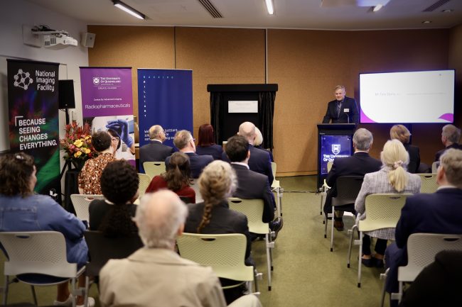
Boosting health innovation and commercialisaton: $2m Queensland Government investment in National Imaging Facility
The Queensland Government Department of Environment, Science and Innovation has announced $2m will be awarded to National Imaging Facility (NIF) capabilities in Queensland through the Research Infrastructure Co-Investment Fund (RICF).
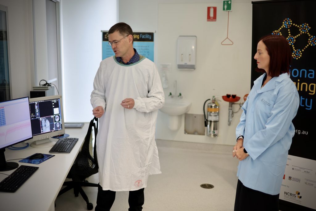
RICF co-invests with universities, research centres and industry to support facilities of national significance, and this grant will contribute to NIF capabilities and operations at the University of Queensland (UQ) Centre for Advanced Imaging (CAI) and Herston Imaging Research Facility (HIRF).
This investment will enhance imaging research infrastructure in Queensland, increasing the leverage of the Australian Government Department of Education’s National Collaborative Research Infrastructure Strategy (NCRIS) funding secured by NIF last year.
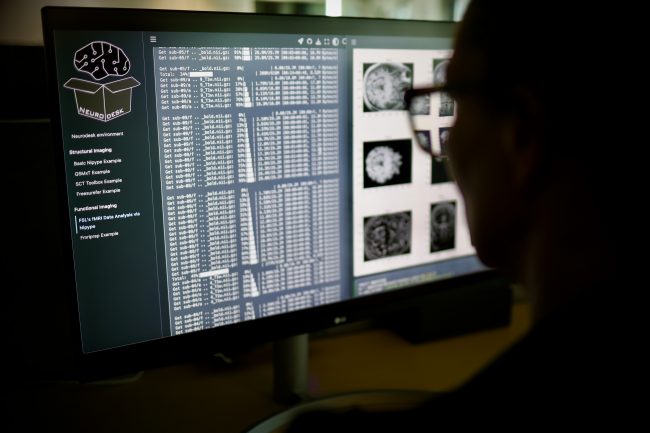
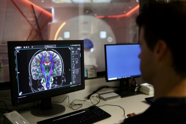
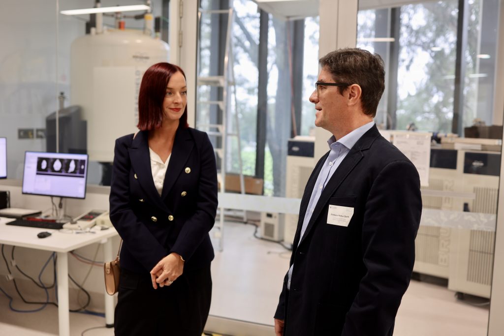
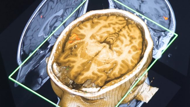
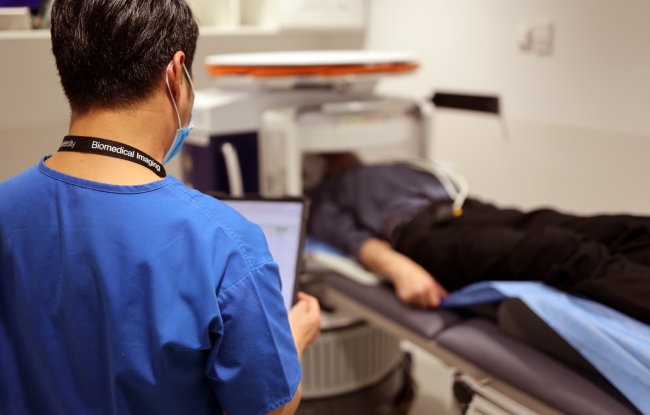
NIF operates Queensland’s most advanced biomedical imaging equipment across CAI and HIRF, and collaborates with research and clinical precinct partners, including QUT, QIMR-Berghofer and Brisbane’s Metro North Hospital and Health Service (MNHHS), bridging the gap between scientific breakthroughs and patient outcomes, to provide a strong research translation pathway.
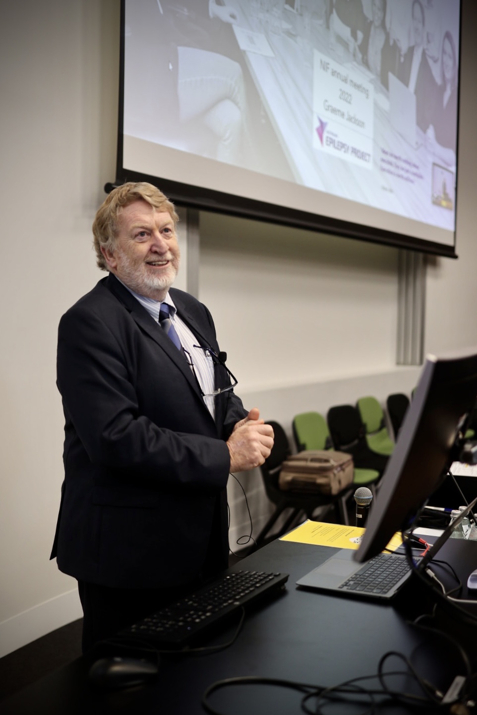
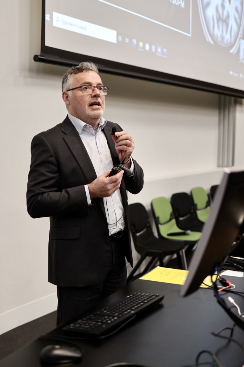
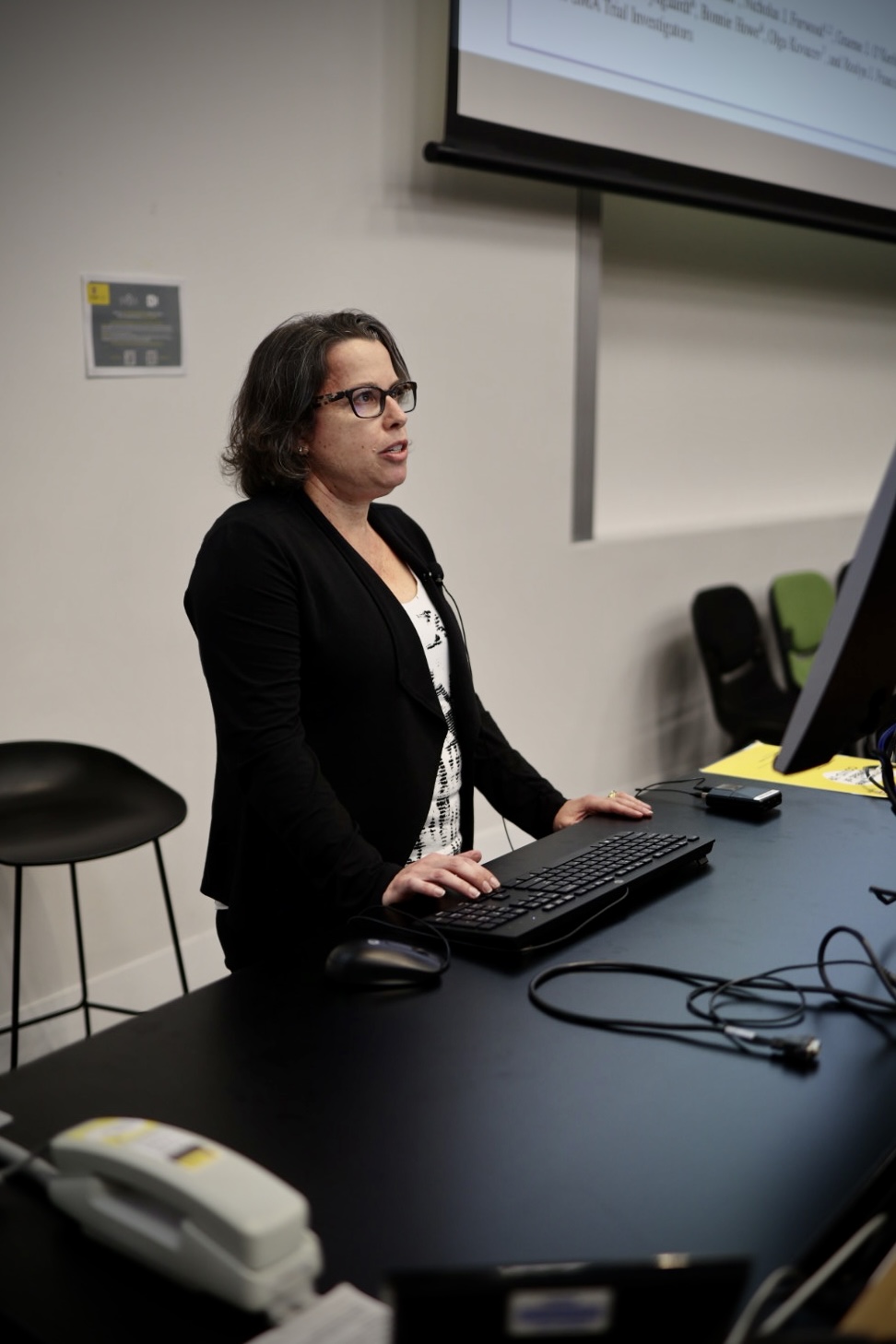
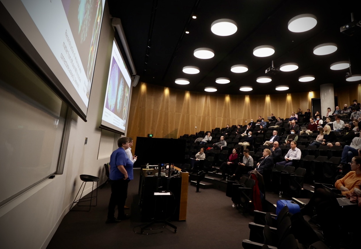
NIF HIRF Node Director, Prof Katie McMahon said RICF’s funding will bolster NIF’s Queensland operations to maintain and expand cutting-edge advanced imaging capabilities and expertise, underpinning the translation of research into tangible health discoveries and commercialisation opportunities.

“NIF’s integrated imaging facilities provide a translational pathway from discovery to preclinical testing through to first-in-human studies and clinical trials,” Prof McMahon said.
“We’re proud to work with pharmaceutical and biomedical industry partners to accelerate the development of the latest scientific discoveries into better outcomes for patients, including creating new medical products and therapies to treat diseases like dementia, cancer and epilepsy.”
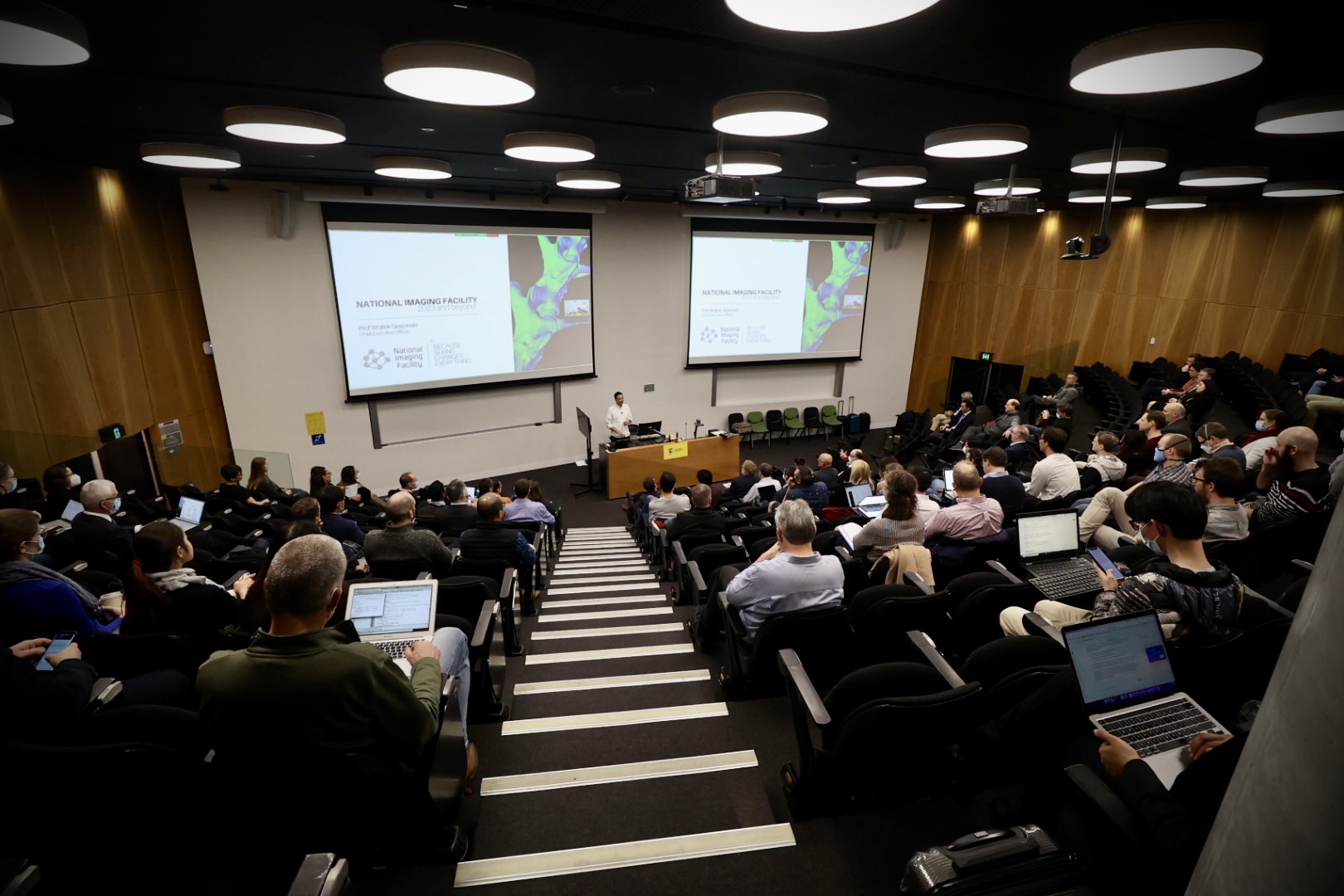
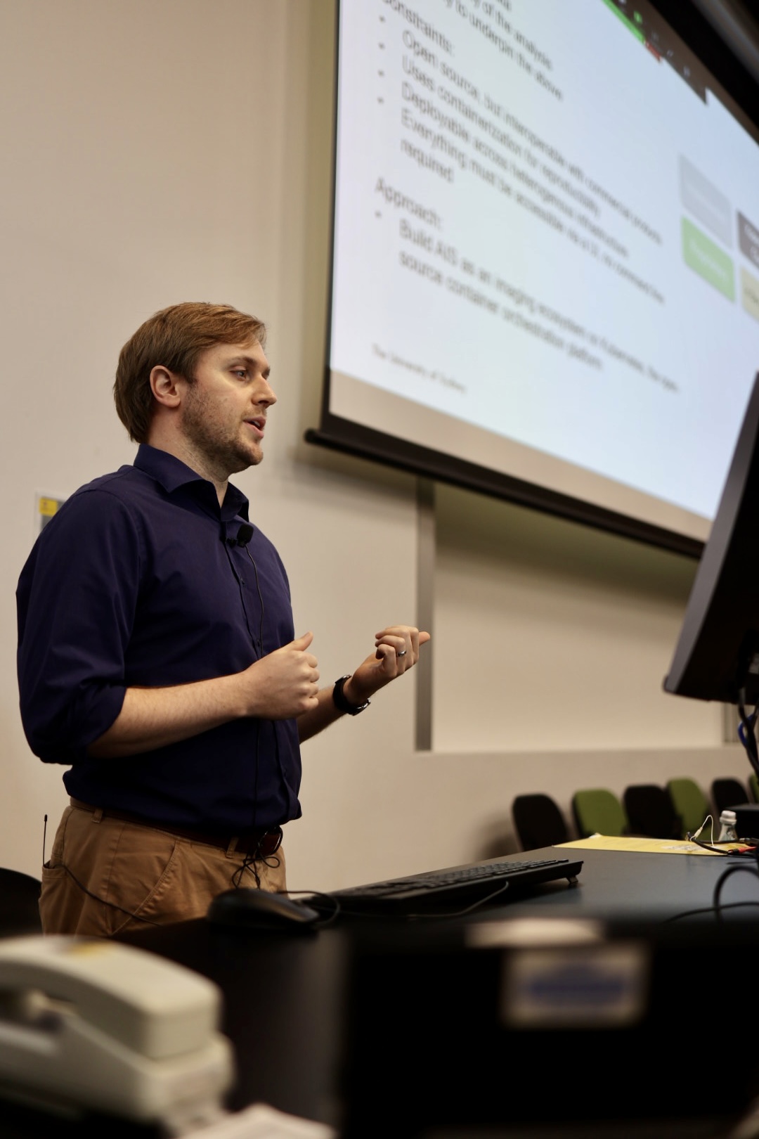
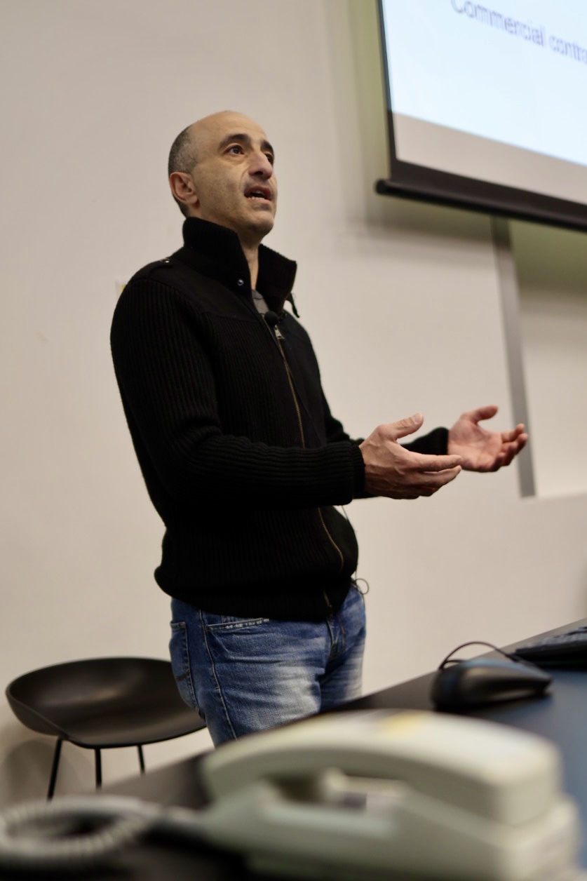
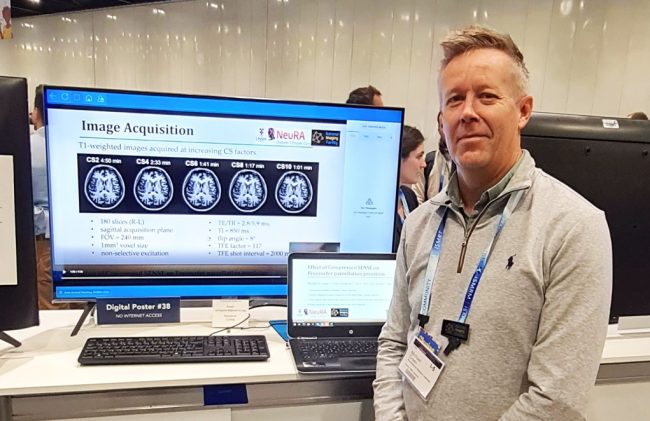
NIF CEO Prof Wojtek Goscinski said this co-funding would ensure scientists have access to the most advanced translational research infrastructure in Australia.
“RICF’s co-investment in NIF will help facilitate the procurement of state-of-the-art advanced imaging equipment. Additionally, it will help attract, train and create jobs for the world-class experts critical to driving innovation through this infrastructure.” Prof Goscinski said.
Read the full media statement from the Department of Environment, Science and Innovation here.