Exploration of the deep foot muscles at ultra-high field
The arches of the human foot are unique structures that are important for functions like walking and running. The deep intrinsic muscles of the foot, such as the adductor halluces and interossei, are thought to play key roles in arch control; yet little is known about how they are controlled during functional tasks. The traditional measurement techniques can only provide information regarding muscle size, which is inadequate to evaluate the force-generating capacity of the muscles, assess the process of force generation by the muscles, and understand the involvement of the neural drive sent by the body to the muscles to regulate force production.
Using the 7T human MRI at the Centre for Advanced Imaging, a research study is currently investigating the muscle architecture of the adductor halluces and interossei. This research aims to quantify the force-generating capacities of these deep-foot muscles by measuring their MRI muscle volumes, estimate their force production by shear wave Elastography and measure their neural drive by using Electromyography.
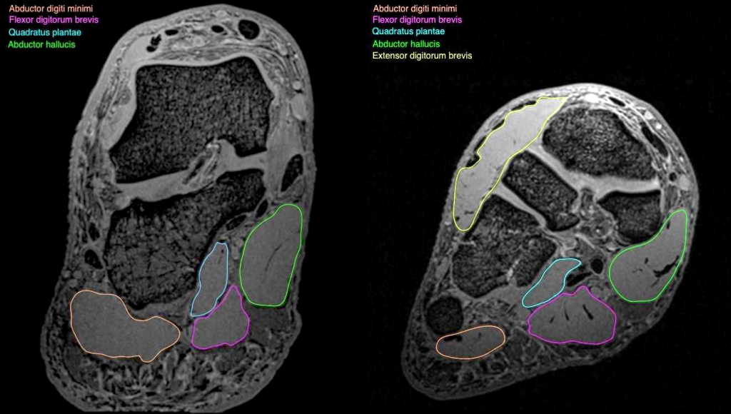
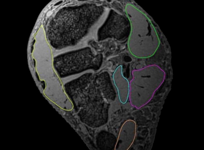
Deep foot muscles outlined in two cross-sections of a structural MRI scan obtained at ultra-high field: abductor halluces (green), abductor digiti minimi (pink), flexor digitorium brevis (magenta), quadrature plantae (cyan) and extensor digitorium brevis (yellow). With thanks to Dr Natalie Collins, University of Queensland
MRI images are obtained from male and female volunteers with no lower limb pain or injury. To date, structural MR images of deep foot muscles have been obtained using T1 VIBE 3D Transverse Oblique sequence and MRI muscle volumes have been measured. In future, diffusion tensor imaging will be performed to measure apparent diffusion coefficient and fractional anisotropy of the deep foot muscles.
This story was contributed by the University of Queensland, with acknowedgements to Dr Natalie Collins of the School of Health and Rehabilitation Sciences, Health and Behavioural Sciences, University of Queensland.
For further information, please contact Dr Tonima Ali.