Imaging enabling nanomedicine to treat aggressive brain cancer
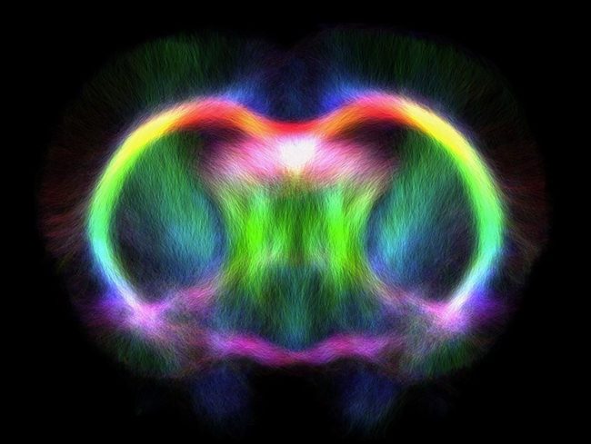
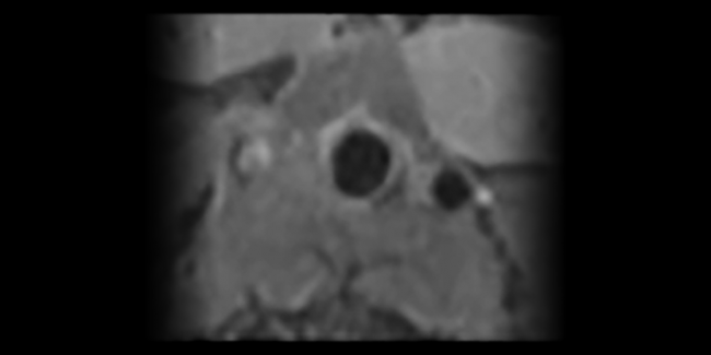
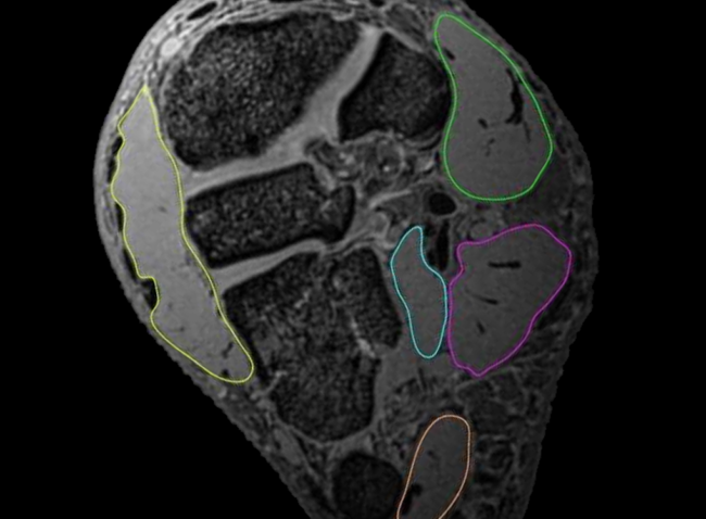
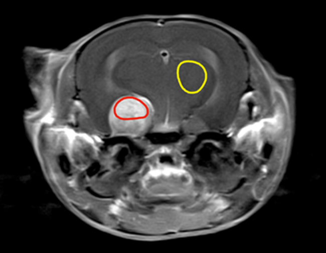
Image: Gadolinium enhanced MRI showing the bright brain tumour (red circle) compared to the normal brain tissue (yellow circle).
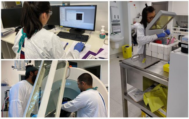
‘Nanomedicine’ sounds like a term you’d hear in a futuristic novel or an episode of Doctor Who, but cutting-edge scientists from the National Imaging Facility’s Node at the University of Queensland’s Centre for Advanced Imaging are already applying it to solve complex health challenges in collaboration with the Australian Institute for Bioengineering and Nanotechnology, and the Australian Research Council’s Centre of Excellence in Convergent BioNano Science and Technology and Training Centre in Biomedical Imaging Technology.
Nanomedicine applies nanoscale materials, such as nanoparticles and nanorobots (wow!) to the prevention and treatment of disease. Nanomedicine is a promising strategy to target tumours with chemotherapy in a safe and controlled manner.
This all sounds great, but within the context of the brain things get a little more complicated. For brain tumours, the integrity of the blood brain barrier (BBB) is central to its effective use as treatment.
The BBB is a protective barrier between the blood vessels and brain tissue, providing a defence against pathogens and toxins that may be present within the blood, while at the same time allowing vital nutrients to reach the brain.
While the BBB protects the brain against pathogens and toxins that could cause infection, it also blocks medicine from crossing the barrier in many cases, which can hamper the (often urgent) treatment of tumours, among other neurological disorders.
This is where imaging comes in…
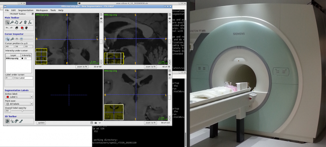
UQ researchers have undertaken studies utilising NIF’s flagship preclinical magnetic resonance –positron emission tomography (MR-PET) system to develop hybrid imaging, combining the imaging of MRI with the information of PET.
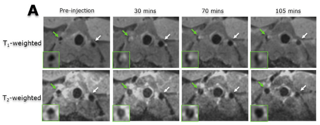
Simultaneous MR-PET imaging enables experts to measure opening of the BBB using Gadolinium (Gd) contrast agents at the same time as delivering novel PET tracers and new theranostic candidates including nanomedicine.
These tools assist in investigating the link between BBB integrity and tumour diagnosis and treatment, and ensure the development of promising new treatments such as nanomedicine that can permeate the BBB.
BBB integrity: Tumour diagnosis and treatment
The rapid growth of brain tumours requires the formation of new vessels to supply increased demands for nutrients. The new vessels are leaky compared to normal brain vessels, where the BBB tightly regulates the transfer between blood and tissue.
The team have used Gd MRI to estimate how leaky the tumour vessels are. This information is vital for understanding how tumours are developing, and when and how diagnostic or therapeutic drugs can enter the tumour tissue.
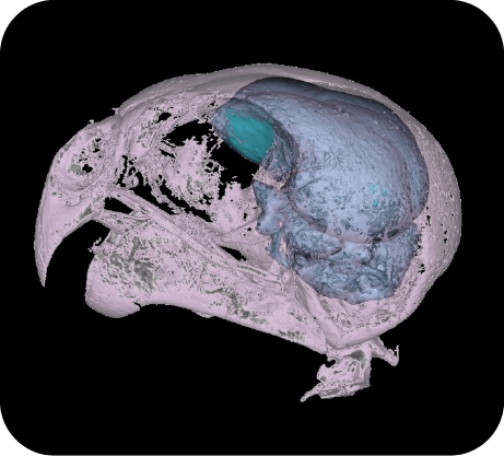
In combination with new nanomedicines, there are also exciting new techniques that allow opening of the brain for treatment without surgery, which can increase risk for patients. These new imaging methods allow the opening of the brain to be accurately monitored to ensure entry of the treatment without any unrelated damage to the brain.
So – our takeaway message? While advances in nanomedicine alone are exciting and important, the integration of nanomedicine and advanced imaging brings opportunity for exponential synergistic innovation in healthcare, that can improve outcomes for patients and ultimately has the potential to save lives.