NIF to demonstrate impact of coordinated data and AI at RANZCR ASM 2022

National Imaging Facility (NIF) will host a session at the Royal Australian and New Zealand College of Radiologists (RANZCR) 72nd Annual Scientific Meeting (ASM) this month.

The NIF Showcase will highlight critical expertise and human imaging projects from across Australia’s advanced imaging network, including regional MRI and life-changing imaging for Australians living with epilepsy.


Point-of-care imaging leveraging AI to grow healthcare equity in regional Australia

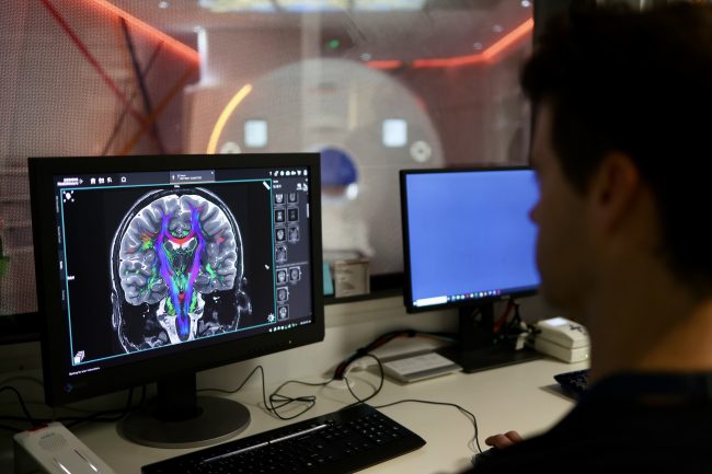
Head of Diagnostic and Interventional Radiology research at Alfred Hospital, Prof Meng Law will present on new technology for point-of-care imaging and regional MRI. Prof Law is an expert on neuroimaging and AI, and his presentation will focus on federated deep learning for signal-to-noise ratio imaging and motion correction, using NIF’s low-field magnetic resonance network.

NIF is deploying four low-field MRI scanners to remote and regional sites to help researchers apply this affordable imaging technology in rural areas. These scanners will be used to understand how this fast-developing technology can be used to diagnose stroke, traumatic brain injury, and other conditions after testing in research laboratories at NIF nodes.


Imaging networks and datasets to support life-changing platform for more than 150,000 Australians living with epilepsy

Clinical Director of The Florey Institute of Neuroscience and Mental Health, Prof Graeme Jackson will present on the Australian Epilepsy Project (AEP), reducing diagnosis uncertainty and fast-tracking optimal treatment by combining advanced imaging, genetics, cognition, and artificial intelligence (AI).

Prof Jackson is the Chief Investigator on the AEP Platform, which will drive major advances in decision support tools for epilepsy, and NIF’s national human MR network is set to enable scanning across Australia.

The data collected by the AEP will provide a rich resource for addressing many other traditional science and mechanistic questions in epilepsy to progress epilepsy research worldwide.


NIF CEO Prof Wojtek Goscinski said the invitation to showcase NIF at the RANZCR ASM was an opportunity to highlight the transformation of imaging through AI and big data, and to underline the unique capabilities that NIF provides.

“We’re privileged to have world-class speakers Prof Meng Law and Prof Graeme Jackson presenting on two projects that are supported by data collections and the AI models around them, which will lead to better treatment and diagnosis for Australians,” Prof Goscinski said.

The impact of imaging in radiology is only increasing, with experts now able to extract quantifiable information from ever larger data collections by applying machine learning methods such as deep learning and convolutional neural networks.

Big data and AI have a transformative effect on radiology, enhancing patient outcomes by distinguishing irregularities and patterns in data collections, and enabling diagnosis with speed and accuracy.

“NIF is focused on keeping Australia at the forefront of imaging technology and imaging data analytics, and is exploring a range of activities to increase uptake of machine learning in imaging, including data infrastructure and imaging quality,” he said.

The NIF Showcase session will also see a panel of experts discuss opportunities for collaboration between NIF and RANZCR for the benefit of medical research.

View the NIF Showcase agenda below:

RANZCR ASM NIF Showcase: Friday 28 October, 08:30-10:00

TIMETOPICSPEAKER
8:30IntroductionA/Prof Sanjay Jeganathan
RANZCR President
8:35Introduction to National Imaging FacilityProf Wojtek Goscinski
NIF Chief Executive Officer
8:45Point of Care Imaging and Regional MRI 
NIF Low Field MR Network
Federated Deep Learning for SNR, Motion Correction
Prof Meng Law
Professor and Director of Radiology, Alfred Health
Director of iBRAIN
Monash University
9:05The Australian Epilepsy Project
MR guided focused ultrasound
Prof Graeme Jackson
Chief Investigator, Australian Epilepsy Project
Clinical Director, The Florey Institute of Neuroscience and Mental Health
9:25Panel discussion
RANZCR and NIF: Opportunities for collaboration for the benefit of Australian healthcare
Chair: Prof Paul Parizel
NIF UWA Node Director
Chair, UWA Medical School
David Hartley Chair in Radiology, UWA Medical School

Prof Wojtek Goscinski

Prof Meng Law

Prof Graeme Jackson

A/Prof Christen Barras
Radiologist
Co-Convenor RANZCR ASM 2022

Dr Lauren Oakden Rayner
Director, Research
Royal Adelaide Hospital Medical Imaging

The RANZCR ASM will take place at the Adelaide Convention Centre on 27–30 October 2022.

Under the theme of Reflect, Revive, Reimagine, the 72nd RANZCR ASM will be the largest meeting to date, with an innovative scientific program of over 250 presentations across 70+ sessions.

The four-day conference has lined up leading international and local radiologists to share best practices and highlight emerging medical advancements.

Advanced imaging collects insights into museum’s birds and their evolution

Using advanced NIF imaging techniques to study bird skulls is helping researchers understand how they see, how they evolved to hunt at night, and the best ways to protect them.

In the process, researchers are also digitising valuable museum collections, connecting communities to nature and science and unlocking possibilities for researchers to investigate our natural world.

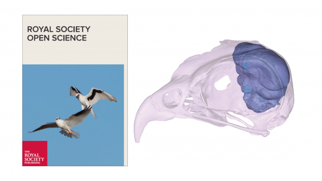
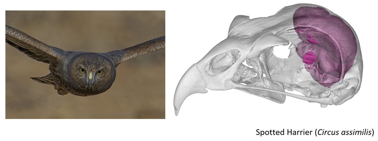
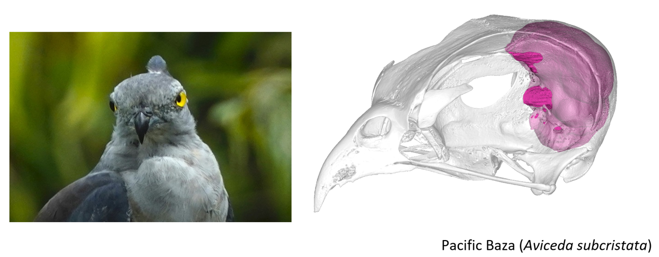
NIF Micro-computed tomography (CT scanning) at the University of Queensland’s Centre for Advanced Imaging has been used to scan 30 raptor skulls from Australian museums, create 3D reconstructions, measure and then study the anatomy for tell-tale signs of a bird’s visual powers.

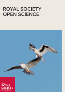
Research published in Royal Society Open Science compared the world’s only nocturnal hawk, the Australian letter-winged kite Elanus scriptus, to other hawks and falcons with differing hunting styles.

Associate Professor in Evolutionary Biology at Flinders University Vera Weisbecker said findings threw into doubt long-held views that changes to skeletal structure were needed for evolution.

The research sought to understand whether evolutionary changes to the eye-area of the skull was evidence of the kite’s adaptation to night-time hunting, Dr Weisbecker said.

“The answer is no. In fact, there are two close relatives of the letter-winged kite that have a similar bony visual system, but both hunt in daylight,” she said.

The findings have implications for the study of evolution, with researchers often deducing that changes in skeletal remains are linked to behavioural changes.

“That’s not necessarily the whole story. In this instance, there’s no difference between the eye regions in the skulls of the night-time and the day-time hunters, so if you were just looking at the skull, you’d never know.”

Dr Weisbecker said different birds had greatly adapted their vision to have excellent visual sensitivity, sharpness, colour discrimination or even UV wavelength detection.

For Australia’s letter-winged kite, it’s possible that the nocturnal bird also picks up odours and movement, as well as adjusting its hunting methods.

CT-scanning the 30 birds of prey was primarily undertaken by CAI’s Dr Karine Mardon, on NIF-funded equipment, with skulls provided by Queensland Museum.

Dr Mardon said the imaging techniques, teamed with recent advances in anatomical understanding, opened the door to a wealth of new knowledge without needing live birds or their tissues.

CAI was the ideal place to undertake the research, enabled through national investment in imaging equipment and expertise, data analysis capability, and existing relationships with the Queensland Museum and Flinders University, she said.

Dr Weisbecker said obtaining eyes and brains of rare species was generally not feasible but some aspects of their anatomy could be estimated from skulls.

“We are extremely lucky to have Australia’s amazing museum collections at our disposal to help us understand this bird without the need to find and disturb the species,” she said.

“The kinds of things you can study closely with CT scanning are the size of their eyes and their position in the skull – are they facing forward or more on the side?”

PhD student Aubrey Keirnan compared 3D reconstructions of the letter-winged kite’s skull and brain with other birds of prey in the Weisbecker lab.

“The diversity among hawks that are active during daylight is possibly the most striking between the Spotted Harrier and the Pacific Baza,” she said.

“Both are incredible predators, but one species resembles owls while the other is much more pigeon-like in appearance.

“These two species really highlight how adaptable and diverse the visual systems of birds are, even amongst species within the same family.

“You can have birds that are anatomically similar but behave differently – and species that are behaviourally similar but anatomically different. Both sides of the coin are true.”

But Dr Weisbecker said the research was not just about insights into evolution.

The Australian letter-winged kite lives in remote, arid Australia, avoids human settlements and is highly elusive. It is listed as near-threatened, with population estimates varying between 670 and 6,700, she said.

“To conserve the species, it is critical that we understand its behavioural needs and capabilities, but these are extremely difficult to observe.

“Think about fences and powerlines potentially posing a greater threat to nocturnal birds than their daytime relatives.

“For example, in an earlier study, we found that the nocturnal night parrot is likely unable to see small objects because it may trade high resolution for higher contrast. This may put it at risk of hitting with thin fence wires.”

Dr Mardon has also scanned the bones of a night parrot, a bandicoot and many Australian marsupial mammals.

“We have an excellent working relationship with Queensland Museum, who trust us with handling some of their precious items,” she said.

Later this year CAI will install a new NIF-funded CT-scanner, a Yxlon FF35CT, along with new software to increase graphics capability, such as accurate reconstructions of soft tissue around the skull.

CAI expects greater demand for scanning which contributes to research on evolution, and Australia’s native flora and fauna.

Read the article on the Australian letter-winged kite Elanus scriptus here: Not like night and day: the nocturnal letter-winged kite does not differ from diurnal congeners in orbit or endocast morphology | Royal Society Open Science (royalsocietypublishing.org)


More about National Imaging Facility (NIF)

NIF is Australia’s advanced imaging network.

We provide open access to flagship imaging equipment, expertise, tools, data and analysis. We address Australia’s strategic science and research priorities, and this benefits Australian industry and helps keep Australians healthy.

NIF provides a full suite of advanced imaging capability including preclinical and clinical, human and animal imaging, radiochemistry and imaging data analysis. We focus on health and medical innovation, and also provide highly specialised capabilities for agriculture, materials science, museums and cultural applications.

NIF assembles partnerships that produce quality-controlled and harmonised data that provides invaluable evidence to make new discoveries, validate new products and demonstrate new therapies.

We partner with people who can translate their discoveries into real-world applications. NIF has helped Australians innovate in fields such as bioengineering, clinical science, biology, medical technology, pharmaceutical and non-pharmaceutical therapies, agriculture, materials, museums and cultural collections.

More about the Centre for advanced Imaging (CAI)

The Centre for Advanced Imaging (CAI) brings together the skills of a critical mass of researchers and ‘state-of-the-art’ research imaging instruments. It is the only facility of its type in Australia, one of only a handful in the world. The 5,500 m2, $55M CAI building was funded by the Federal Education Investment Fund in 2010 and contains over $50M of imaging and spectroscopy equipment, putting The University of Queensland’s researchers at the forefront of a field that is advancing swiftly.

Our researchers work on innovations in spectroscopic and imaging technology, imaging biomarker development and in biomedical research disciplines, frequently in collaboration with clinical research sites and other local, national, and international research institutes.  Find out more here

#WorldHealthDay: Imaging unlocking research to keep people healthy

#WorldHealthDay: As Australia’s advanced imaging network, we’re focused on addressing national science and research priorities to help keep people healthy. Our expertise, equipment and services are critical to Australia’s ability to translate health discoveries, undertake clinical trials and commercialise medical products.

The importance of protecting Australians from health threats is critical, as is Australia’s strong medical research capability and reputation for quality and standards.

The National Imaging Facility is unlocking solutions to the world’s biggest imaging challenges across commercial, clinical and research fields. We have helped Australians innovate in fields such as bioengineering, clinical science, biology, medical technology, pharmaceutical and non-pharmaceutical therapies.

Thousands of scientists, doctors, and professionals across hundreds of Australian institutions, companies and research organisations use our work to help answer their medical research questions. We also work with engaged volunteers and patients who make a valuable contribution to health and discovery by being part of research.

We’ve included some examples of the medical projects we’re proud to have partnered with to keep people healthy below:

Dr Ciara Duffy from Western Australia’s Harry Perkins Institute of Medical Research imaging the investigation of honeybee venom to treat breast cancer cells at the University of Western Australia’s Centre for Microscopy, Characterisation and Analysis in collaboration with Microscopy Australia

Associate Professor David Parsons and Dr Martin Donnelly performing preclinical testing of a ground-breaking and simple to use ‘field ventilator’ that can be locally produced at a low cost from easily acquired parts at SAHMRI, in collaboration with 4DMedical, and the University of Adelaide

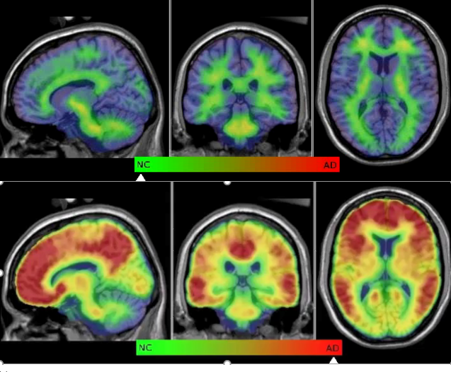
Supporting Australian trials of Biogen’s Aducanumab (Aduhelm), the first disease modifying therapy for Alzheimer’s disease approved by the United States Food and Drug Administration (FDA) with the University of Melbourne, Herston Imaging Research Facility, the Hunter Medical Research Institute, Australian Imaging Biomarkers and Lifestyle Study of Ageing at The Florey Institute of Neuroscience and Mental Health and Austin Health

#ImagingTheFuture Week: Unlocking solutions to major health challenges

#ImagingTheFuture Week: Unlocking solutions to major health challenges


Chan Zuckerberg Initiative’s (CZI) Imaging the Future Week puts a spotlight on the significance of imaging science in biomedicine, and the importance of building a vibrant imaging community across the world to tackle these challenges at scale.

Imaging science and the highly skilled researchers behind it are vital to addressing global health challenges, and driving innovation in disease management, prevention, and cure.

The National Imaging Facility (NIF) invests in state-of-the-art equipment and partners with world-class experts to process and interpret data and apply imaging to solve challenging health problems.

CEO Prof Wojtek Goscinski said he was proud of the NIF’s partnerships which enable the translation of discoveries through to real world applications to improve the health of the population.

“Advanced imaging techniques make it possible to deepen our understanding of health and disease in the human body through visualisation,” Prof Goscinski said.

“Imaging already plays a critical role in healthcare, and the acceleration of its advancements in biomedicine are positioning us, and our colleagues world-wide to continue this work well into the future.”

“We are supportive of the efforts of CZI and I’m excited for NIF to work alongside them and our other international imaging colleagues, building a cutting-edge imaging community at the forefront of global imaging research,” Prof Goscinski said.

You can find out more about Imaging the Future Week here.

Keep scrolling to check out some of the impressive imaging work from a few of the Australian National Imaging Facility’s Nodes.

Time-of-flight angiography of the human brain using 7 Tesla MRI – courtesy of the Centre for Advanced Imaging, University of Queensland

Human Tooth CT scan – courtesy of Diana Patalwala, University of Western Australia

Angiogram scanned on the Siemens 3T Skyra magnet – courtesy of the Large Animal Research and Imaging Facility, South Australian Health and Medical Research Institute

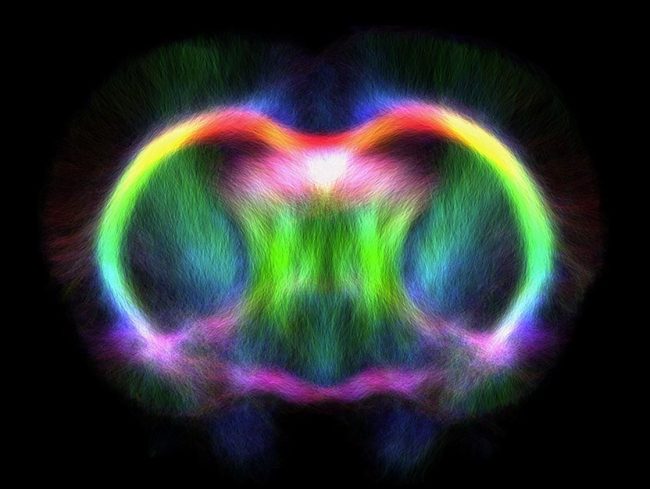
Tractography template image of a sham rat – courtesy of David Wright, The Florey Institute of Neuroscience and Mental Health

fMRI Short Course at UQ

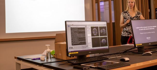
From Friday 20th – Sunday 22nd November 2020, a broad audience of PhD students, postdocs, associate professors, a radiographer and a clinician attended the University of Queensland (UQ) Centre for Advanced Imaging (CAI) functional Magnetic Resonance Imaging (fMRI) short course.

Read More

Creating a Lizard Brain Atlas

Until recently, reptilian evolutionary studies lacked an important resource – a lizard brain atlas. As the subject of numerous ecological and behavioural studies, the Australian tawny dragon (Agamidae: Ctenophorus decresii) was an ideal candidate for creating a high-resolution MRI atlas of a representative scaled reptile (squamata). Such data is not only a resource for studies of the genus but also informs environmental decision making through an improved understanding of animal adaptation and evolution.

Read More

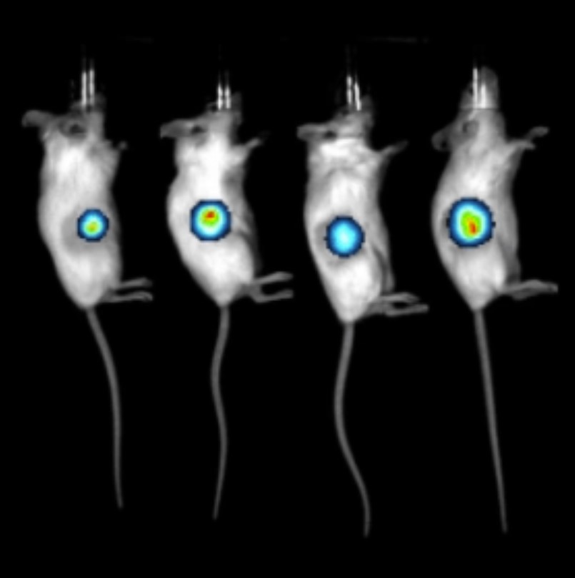
National Preclinical PET QA

The NIF Molecular Imaging & Radiochemistry (MIR) Theme is a group of NIF Fellows, Directors, and users of NIF facilities that focus on state-of-the-art radiochemistry and molecular imaging applications using PET, SPECT, and MRI.

Integrating preclinical PET systems into a national resource requires the development of defined QA programs to monitor and integrate the data from individual systems. Hence, the MIR Theme initiated a national quality assurance (QA) program for the NIF preclinical PET instruments.

Read More

PET training for HDR

From July 14 – 17, four CIBIT HDR students, Saikat Ghosh, Vanessa Soh, Pragalath Sadasivam and Ting Xiang Lim, attended an in-depth training session on PET imaging. Run by Dr Karine Mardon, NIF Facility Fellow and Molecular Imaging Facility Manager at the Centre for Advanced Imaging, the course covered both molecular imaging theory and practical hands-on training relevant to the students’ research projects. 

Read More
Privacy Settings
Youtube
Vimeo
Google Maps