ISMRM and ISMRT ANZ Chapters’ Annual Meetings shine a light on national imaging expertise and infrastructure
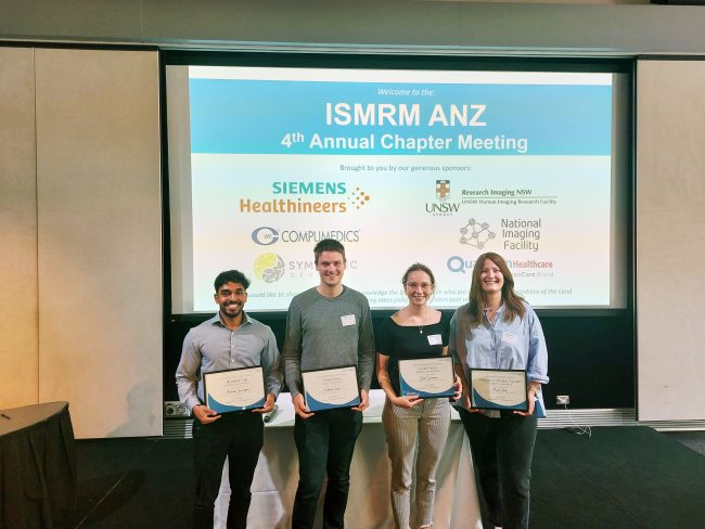
[Image: Presentation award winners at ISMRM ANZ, Honours student, Arunan Srirengan, Dr Ed Green, Dr Gwen Schroyen and Dr Myrte Strik. Photo credit: Dr Adam Clemente]
The Australian and New Zealand Chapters of the International Society for Magnetic Resonance in Medicine (ISMRM) and the International Society for Magnetic Resonance Radiographers and Technologists (ISMRT) held their Annual Meetings in Sydney last month, highlighting the work of leading national researchers and clinicians, including members of the NIF network.
NIF enables coordinated open access to magnetic resonance expertise and infrastructure to support leading national researchers and clinicians, and proudly supported the events.
ISMRM ANZ Joint Chapter Annual Meeting 9-10 Nov
ISMRM ANZ hosted sessions on revolutionising MRI technology, advances in neuroimaging, and clinical applications of advanced MRI, in addition to keynote speakers neurologist and leader in stroke medicine, Prof Mark Parsons and Director of the Institute of Medical Physics at the University of Sydney, Prof Annette Haworth.
Dr Zhaolin Chen was a key speaker in the Revolutionising MRI technology session, presenting the NIF Point-of-Care project, a collaboration between NIF, Australian hospitals, and US medical device manufacturer, Hyperfine, to build the usability of low-field MRI and bring critical imaging to remote Australia and deploy imaging in challenging clinical environments such as COVID wards.
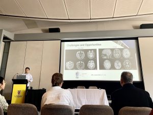
[Image: Dr Zhaolin Chen presenting the NIF Point-of-Care Magnetic Resonance project at ISMRM ANZ]
A number of other NIF users spoke at ISMRM ANZ, including:
- Rebecca Glarin from the University of Melbourne, presenting findings from her PhD on ‘Optimising functional brainstem imaging of sympathetic drive with ultra-high field MRI’.
- Dr Shahrzad Moinian from the University of Queensland Centre for Advanced Imaging, presenting ‘In vivo microstructural border delineation between areas of the human cerebral cortex using magnetic resonance fingerprinting (MRF) residuals’.
- Honours student Arunan Srirengan presenting ‘Early identification of cerebral small vessel disease in obstructive sleep apnoea patients using magnetic resonance spectroscopy: a pilot study’, featuring data obtained on the NIF 3T MRI at NeuRA. This session was awarded second prize in the oral presentation awards.
- Dr Myrte Strik from the University of Melbourne, presenting ‘Altered network topology in patients with visual snow syndrome: a resting-state 7 Tesla MRI study’, winning the award for best Early Career Researcher Data Blitz presentation.
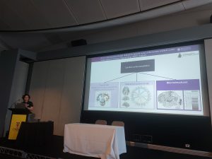
[Image: Dr Shahrzad Moinian from the University of Queensland Centre for Advanced Imaging. Photo credit: Dr Adam Clemente]
Congratulations to University of Melbourne NIF Fellow, Prof Brad Moffatt as ANZ ISMRM Chapter President on the success of the 2022 meeting hosted at UNSW.
ISMRT ANZ Joint Chapter Annual Meeting 12-13 Nov
The ISMRT ANZ 2022 joint meeting program theme was MRI: Past, Present and Future, and featured a range of internationally renowned speakers demonstrating future technologies and cutting-edge imaging techniques.
Keynote presenters included Medical physicist and human brain imaging academic researcher Dr Samantha Holdsworth, Chief of the Quantitative Medical Imaging Laboratory, USA National Institute of Biomedical Imaging and Bioengineering, Dr Carlo Pierpaoli, and founding member of the Society of Cardiovascular Magnetic Resonance and Principal Investigator for the Cardiac Atlas Project, Prof Alistair Young.
NIF Senior Manager and Senior Research Scientist – National Magnetic Resonance Capability, Dr Shawna Farquharson was a key speaker at the Diffusion Weighted Imaging (DWI) Forum, presenting on ‘DWI: Principles and practical applications’.
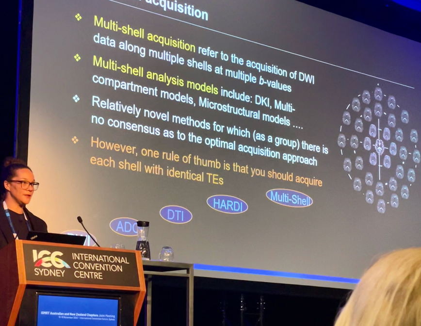
[Image: Dr Shawna Farquharson, National Imaging Facility]
NIF users showcased at ISMRT ANZ included:
- Prof Lynne Bilston from NeuRA, presenting ‘Brain Elastography’.
- Sarah Daniel from the University of Queensland Centre for Advanced Imaging, presenting ‘Image quality enhancement using deep learning for in vivo human kidney MRI’.
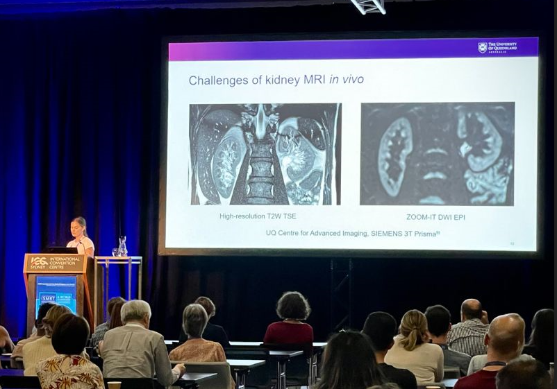
[Image: Ms Sarah Daniel from the University of Queensland Centre for Advanced Imaging]
Congratulations to all presenters at ISMRM and ISMRT ANZ.

")


