Because Seeing Changes Everything: Placing Australian imaging at the global forefront

Today the National Imaging Facility (NIF) released Because Seeing Changes Everything, its roadmap for Australian imaging.

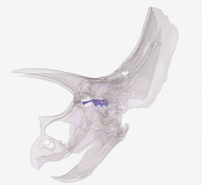
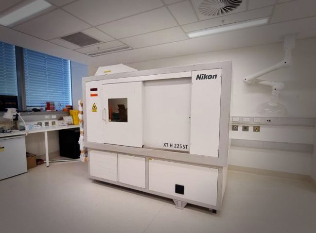
NIF is Australia’s advanced imaging network, providing open access to flagship imaging equipment, expertise, tools, data and analysis. NIF enables a full suite of advanced imaging capability including preclinical and clinical, human and animal imaging, and radiochemistry.

Because Seeing Changes Everything provides a plan to enable Australian researchers to unlock solutions to major health, agriculture and materials challenges.

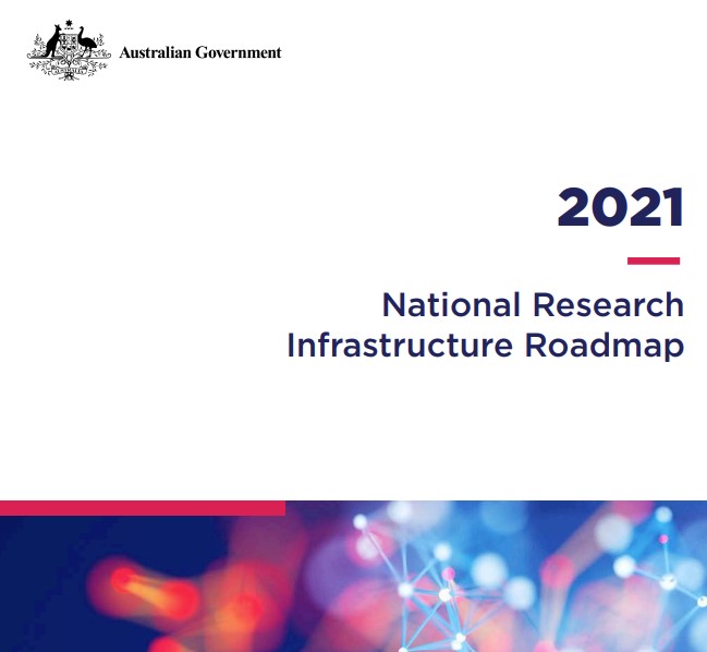
NIF Governing Board Chair, Professor Margaret Harding said Because Seeing Changes Everything demonstrates the way NIF contributes to Australian wellbeing, and outlines NIF’s future priorities, which deliver to the Australian Government’s 2021 National Research Infrastructure Roadmap.

“NIF’s imaging infrastructure, data and leading expertise support Australian research and innovation to help improve our standard of living and strengthen our economic standing,” Prof Harding said.

“Scientists, clinicians and professionals from a range of industries across Australia use NIF capabilities to unlock solutions to their research questions.”

NIF has identified impacts where imaging plays a key role, and will prioritise investments that address these areas:

  • Better evidence for decision-making in health
  • New diagnostics and therapies combined
  • Better health for the young and older Australians
  • Equitable regional and rural health
  • Growing use of imaging in agriculture and ecology 
  • Critical contributions to materials, engineering and culture

NIF Chief Executive Officer, Professor Wojtek Goscinski said Because Seeing Changes Everything outlines NIF’s roadmap supporting innovation and ensuring Australia’s world-class imaging capability remains at the global forefront.

“NIF will deliver to the priorities outlined in the 2021 National Research Infrastructure Roadmap, helping Australians to apply state-of-the-art imaging to address emerging challenges,” Prof Goscinski said.

“Our expertise and infrastructure will ensure Australia is at the cutting-edge of advanced imaging, now and into the future as we continue to meet the evolving needs of modern research.”

The document highlights some of the future capabilities NIF is planning to expand, including:

  • Accelerating next-generation imaging technologies
  • Furthering critical magnetic resonance technology
  • Translating portable biomagnetic imaging
  • Pioneering full-colour x-ray scanners
  • Applying new-generation ultrasound for treatments and diagnostic techniques
  • Advancing molecular imaging to visualise whole-body processes


You can read Because Seeing Changes Everything here.

NIF Molecular Imaging and Radiochemistry Showcase to be presented at ANZSNM

National Imaging Facility enables access to imaging capabilities across the country and will present a Molecular Imaging and Radiochemistry Showcase at ANZSNM 2022, featuring presentations from a range of research leaders from Australia’s advanced imaging network.

See the full ANZSNM program here.

Register to attend ANZSNM 2022.

National Imaging Facility: Molecular Imaging and Radiochemistry Showcase
Saturday 14 May 2022, 3:15pm – 4:15pm
Session Chair: Prof Wojtek Goscinski, CEO National Imaging Facility

TimeSpeakerTopic
3:15 – 3:20Professor Wojtek Goscinski

Chief Executive Officer
National Imaging Facility

Introduction to NIF Molecular Imaging and
Radiochemistry Showcase

3:20 – 3:30Professor Steven Meikle

Head of the Imaging Physics Laboratory, Brain and Mind Research Institute, University of Sydney

Total Body PET
3:30 – 3:40Associate Professor Roslyn Francis

Head of Department of Nuclear Medicine and WA PET Service, Sir Charles Gairdner Hospital, University of Western Australia

Radiochemistry activities in Western Australia
3:40 – 3:50Professor Gary Egan

Professor and Foundation Director, Monash Biomedical Imaging

Director, ARC Centre of Excellence for Integrative Brain Function

Australian Precision Medicine Enterprise
3:50 – 4:00Prof Kristofer Thurecht

Acting Deputy Director (Research Technologies) and Group Leader – Principal Research Fellow,

Centre for Advanced Imaging, University of Queensland

Affiliate Principal Research Fellow and Group Leader,

Australian Institute for Bioengineering and Nanotechnology

Alpha therapies and activities
4:00 – 4:10Dr John Bennett

Research Infrastructure Platform Leader – Biosciences,
ANSTO

ANSTO’s new NIF Alpha Radioisotopes and
Radiopharmaceuticals Facility

World class imaging expertise empowers a sight for (dino)saur eyes

ICYMI, Horridus, the world’s most complete and finely preserved Triceratops was unveiled to the public (for the first time in 67 million years) last month at Melbourne Museum, as part of their new exhibition, Triceratops: Fate of the Dinosaurs.

Advanced Imaging experts from Monash Biomedical Imaging (MBI) and the National Imaging Facility (NIF) worked with Melbourne Museum to create a digital record of Horridus and further examine how the dinosaur would have walked the earth back-in-the-day for the exhibition’s immersive digital experience.

Longstanding research collaborations exist between the Melbourne Museum palaeontology team and the Biomedicine Discovery Institute and School of Biological Sciences at Monash University, with MBI facilitating imaging of many important fossil specimens for collaborative scientific study, teaching and outreach.

Imaging is critical to a broad variety of research problems including environmental and ecosystems research, palaeontology and preservation. The National Imaging Facility (NIF) makes cutting edge imaging capabilities accessible to Australian researchers and companies, and NIF’s world-class network of Fellows provides expertise in processing and interpreting imaging data and applying imaging to solve complex challenges.

As the CT scanner can penetrate plastic and foam, the team were able to keep the bones in their protective stillages during scanning


Why make a digital record of Horridus?

As the most complete real dinosaur fossil in any Australasian Museum, Horridus was scanned in MBI’s large bore Siemens CT scanner before it was assembled for display, to enable further study of the Triceratops by the global scientific community.

The imaging of rare and high-value specimens such as Horridus allows the preservation of information from fragile objects, in addition to the non-destructive exploration of the interior of specimens.

Monash University PhD student Hazel Richards conducted the scans and created 3D models of the Triceratops bones for the exhibition as part of her role as research assistant on the project.

“When we combine the internal images produced by these CT scans with the external surface scan images, we can create a complete 3D model of the Triceratops bones that allows us to research a range of exciting biological and evolutionary questions,” Ms Richards said.

“The team at MBI are always enthused and accommodating when we come to them with proposals for scanning weird and often unwieldy objects like these Triceratops fossils.”

“With their support and expertise, we have been able to maximise the scientific potential of these remarkable pieces of natural history,” Ms Richards said.

Monash University PhD student Hazel Richards conducted the scans and created 3D models of the Triceratops bones for the exhibition as part of her role as research assistant on the project.


Applying imaging expertise to solve challenges

NIF Facility Fellow, and MBI’s Head of Pre-Clinical Imaging, Dr Michael de Veer worked with Ms Richards to provide training and operational guidance on the optimal use of the instrument to generate the data.

“Fossilised bone is very dense, so our scanning challenge was to manipulate the CT settings so that the X-rays would penetrate the bones, allowing visualisation of internal structures such as the dinosaur’s brain case,” Dr de Veer said.

“Different parts of the dinosaur fossil were scanned over a number of visits, and the bones were transported in special crates to reduce the possibility of damage.

“As the CT scanner can penetrate plastic and foam, we were able to keep the bones in their protective stillages during scanning, a capability that made Museums Victoria palaeontologist Tim Ziegler very happy” Dr de Veer said.

As Collection Manager Vertebrate Palaeontology, Tim Ziegler manages the preservation of Victoria’s fossil collections of backboned animals, plants, and microfossils, including dinosaurs.

“Fossils are surprisingly fragile once they are uncovered and brought out of the ground,” Mr Ziegler said.

“We take any opportunity to improve the safety of specimens under research. As part of Victoria’s State Collection, this skeleton will be kept and preserved in perpetuity, and will offer scientific potential for years to come.”

Museums Victoria palaeontologist Tim Ziegler, Monash University PhD student Hazel Richards and NIF Facility Fellow, and MBI’s Head of Pre-Clinical Imaging, Dr Michael de Veer. 


What can a CT tell us about life 67 million years ago?

Visitors to the Triceratops exhibition will see the CT data captured first-hand – both virtually –through animated projections from the scans, and physically – as life-size touchable resin casts made from the 3D printed models.

This information can be used to tell deeper stories about Triceratops, including its evolution, behaviour and how it sensed its Cretaceous world.

The data of the upper and lower jaws reveal Horridus had more than 800 teeth. These CT scans can contribute to investigations of feeding biomechanics and diet in Triceratops and other highly specialised herbivorous dinosaurs.  

Scans of the dinosaur’s well-preserved braincase provided 3D models of the internal cranial cavity, allowing the team to examine the size and shape of regions of the brain and inner ear.

These provide important data for research reconstructing what sorts of sounds Triceratops was adapted to hear, and the relative importance of vision, smell and hearing in the daily lives of these long-extinct beasts.

The data of the upper and lower jaws reveal Horridus had more than 800 teeth. These CT scans can contribute to investigations of feeding biomechanics and diet in Triceratops and other highly specialised herbivorous dinosaurs. Image copyright Museums Victoria.


Say hi to Horridus

You can visit Horridus in the flesh (er.. bones?) at Melbourne Museum’s exhibition, Triceratops: Fate of the Dinosaurs.

View Horridus in 3D here.

For more information, contact Dr Michael de Veer, Head of Pre-Clinical Imaging and NIF Facility Fellow.

Images courtesy of Museums Victoria and Monash Biomedical Imaging.

NIF’s newest capability: Medical industry, manufacturers, and museums set to benefit from WA’s first high-power research-dedicated CT scanner

National Imaging Facility’s (NIF) University of Western Australia (UWA) node located at the Centre for Microscopy Characterisation and Analysis (CMCA) will grow its capacity with the arrival of a new computed tomography (CT) scanner to expand capabilities for industry, manufacturing and museums who require imaging of large samples.

The Nikon XT H 225 ST will increase NIF’s scope to cater for specimens that require a large field of view, including medical implants, additive manufacturing and industrial components, and environmental or historical artefacts.


Research applications of the new CT scanner will extend from medical material testing, industrial material including castings, turbine blades, plastics, packaging, dispensers, to precious palaeontology and archaeology articles.

Diana Patalwala, NIF’s Facility Fellow at UWA’s CMCA said the CT will enable engagement with the biomedical, agriculture, environmental, renewable resource, advanced manufacturing, electronics and defence industries.

“Our new CT capabilities will have increased applications in pre-clinical and clinical research involving medical prosthesis, dental implants, critical assemblies of medical devices and drug delivery systems,” she said.

“It is vital for components such as patient-specific medical implants manufactured through additive manufacturing technologies to be of outstanding quality, and an X-Ray CT can play an important role in this process from start to end.”

Other medical applications include verifying the dimensions of drug delivery systems’ inhaler chambers or dispenser mechanisms, syringes, stents, pacemakers and more.

“Industry will greatly benefit from the Nikon XT H 225 ST as it is the only CT technology of its kind to provide a 225kV (450W) rotating target X-ray source, this means we can image larger and denser samples with increased accuracy than previously possible,” Ms Patalwala said.  

“This makes it ideal for industry users involved in materials testing, inspection and quality control applications.

“This CT scanner would also be ideal for examining archaeological samples, museum specimens and fossils as well, enabling us to get the detailed inside picture without destroying these precious artefacts!” Ms Patalwala said.


With an X-ray source as powerful as 225kV/450W, it is the only high-power research-dedicated CT system in WA.

The unique and versatile scanner can examine specimens ranging in size from small rock cores, which are important for minimising the risk associated with the planned drilling operations in mining and increase the probability of meeting the target yield, to large industrial manufacturing components, such as casting moulds parts, batteries, fuel cells and electronic circuits.

The Nikon XT H 225 ST has an impressive maximum field of view (35cm x 35cm x 35cm), a sample height that can accommodate up to 65cm and a sample weight of 50kg – which will allow for greater capacity in imaging larger samples.


Its large field-of-view, makes it capable of CT scanning the internal tomography of an object non-destructively

The CT uses multiple axial scans to generate cross-sectional information or three-dimensional reconstructions. The X-ray CT has the typical mechanism for taking ‘slices’ which are then digitally reconstructed into 3D volumes.

The Nikon CT has an extremely high-powered X-ray source (450W) for penetrating geological, marine and industrial objects as well as the capability of producing lower energy X-rays (20W) for bio-medical applications.

With resolutions down to the 10um range, academia and industry will have access to 2D cross-sectional slices and 3D volume rendered models, as well as access to advanced quantitative analysis software packages capable of characterising material properties involving cracks, pores, and fibres – just to name a few.


The new Nikon XT H 225 ST CT scanner was delivered at the end of March, with installation commencing from April, and a view to opening to users in May.

This instrument has been funded by National Imaging Facility, enabled by the National Collaborative Research Infrastructure Strategy, with the Government of Western Australia and supporters of the Western Australia National Imaging Facility.

For further information about the instrument, contact NIF Facility Fellow, Diana Patalwala diana.patalwala@uwa.edu.au.

National Roadmap highlights advanced imaging capabilities to support leading research and innovation

The National Research Infrastructure (NRI) Roadmap has been unveiled by the Commonwealth Government’s Department for Education, Skills and Employment and Department of Industry, Science, Energy and Resources, recommending continued investment in NRI, which enables Australia to maintain its research excellence, increase innovation and address emerging research challenges.

The Government has announced it will invest $900 million over five years on the tools, technology and skills to ensure Australian research remains at the global forefront.

The application of advanced imaging will play a critical role for Australia in underpinning fundamental and applied research across disciplines to address domestic and global challenges.

The Roadmap highlights key priorities for imaging capabilities in supporting leading-edge research and innovation.

Advanced imaging is essential to addressing challenges in the focus areas of medical products and frontier technologies and modern manufacturing. It also plays an important role in supporting the focus areas of food and beverage and developing physical collections and biobanking.

Medical products

The importance of protecting Australians from health threats is critical, as is Australia’s strong medical research capability and reputation for quality and standards. Human imaging technology is highlighted as playing a vital role in:

  • Improving health outcomes through novel medical products, platforms, technologies and practices to improve quality of life and decrease health-related costs.
  • Supporting nuclear medicine and radiotherapy innovation, including the development of new radiopharmaceuticals for diagnosis and therapy (e.g. theranostics).
  • Supporting preclinical and clinical research, including modelling for both disease mechanisms and early drug candidate testing.

Frontier Technologies and Modern Manufacturing

Developing and translating critical technologies required to support modern manufacturing and secure supply chains is of significant priority to Australia.

Advanced imaging facilities will enable success through research and commercialisation of critical technologies:

  • Enhancing outcomes and visualising essential components through a wide array of preclinical and clinical studies.
  • Representing fundamental research technology for understanding the effect of pandemics and unique environmental events (e.g. floods and bushfires) on the human body.
  • Applying imaging to a broad variety of industrial, research problems including chemical processes, materials science, security, palaeontology and cultural preservation.

Food and beverage

Australia has an international reputation for premium, safe and high-quality food and beverage products, strong production capabilities, research expertise and market proximity, and imaging enables researchers to capture data to increase agricultural and economic productivity.

Physical collections and biobanking

Biodiversity and environmental sample biobanks have significant potential as baseline infrastructure to support environmental monitoring and management, biosecurity, biodiscovery and bioprospecting and advanced imaging will enhance the value of biological and environmental sample collections.

Rising to meet the research challenges of the future

National Imaging Facility (NIF) continues to work in partnership with National Collaborative Research Infrastructure Strategy (NCRIS) capabilities to support and enable the translation of research outcomes to benefit Australians.

NIF already provides access to national capabilities supporting these areas of importance, including:

  • preclinical and clinical research, as well as nuclear medicine and radiotherapy to keep Australians healthy
  • developing and translating critical technologies required to support modern manufacturing
  • supporting Australia’s biodiversity, agricultural and economic productivity.

Planning is well underway for the next stages of growth and integration for NIF, as we work alongside our NRI colleagues to provide infrastructure that helps improve Australia’s standard of living and strengthen our economic standing.

You can read the NRI Roadmap and the recommendations made here.

Meet our new Fellows

The National Imaging Facility provides state-of-the-art facilities and services that support critical leading-edge innovation and research, but our capability is much more than instruments and equipment.

The NIF network is privileged to comprise of a range of highly skilled experts across our Nodes, making cutting-edge imaging capabilities accessible to Australian medical researchers, materials and agriculture scientists, enabling them to solve challenges across research and industry.

We’re proud to extend our welcome to, and introduce, the newest members of Australia’s advanced imaging network, joining our team of experts enabling Australian imaging science to unlock solutions to major challenges.

Muneer Ahamed
South Australian Node
Maggie Aulsebrook
Monash University Node
Robert Brkljaca
Monash University Node
Ingrid Burvenich
La Trobe University – ONJCRI Node
Yaser Gholami
Yaser Gholami
University of Sydney – ANSTO Node

Joseph Ioppolo
University of Western Australia Node
Edward Green
University of Melbourne Node
Ekaterina Salimova
Monash University Node
Sjoerd Vos
University of Western Australia Node

#WorldHealthDay: Imaging unlocking research to keep people healthy

#WorldHealthDay: As Australia’s advanced imaging network, we’re focused on addressing national science and research priorities to help keep people healthy. Our expertise, equipment and services are critical to Australia’s ability to translate health discoveries, undertake clinical trials and commercialise medical products.

The importance of protecting Australians from health threats is critical, as is Australia’s strong medical research capability and reputation for quality and standards.

The National Imaging Facility is unlocking solutions to the world’s biggest imaging challenges across commercial, clinical and research fields. We have helped Australians innovate in fields such as bioengineering, clinical science, biology, medical technology, pharmaceutical and non-pharmaceutical therapies.

Thousands of scientists, doctors, and professionals across hundreds of Australian institutions, companies and research organisations use our work to help answer their medical research questions. We also work with engaged volunteers and patients who make a valuable contribution to health and discovery by being part of research.

We’ve included some examples of the medical projects we’re proud to have partnered with to keep people healthy below:

Dr Ciara Duffy from Western Australia’s Harry Perkins Institute of Medical Research imaging the investigation of honeybee venom to treat breast cancer cells at the University of Western Australia’s Centre for Microscopy, Characterisation and Analysis in collaboration with Microscopy Australia

Associate Professor David Parsons and Dr Martin Donnelly performing preclinical testing of a ground-breaking and simple to use ‘field ventilator’ that can be locally produced at a low cost from easily acquired parts at SAHMRI, in collaboration with 4DMedical, and the University of Adelaide

Supporting Australian trials of Biogen’s Aducanumab (Aduhelm), the first disease modifying therapy for Alzheimer’s disease approved by the United States Food and Drug Administration (FDA) with the University of Melbourne, Herston Imaging Research Facility, the Hunter Medical Research Institute, Australian Imaging Biomarkers and Lifestyle Study of Ageing at The Florey Institute of Neuroscience and Mental Health and Austin Health

#IWD | PODCAST: In conversation with Professor Leigh Johnston

The United Nations International Women’s Day (IWD) is an opportunity to reflect on progress made, call for change and celebrate acts of courage and determination by ordinary women who have played an extraordinary role in their communities. 

The National Imaging Facility’s (NIF) mission is to make cutting-edge imaging capabilities accessible to Australian researchers, and we envision a society that provides equal opportunity for people of all genders to learn, work and engage in science, technology, engineering, and mathematics (STEM). 

Today we highlight the exceptional work of women leading the way in these fields and thank them for the impacts of their life-changing research. 


Professor Leigh Johnston is the NIF Node Director at the Melbourne Brain Centre Imaging Unit within the Department of Medicine and Radiology, and is also the Head of the Department of Biomedical Engineering, at the University of Melbourne.  

Professor Johnston started out as an Engineer, but a unique skill set, passion for collaboration, and drive to pursue challenges have led her to have a great impact on the imaging community. 

Listen to our podcast here.


Professor Johnston talks us through some standout imaging projects: 

The baby mummy 

#JoinNIF: Three National Capability Managers to facilitate enhanced accessibility to cutting-edge imaging capabilities

Today the National Imaging Facility (NIF) launched an Australia-wide recruitment campaign seeking three Senior Managers to lead the implementation of a national coordination strategy across Australia’s advanced imaging network.

NIF provides capabilities that underpin nationally significant and impactful research, which translates to products and benefits for Australians in health, agriculture and new materials.

Appointees will enhance the accessibility of NIF’s network of state-of-the-art imaging capabilities Australia-wide for researchers, health professionals and industry to utilise imaging to unlock solutions to major challenges.

The enhanced accessibility will enable increased engagement with multi-site, large scale projects which will translate to collaborative improved outcomes for major health challenges such as brain injuries, cancer, cardiovascular diseases, and neurological disorders including epilepsy and dementia.

It will also allow the refinement of and accessibility to critical data, including large scale national repositories and digitised libraries.

NIF is at the forefront of imaging technology, enabling access to more than 80 imaging instruments, with multiple large-scale projects planned across capabilities.

The Senior Managers will be responsible for leading the coordination, harmonisation and project management of three different areas of NIF’s capabilities:

  • Magnetic Resonance Imaging (MRI)
  • Molecular Imaging and Radiochemistry
  • Data

The ideal candidates will possess qualifications and experience in relevant imaging, science, engineering, physics, data or health related fields. They will also be skilled in managing large and complex projects, and excellent influential, interpersonal and communication skills.

For more information and to apply visit the relevant job ad:

To discuss this role please contact NIF Chief Operating Officer, Ms Saba Salehi.

Imaging enabling nanomedicine to treat aggressive brain cancer

Image: Gadolinium enhanced MRI showing the bright brain tumour (red circle) compared to the normal brain tissue (yellow circle).

‘Nanomedicine’ sounds like a term you’d hear in a futuristic novel or an episode of Doctor Who, but cutting-edge scientists from the National Imaging Facility’s Node at the University of Queensland’s Centre for Advanced Imaging are already applying it to solve complex health challenges in collaboration with the Australian Institute for Bioengineering and Nanotechnology, and the Australian Research Council’s Centre of Excellence in Convergent BioNano Science and Technology and Training Centre in Biomedical Imaging Technology.

Nanomedicine applies nanoscale materials, such as nanoparticles and nanorobots (wow!) to the prevention and treatment of disease. Nanomedicine is a promising strategy to target tumours with chemotherapy in a safe and controlled manner.

This all sounds great, but within the context of the brain things get a little more complicated. For brain tumours, the integrity of the blood brain barrier (BBB) is central to its effective use as treatment.

The BBB is a protective barrier between the blood vessels and brain tissue, providing a defence against pathogens and toxins that may be present within the blood, while at the same time allowing vital nutrients to reach the brain.

While the BBB protects the brain against pathogens and toxins that could cause infection, it also blocks medicine from crossing the barrier in many cases, which can hamper the (often urgent) treatment of tumours, among other neurological disorders.

This is where imaging comes in…

UQ researchers have undertaken studies utilising NIF’s flagship preclinical magnetic resonance –positron emission tomography (MR-PET) system to develop hybrid imaging, combining the imaging of MRI with the information of PET.

Simultaneous MR-PET imaging enables experts to measure opening of the BBB using Gadolinium (Gd) contrast agents at the same time as delivering novel PET tracers and new theranostic candidates including nanomedicine.

These tools assist in investigating the link between BBB integrity and tumour diagnosis and treatment, and ensure the development of promising new treatments such as nanomedicine that can permeate the BBB.

BBB integrity: Tumour diagnosis and treatment

The rapid growth of brain tumours requires the formation of new vessels to supply increased demands for nutrients. The new vessels are leaky compared to normal brain vessels, where the BBB tightly regulates the transfer between blood and tissue.

The team have used Gd MRI to estimate how leaky the tumour vessels are. This information is vital for understanding how tumours are developing, and when and how diagnostic or therapeutic drugs can enter the tumour tissue.

In combination with new nanomedicines, there are also exciting new techniques that allow opening of the brain for treatment without surgery, which can increase risk for patients. These new imaging methods allow the opening of the brain to be accurately monitored to ensure entry of the treatment without any unrelated damage to the brain. 

So – our takeaway message? While advances in nanomedicine alone are exciting and important, the integration of nanomedicine and advanced imaging brings opportunity for exponential synergistic innovation in healthcare, that can improve outcomes for patients and ultimately has the potential to save lives.

For more information, contact Dr Gary Cowin, Queensland Facility Fellow, UQ Centre for Advanced Imaging.

Privacy Settings
Youtube
Vimeo
Google Maps