NIF investment builds understanding of Australia’s unique paleontological collections

A 105-million-year-old shark vertebrae fossil and a 4,000-year-old thylacine skull are amongst the first items to be scanned on NIF’s materials investment at the University of Western Australia (UWA), building a valuable digital collection of unique national artefacts with the Museum of Western Australia.

UWA NIF Fellow, Diana Patalwala said computed tomography was an invaluable tool for imaging materials providing high resolution 3D data, and preserving accurate anatomical information on the relative shape, size and location of different structures that would not be attainable by physical dissection.

“For each specimen, our materials-dedicated CT scanner takes approximately 1800–3000 x-ray images as the sample is rotated in the x-ray beam, at a level of resolution 100 times that of a typical medical CT scanner used on humans,” Ms Patalwala said.

“These images are then used to create a 3D model of the entire specimen, which is in essence a stack of virtual dissection slices that can be manipulated, rotated, and studied from every angle, revealing unprecedented details of the internal structure of the specimen.”

NIF is supporting the capture of this valuable data to create permanent digital records of specimen collections and enabling its reuse by multiple researchers to minimise duplication of efforts and resources.

Western Australian Museum Curator of Entomology, Collections and Research, Dr Nikolai Tartanic said the data captured could serve several different purposes.

“We’ve used scans to generate 3D models of items that can be printed and put on display. Sometimes these are scale models of minute organisms that are otherwise too small to observe and appreciate, other times we use 3D models to replace fragile or rare specimens,” Dr Tartanic said.

“The 3D datasets can also be shared with colleagues electronically, which in some cases replaces the need to send the physical specimen without putting the specimen at risk.”

Western Australian Museum Head of Earth and Planetary Sciences Dr Mikael Siversson said prior to the availability of this technology, some information about specimens could only be found by physical dissection, which could result in damage.

“In the old days, palaeontologists studying internal structures of vertebrate fossils would sometimes cut up the fossil slice-by-slice, and build a 3D model using clay,” Dr Siversson said.

“Printed 3D models enable palaeontologists to hands-on examine the morphology of primary type specimen without risking damaging the actual specimen.”

Scroll on to see some of the scans captured for the Museum’s collection.


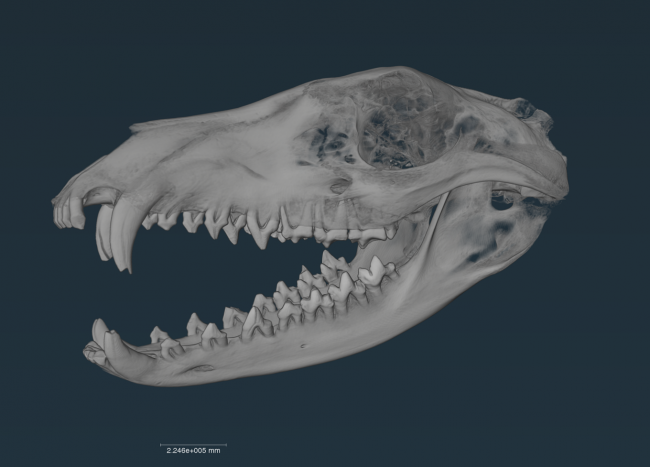
Nullarbor Thylacine Skull sub-fossil: (Scan resolution: 105um; 185kV; 100uA)
Thylacinus cynocephalus (Tasmanian Tiger)
Murra-El-Elevyn Cave, Nullarbor

This subfossil skull from the Nullarbor was scanned in addition to a modern skull from Tasmania, enabling the team to capture data that can be used for morphometric (shape variation and comparison) studies.

This fossil is about 4,000 years old (carbon dated – 3,885 carbon years) and there’s a hole in the jaw where they took a sample for the carbon dating. It is thought to be a female, based on the size of the skull. 


Shark vertebrae fossil: (Scan resolution: 56um; 200kV; 100uA)
Anacoracidae sp. (undescribed species of anacoracid shark)
Toolebuc Formation, Richmond, Queensland

This shark vertebra fossil is about 105 million years old (Albian Stage, Cretaceous Period), and belongs to a group of lamniform sharks called the anacoracids which were the Cretaceous ecological equivalents to modern whaler sharks.

Dr Siversson said the fossil represents a new species.

“It is exceptionally well preserved – anacoracid vertebrae are notoriously fragile – and this particular vertebra is surprisingly large considering the small size of anacoracid teeth in the same geological formation,” Dr Siversson said.

The team suspects this species was a plankton feeder, and based on its size, the animal would have been about four metres in length.


Aplysinopsis sponge: (Scan resolution: 91um; 850V; 250uA)

This species of sponge belongs to the order Dictyoceratida, which are sponges with spongin fibres. Some of them incorporate sand granules in their fibres, and small crustanceans are often found inside the sponge cavities.

Scanning revealed this sponge had several brittle stars living within it.


Echinodictyum Clathroides: (Scan resolution: 121um; 180kV; 100uA)

This species of sponge was first discovered in Shark Bay area, making its location a type locality. The sponge has three different spicules found in its fibres, some densely covered with spines.


Amber resin (Scan resolution: 78um; 190kV; 100uA)

These are specimens of amber-like natural resin of paleobotanical origin collected from WA beaches. The ultimate origin of this amber-like resin is likely South East Asia and it is thought these samples may have floated down to Rottnest Island where they were collected all the way from Indonesia.

Chemical analysis of similar amber found on Cape York in Queensland identified that it was produced by the Dipterocarpaceae, a family of lowland tropical rainforest trees.

Dr Tatarnic said the research team were looking for trapped insects using the CT scanning, but unfortunately did not find any.

“Scans of amber can detect the presence of now extinct insects. These may be new to science, or they may help us reconstruct past ecosystems, or identify from where the piece of amber originates,” Dr Tatarnic said.


The Nikon XT H 225 ST CT scanner was delivered to UWA in 2022 and is funded by National Imaging Facility, enabled by the National Collaborative Research Infrastructure Strategy, with the Government of Western Australia and supporters of the Western Australia National Imaging Facility.

For further information, contact NIF Facility Fellow, Diana Patalwala diana.patalwala@uwa.edu.au.

NIF’s SAHMRI Node becomes second Hub of the Australian Epilepsy Project

[Image: Prof Graeme Jackson, AEP Chief Investigator, Dr Michelle Kiley AM, Director of Epilepsy Services, CALHN and Lead Epileptologist AEP South Australia, Martin Adams, Chair of the Florey Board, The Florey Institute of Neuroscience and Mental Health and Prof Steve Wesselingh, Executive Director SAHMRI]

ICYMI, an Australian Epilepsy Project (AEP) Hub has opened at SAHMRI – South Australia’s independent not-for-profit health and medical research institute, and National Imaging Facility’s (NIF) second node to join the national multidisciplinary collaboration to improve epilepsy outcomes.

AEP’s first Hub was established at NIF’s Node at the Florey Institute of Neuroscience and Mental Health in Melbourne and the SAHMRI Hub will provide magnetic resonance imaging (MRI) scanning facilities and expertise to support this important initiative in Adelaide.

Epilepsy affects over 150,000 Australians, and its expenditure burden on the national health system is around $333M each year.

[Image: Ned Travers, AEP Lived Experience Ambassador South Australia, Dr Michelle Kiley AM, Director of Epilepsy Services CALHN and Lead Epileptologist AEP South Australia, Amanda Anderson, AEP Lived Experience Ambassador and Participant Lead and Carolyn Travers, AEP Lived Experience Ambassador South Australia]

The AEP aims to develop a critical resource to progress epilepsy research to reduce diagnosis uncertainty and facilitate fast-tracking of optimal treatment by combining advanced imaging, genetics, cognition, and artificial intelligence (AI).

AEP’s Chief Investigator, Professor Graeme Jackson said the ultimate aim of the AEP is to improve the standard of care and change the lives of people with epilepsy.

“Epilepsy is life-long condition and we need life-long solutions. Using algorithms, imaging and rich data we can extract insights to predict patterns in epilepsy and create individualised treatment plans for patients. This is an exciting new standard of care that we’ll be able to offer people living with epilepsy,” Prof Jackson said.

The AEP program has been developed using novel advanced imaging techniques with AI and machine learning, supported by NIF’s world-class infrastructure, which will provide the highest quality of data to epilepsy research.

NIF CEO Prof Wojtek Goscinski said the geographical expansion of the project will drive major advances in decision support tools to guide diagnosis and highlight opportunities for precision treatment for epilepsy, while addressing the disparity in epilepsy research in Australia’s diverse population.

“NIF’s national network of world-class human MR expertise and infrastructure will enable scanning across the country, in alignment with our impact goals addressing health equity for all Australians” Prof Goscinski said.

“It’s a privilege for NIF to support this life-changing project at our node at the Florey for patients in Victoria, and now SAHMRI for patients in South Australia,” Prof Goscinski said.

[Image: Dr David Vaughan, AEP Imaging Lead and Clinician, Paul Lightfoot, AEP Operations Lead, Jemima Gore, Operations Officer, SAHMRI Clinical Trials Platform, Lisa Carne, SAHMRI Operations Manager and Dr Karen Best, Director, SAHMRI Clinicial Trials Platform]

SAHMRI Clinical Trials Platform Director Dr Karen Best said enabling Adelaide’s medical research sector to engage with national initiatives like the AEP is a key reason that SAHMRI’s Clinical Trials Platform exists.

“We’re proud to be able to help at all stages of the project’s SA-based activities, from coordinating patient enrolment to making connections for diagnostic testing at facilities like the SAHMRI Clinical Research Imaging Centre.”

In addition to the AEP’s expansion to South Australia, Hubs in Queensland and New South Wales are set to launch in mid-2023.


Want to join the Australian Epilepsy Project? Ask your neurologist for a referral.

People in South Australia as well as Victoria living with epilepsy can be referred into the hub for advanced testing, free of charge, as part of their participation in the AEP. Find out more at epilepsyproject.org.au

NIF imaging to underpin research building complete picture of concussion

Concussion is a form of mild traumatic brain injury (mTBI). Across Australia each year, an estimated 35,000 people with reported mTBI experience symptoms that persist for monthsor even years. There is a pressing need to develop improved clinical and imaging tools to aid early diagnosis and better monitor ongoing recovery for patients. NIF advanced imaging technology will be used to establish a national imaging data resource of people experiencing mTBI across Australia. 

NIF is partnering with AUS-mTBI, a national consortium of clinicians, researchers, industry partners and decision-makers working to build Australia’s first clinical and imaging data resource of people experiencing mTBI. 

The world-leading initiative aims to help provide a better understanding of normal patterns of recovery and to identify the risk factors associated with delays or persistent post-concussion symptoms. 

Curtin University neuroscientist Professor Melinda Fitzgerald leads AUS-mTBI, with funding from the Federal Government’s Medical Research Future Fund, in collaboration with experts in brain biology, trauma, human behaviour, risk assessment, software design and development, support and patient care. 

NIF imaging will play an important role in helping the consortium in realising its aims of better understanding – and ultimately treating – concussion. 

NIF’s MRI technology will be used to scan the brains of people to be recruited to the research project in coming months. 

This will improve understanding of each person’s brain biology to accurate predict their outcomes and guide personalised treatment. 

Professor Fitzgerald says the imaging will validate the complex picture of concussion that AUS-mTBI is building, including each person’s unique biology, background and behaviour. 

She says AUS-mTBI will build this complex picture of concussion as it expands upon the HeadCheck app to gather a range of information including demographic data and factors that will have a bearing on a person’s outcome. 

The database will provide the information to people with concussion and clinicians to improve treatment recommendations. 

“We aim to have an evidence-based resource for everyone who may come across someone with a concussion, especially GPs, physiotherapists and trainers,” Professor Fitzgerald says. 

“The resource will also be for people with concussion who may not have sought access to clinical care.” 

Professor Fitzgerald says concussion is complex and a detailed picture is needed to predict a patient’s outcome, ahead of personalising the treatment. 

“In order to predict whether people will have continuing symptoms following a concussion, it’s important to have information about the type of injury, such as if the person had amnesia after the initial trauma, their mental health, previous concussions and even social factors such as family support and access to healthcare. 

“The research will find out whether all this information helps with predicting which people may be likely to have long-lasting negative impacts or a delayed recovery.” 

Up to 200,000 TBIs are reported each year in Australia, typically resulting from traffic accidents, falls, contact sports or acts of violence. 

While about 180,000 are considered mild, an estimated 35,000 people can have long-lasting symptoms, such as headaches, dizziness, fatigue, irritability, anxiety, trouble sleeping, ringing in the ears and loss of concentration and memory. 

AUS-mTBI will also develop programs designed with Aboriginal and Torres Strait Islander people and those in who live in rural and remote areas. 

The research is starting with recruitment of people who will undergo brain scans using NIF capabilities at the University of Western Australia and University of Queensland Nodes. 

Consortium members include Curtin, Monash, Edith Cowan, Griffith, Macquarie and Deakin universities, software company Curve Tomorrow, the Queensland Brain Institute, Poche Centre for Indigenous Health and support organisation Synapse Australia. 

“This research is valuable because it will determine the best information to collect to predict the outcome of mild TBI, analyse that information to guide treatment, make treatment more consistent across Australia – and possibly the world – and provide personalised care plans,” Professor Fitzgerald says.   

“We have an opportunity, through better healthcare, to improve quality of life for people with mild TBI and reduce the impacts on their families, our society and the healthcare system.” 


The NCRIS Health Group 

This cross-disciplinary research project is supported by National Imaging Facility and the Population Health Research Network as part of the NCRIS Health Group, assisting Australian researchers to leverage access to world-leading facilities for impactful outcomes.  

The NCRIS Health Group enhances collaborative opportunities between infrastructure capabilities, enabling support across the whole research translation cycle. It includes Bioplatforms Australia (BPA), National Imaging Facility (NIF), Phenomics Australia, Population Health Research Network (PHRN) and Therapeutic Innovation Australia (TIA). Click here for more information. 

Researchers gain unique insights, setting aim for the stars through southern hemisphere’s only paediatric MEG

National Imaging Facility’s (NIF) node at Macquarie University is home to Australia’s only paediatric magnetoencephalography (MEG) system, allowing children to undergo scans to discover how the brain develops normally as well as what might be different in epilepsy and autism.  

MEG enables highly precise, real-time study of brain activity and it is one of the most advanced methods of recording and evaluating the brain while it is actively functioning.  

It non-invasively measures the magnetic fields produced by the brain’s electrical currents through sensors in a helmet to help identify sources of activity in the brain. 

NIF’s Macquarie University Node Co-Node Director Professor Paul Sowman oversees the KIT-Macquarie Brain Research Laboratory, where he has worked since 2009 after being awarded National Health and Medical Research Council training fellowship to join the lab in its early development. 

At the time, the lab was the only MEG facility in the southern hemisphere and had the only whole-head child MEG system in the world. Over ten years later, it is still the only paediatric MEG facility in the southern hemisphere.  

He led the first investigation to longitudinally track the changes in young children’s auditory function as they grow older; providing much needed insight into typical brain development and an important first step towards understanding neurodevelopmental disorders such as autism.  

Prof Sowman said the preschool years are a time of huge change in children’s cognitive abilities although little is known about the corresponding changes in brain function. 

“Using MEG, we’ve been able to show that normal cognitive development is characterised by an increasing tendency for the brain to make predictions about its sensory environment,” Prof Sowman said.  

“This ‘predictive-coding’ theory of cognition is thought to be a key site of cognitive difference in schizophrenia and autism, and hence developing objective brain-based measures of this might be key to a better understanding of those neurodiverse states,” he said. 


Brain scans can be scary, especially if you’re a preschooler, but the team at Macquarie have created an environment to put kids at ease with the MEG set up as an outer space adventure.  

Intrepid mini astronauts (the kids) are put through a fun space cadet preparation, including taking measurements, training and simulation to make them comfortable and confident about their ultimate mission. 

This astronaut training process prepares the cadets so that when it’s time to launch the real MEG spaceship, the kids are well versed and ready to follow instructions from ground control (the scanner control room), ensuring accurate results. 

NIF Macquarie Node Facility Fellow Dr Judy Zhu supports research projects at the MEG laboratory and is the first point of contact for potential new users.  

“I love the opportunity to work with researchers on different projects,” Dr Zhu said. 

“We are very excited to be part of NIF so we can enable more collaborations and make the MEG facility available to a broader research community.” 

For more information, and to access the paediatric MEG as well as other NIF imaging capabilities at Macquarie University, contact us. 

International partnership launches Monash University-Helmholtz lab to solve global oncology, cardiology and infectious disease challenges

Monash University has partnered with the Helmholtz-Zentrum Dresden-Rossendorf (HZDR), a member of the Helmholtz Association of German Research Centres to establish the Monash-Helmholtz Laboratory for Radio-Immuno-Theranostics (MHELTHERA) to enable clinical translation at a launch event in Melbourne today.

The MHELTHERA Lab is a collaboration to optimise non-invasive imaging techniques and personalised therapy, drawing on diagnostic and therapeutic expertise in cancer, infectious and cardiac disease.

HZDR and Monash are developing next-generation biomedical imaging platforms and the MHELTHERA Lab will specialise in molecular imaging through nuclear techniques such as Positron Emission Tomography (PET) and Single Photon Emission Computed Tomography (SPECT), as well as optical techniques including Fluorescence Imaging (FLI).

Prof Sebastian M. Schmidt, Scientific Director of HZDR, Mr Simon McKeon AO, Chancellor of Monash University, Prof Otmar Wiestler, President of Helmholtz Association of German Research Centres and Prof Christoph Hagemeyer, National Imaging Facility Monash Biomedical Imaging Node Director opened the MHELTHERA Lab.


What are radio-immuno-theranostics?
Radio-immuno-theranostics pair diagnostic radiopharmaceuticals, applied through molecular imaging to identify tumours, with combined radiation therapy and immunotherapy to target cancer and enable clinicians to ‘see what they treat’.
Nuclear theranostics are a new and highly impactful field, revolutionising cancer therapy through a personalised approach.

The MHELTHERA Lab was officially opened at its Australian site, Monash Biomedical Imaging (MBI), a node of the National Imaging Facility (NIF) by Mr Simon McKeon AO, Chancellor of Monash University, Prof Otmar Wiestler, President of Helmholtz Association of German Research Centres, and Prof Sebastian M. Schmidt, Scientific Director of HZDR.

Mr McKeon said the partnership is an excellent example of the power of international collaborations.

“No single institution or nation has all the answers when it comes to addressing the most pressing health challenges of our time. By partnering with institutions such as Helmholtz, we can bring together the best talent, knowledge, and resources to tackle these challenges together,” Mr McKeon said.

Prof Wiestler said the Helmholtz Association is Germany’s largest research organisation, developing solutions and innovative technologies to preserve the foundations of human life.

 “By supporting MHELTHERA, we are bringing together leading international researchers to develop a highly original class of radiopharmaceuticals that will provide new treatment prospects for cancer patients in particular,” Prof Wiestler said”.

Prof Schmidt said MHELTERA will offer excellent opportunities, especially for young researchers.

“The cooperation is particularly attractive for PhD students, who will have the opportunity to improve their skills through joint research training programs of HZDR and Monash University,” Prof Schmidt said.

MHELTHERA builds upon a successful long-standing collaboration between the two organisations and is led by Prof Christoph Hagemeyer from Monash University, who is also the NIF MBI Node Director, and Prof Michael Bachmann from the HZDR Institute of Radiopharmaceutical Cancer Research. 

“The joint laboratory will leverage the research infrastructure capabilities at both sites to accelerate the development of promising technologies in the field of precision medicine,” Prof Hagemeyer said.

NIF CEO Prof Wojtek Goscinski said there are ten Helmholtz International Labs dedicated to innovative research located around the world, and it was a privilege for NIF co-funded infrastructure and staff to support the first of its kind in Australia.

“NIF has invested in world-class radiochemistry expertise and capabilities at MBI and this is an exciting opportunity for us to support a unique Helmholtz lab at the global cutting-edge of cancer research,” Prof Goscinski said.

“The application of radiopharmaceuticals and nuclear theranostics through PET imaging is revolutionising cancer treatment, and NIF is committed to improving health outcomes by supporting these innovative medical products,” Prof Goscinski said.

NIF has committed $4.95 million to a new cyclotron, enabling critical radioisotope production, and an expanded radiochemistry facility at MBI which is central to the infrastructure supporting the collaboration.

This critical infrastructure will complement the lab, providing a platform for manufacturing capability, drug innovation, treatment and advanced training.

Click here for more information about MHELTHERA.


Monash University Contact
Professor Christoph Hagemeyer
Director (Acting) of Monash Biomedical Imaging
Tel: +61 3 9905 0100
Email: Christoph.Hagemeyer@monash.edu

HZDR Contact
Professor Michael Bachmann
Director Institute of Radio­pharma­ceutical Cancer Research
Tel: +49 351 260 3170
Email: m.bachmann@hzdr.de

#ImagingTheFuture Week: Enabling breakthroughs in biomedical science and technology

Chan Zuckerberg Initiative’s (CZI) Imaging the Future Week puts a spotlight on the importance of imaging science in biomedicine, and the value of the global imaging community in translating health research.

Imaging is unlocking solutions to the world’s biggest challenges across commercial, clinical and research fields and has helped innovate in bioengineering, biology, medical technology and science, pharmaceutical and non-pharmaceutical therapies.

National Imaging Facility (NIF) supports the Imaging the Future Week initiative, and the 2023 event is focused on highlighting advances in technology and the impact this has on our understanding of health and disease.

As we continue to meet the evolving needs of modern research, NIF is accelerating new technology, enabling experts to develop protocols, tools, imaging data, and the application of imaging to solve complex problems – scroll on to find out more.


Better evidence for decision-making in health

Advanced imaging methods and analysis provide critical evidence for decision-making across all aspects of health and clinical science to keep Australia healthy.


Australia’s largest investment in molecular imaging
Australia’s first open access research Total Body Positron Emission Tomography scanner is NIF’s largest investment to date, and it will deliver a transformative understanding of complex health problems. Next-generation molecular imaging and radiopharmaceuticals are revolutionising how we see biological processes, paving the way for better diagnosis and treatment of chronic, systemic adult and childhood diseases. The instrument will produce high quality data at lower doses of radiation. It can be used to capture information from all body organs simultaneously to build a better picture of complex processes such as ageing, metabolism, brain signalling, behaviour, cognition and drug interactions.

Multidisciplinary collaboration to improve epilepsy outcomes
MRI imaging technology, AI, machine learning and data analysis are helping improve the lives of 150,000 Australians with epilepsy. The Australian Epilepsy Project will combine neuroimaging with cognitive and genetic data, and integrate them using AI, to develop predictive tools that will guide diagnosis and highlight opportunities for precision treatment. Expertise from the Florey Institute of Neuroscience and Mental Health, the University of Melbourne, Monash University and Austin Health drives the project, aiming to reduce seizure frequency and the risk of injury or death.


Better health for the young and older Australians

Imaging studies that look at conditions in younger and older Australians are essential for understanding and promoting healthy development and ageing.


Understanding the development of cerebral palsy
NIF is contributing to valuable data assets, including the first collection to show the way that muscles grow in children with cerebral palsy. The MUGgLE Study is the first longitudinal study comparing muscle growth in the development of children with cerebral palsy and typically developing children. The study is a partnership between Neuroscience Research Australia, the University of NSW and the Cerebral Palsy Alliance Research Institute. Imaging is being used to study muscle tightening and shortening as it happens, with high-resolution measurements of the architecture of whole muscles, giving researchers detailed, anatomically accurate, three-dimensional reconstructions to understand disordered muscle growth. The project has included the development of imaging methods and algorithms to be able to study this, adapting the acquisition protocols as well as the imaging analysis techniques to accommodate measurement of the specific features of muscles.

Brain-computer interface restoring independence after paralysis
An implant the size of a paperclip is allowing people who are paralysed to operate technological devices using their thoughts without open brain surgery. NIF expertise and the 7T MRI at the University of Melbourne enabled early developments of the device which can translate brain signals from the inside of a blood vessel into commands on a computer.

The Synchron Stentrode is a world first brain-computer interface designed to restore functional independence in patients with paralysing conditions like ALS. The device was named one of TIME Magazine’s best inventions of 2021, and is currently undergoing expanded human clinical trials in preparation for submission to the FDA.


Equitable regional and rural health

Crucial to societal equity and research quality, delivering a geographically distributed network of advanced imaging to support research and personalised medicine, and taking part in medical trials, is a major national challenge.


Bringing health equity to regional and rural Australia
NIF is deploying four low-field portable MRI scanners to remote and regional sites to help researchers apply this affordable imaging technology in rural areas. The national mobile magnetic resonance (MR) network will be the first project of its kind world-wide and is a collaboration with partners including Monash University, University of Queensland, South Australian Health and Medical Research Institute (SAHMRI), the Alfred Hospital, Royal Perth Hospital, University of Western Australia and MedTech company, Hyperfine. These portable scanners will be used to understand how this fast-developing technology can help diagnose stroke, traumatic brain injury, and other conditions after testing in research laboratories at NIF nodes to build the usability of low-field MR, including developing techniques to maximise data quality and improve image processing.

Imaging mobilises ground-breaking field ventilator for deployment in the COVID-19 crisis
NIF provided critical support in preclinical testing to mobilise the now commercialised ventilator, 4DMedical ‘XV technology’ at the LARIF multipurpose fluoroscopy laboratory. A team of Australian collaborators, including biomedical company 4DMedical and University of Adelaide scientists created the ground-breaking, simple to use ‘field ventilator’ that can be locally produced at a low cost from easily acquired parts. It was developed in response to the global COVID-19 crisis, which identified potential shortages in essential medical equipment.

NIF collaborators and users funded and recognised among top researchers

NIF is enabling research that has attracted national recognition and funding, with collaborators listed among last year’s top medical researchers in The Australian, and as recipients of competitive Federal Government support. 

Top researchers in health and medical sciences 

In case you missed it, NIF collaborators have been listed in The Australian among the top researchers in 2022, covering expertise in areas including neurodegenerative disease, neuropsychiatry, theranostics and nuclear medicine. 

The list features researchers in 250 fields of academic endeavour, spanning the sciences, the social sciences through to the humanities and the arts. It aims to shine a light on what they do and the benefits they bring to the country. 

The weight of a researcher’s contribution was judged through citations from other researchers in their publications. 

NIF collaborators and users included: 

  • University of Melbourne Professor Christopher Rowe, for research in gerontology and geriatric medicine, covering dementia research, patient care and leadership in molecular imaging research at Austin Health and at the Florey Department of Neuroscience and Mental Health
  • University of NSW Professor Perminder Sachdev, recognised for work in neurology, with a focus on conditions such as drug-induced movement disorders, Tourette syndrome, secondary psychosis, healthy brain ageing and dementia, in particular Alzheimer’s disease and vascular dementia, and research into new interventions such as brain stimulation for neuropsychiatric disorders
  • Professor Louise Emmett, a keynote speaker at National Imaging Facility (NIF) Scientific Symposium, listed for work in nuclear medicine, radiotherapy and molecular imaging at the University of NSW and St Vincent’s Hospital in Sydney.

Australian Research Council support for Centre of Excellence 

Professor Michelle Watt from the University of Melbourne is part of a collaboration that has secured $35 million in funding as part of a new Australian Research Council Centre of Excellence (CoE) in Plants for Space. 

The centre aims to create on-demand, zero-waste, high-efficiency plants for better sustainability for space habitation, with the team covering aspects such as process and systems engineering, law, policy and psychology. 

It will make use of imaging equipment at the University of Melbourne Brain Imaging Centre Unit, where NIF support and work from NIF Node Director Professor Leigh Johnston and NIF Fellow Dr Edward Green have contributed to a Functional Plant Imaging Capability, in collaboration with Professor Watt. 

Professor Watt combines imaging and sensor technologies with modelling to understand how roots function in increasingly dynamic climates – and how to increase productivity and decrease environmental impacts. 

Message from the CEO: 2022 year in review

Dear National Imaging Facility partners, users and stakeholders,

Over the past year, the impact of National Imaging Facility, Australia’s advanced imaging network, has been remarkable.

NIF underpins over 1,000 research, clinical and industry chief investigators from over 140 organisations to unlock solutions to research challenges across more than 1,300 projects. We are currently supporting 120 trials in a range of cancer types, and more than 100 studies on diagnosis and therapy for neurological conditions such as dementia, epilepsy and Parkinson’s disease.

We have continued to partner with clinicians, industry and researchers, to design and test future medical products including new pharmaceuticals. We have helped researchers develop innovative future nanomedicines to treat brain cancer, investigate a possible therapeutic to prevent a common cardiac condition affecting children, supported industry studies to develop treatments that allow doctors to better see the cancer that they treat, and revealed insights that could enable better cancer therapy for children with Down Syndrome. We have contributed to valuable data assets, including the first collection to show the way that muscles grow in children with cerebral palsy.  

In 2022, we deployed our first of two plant imaging facilities which will help Australia improve agricultural resilience by investigating how plants respond to new environmental conditions such as temperature and salinity. NIF has also supported some less common, but equally important environment challenges, such as the treatment of an endangered orangutan called Puspa.

A particular highlight of 2022 has been the commencement of the national-scale Point-of-Care Magnetic Resonance project, including the delivery of four portable scanners. The project is a partnership between NIF, partner hospitals and US medical device manufacturer, Hyperfine. It will support health equity by investigating how to make MRI accessible to remote and disadvantaged communities and to make imaging easier to deploy in complicated clinical environments, such as COVID wards.

In May, we published Because Seeing Changes Everything, our strategic plan outlining NIF’s contributions to Australian wellbeing and our future priorities. This document illustrates the way NIF addresses the challenges identified in the Australian Government’s National Research Infrastructure Roadmap 2021. In 2023 we will continue to respond to the Commonwealth Government’s investment planning process and we are well prepared to deliver to national challenges, help accelerate system-wide enhancements and work with our NCRIS colleagues to deliver step change.

It was a privilege to host experts from across Australia whose work is at the leading edge of imaging globally at our inaugural Scientific Symposium in August.

In September, NIF joined Bioplatforms Australia, Phenomics Australia, Population Health Research Network and Therapeutic Innovation Australia, to form the NCRIS Health Group, enabling researchers to seamlessly access collaborative health research expertise, instruments and infrastructure.

The next twelve months will be an exciting time for NIF as we begin transitioning to a new structure that has been developed with our Partners. Our activities will be organised into national translational networks, a structure to foster and accelerate translation across the research, health, innovation and industry sectors.

Thank you to the NIF Central Office team for your operational support and contributions to NIF’s successes this year, and welcome to NIF’s new Chief Operating Officer, Dr Sarah Flaim, who joined us at the end of November.

I’d also like to thank the NIF Board and its Chair, Prof Margaret Harding, Partner Advisory Committee, Scientific Advisory Committee and Fellows for your expertise and impactful work to maintain Australia’s world-leading role in applying advanced imaging technology.

Lastly, thank you to our community of Partners who deliver the NIF program, our users who lead impactful projects, and our engaged research human imaging subjects and patient volunteers who make such a valuable contribution to research.  

The NIF Central Office will shut down from 24 December to 2 January and I’d like to extend my best wishes for an enjoyable and safe holiday break. I look forward to working with you all in the new year.

Best wishes

Prof Wojtek James Goscinski, CEO, and the NIF team,
National Imaging Facility

Communities in regional Australia to benefit from world’s first mobile magnetic resonance imaging network

National Imaging Facility (NIF) is deploying four low-field portable MRI scanners to remote and regional sites to help researchers apply this affordable imaging technology in rural areas.

Australians living in regional and rural areas unduly suffer lower life expectancy and a higher burden of diseases because of poorer access to health services, including reduced screening, late detection and barriers to treatment compared with people living in metropolitan areas.

The national mobile magnetic resonance (MR) network will be the first project of its kind world-wide and is a collaboration with partners including Monash University, University of Queensland, South Australian Health and Medical Research Institute (SAHMRI), the Alfred Hospital, Royal Perth Hospital, University of Western Australia and MedTech company, Hyperfine.

The Hyperfine Swoop is the world’s first highly portable MR imaging system capable of providing neuroimaging at the point-of-care, designed to fit inside elevators and through doorways to be manoeuvred directly to a patient’s bedside, and plug into a standard electrical outlet.

These portable scanners will be used to understand how this fast-developing technology can help diagnose stroke, traumatic brain injury, and other conditions after testing in research laboratories at NIF nodes in Victoria, Queensland, South Australia and Western Australia.

The scanners will enable real time tele-reporting and either remote operation or point-of-care use with low training requirements.

NIF is uniquely positioned to support work to build the usability of low-field MR technology, including developing techniques to maximise data quality.

Researchers at these sites will scan subjects on low-field mobile MR and high-field 3T MRI instruments to build a unique database that can be used to bridge the gap in outputs.

This valuable data will be made available by NIF to researchers to develop techniques to improve image processing and better understand how low-field scans can be interpreted.

Head of the Imaging Analysis Team at Monash Biomedical Imaging and Project lead Chief Investigator, Dr Zhaolin Chen said the collaborative work across NIF Nodes was critical to the success of the project.

“This nationwide network is critically important to identify a viable pathway for point-of-care MRI technology to be used in Australia,” Dr Chen said.

“Multi-site data acquisition is already underway and AI-based solutions to expand utility in regional Australia are in development.

“The network enables our project team to share knowledge, cross-validate findings, optimise resources and plan the next steps, which ultimately provides a route from research into clinics,” Dr Chen said.

NIF Chief Executive Officer, Prof Wojtek Goscinski said there were additional long-term advantages to deploying the national mobile MR network to regional Australia.

“NIF is focused on keeping Australia at the forefront of imaging, and the national mobile MR network is an innovative application of new technology to improve accessibility,” Prof Goscinski said.

“We hope these data collections and the AI models researchers build using them will lead to better technology that will improve treatment and diagnosis for Australians.

“This work will provide the foundation for the development and application of AI in clinical practice for low-field MR scanners, with experts optimising image quality for clinical data usability with reduced noise and improved resolution.

“The national mobile MR network and NIF’s increased national human imaging reach will enable innovative health research in remote populations, improve low-field MR technology, and over the long run will help increase access to better healthcare, professional training and socio-economic equity,” Prof Goscinski said.

Australian cancer patients to benefit from state-of-the-art ACRF Centre for Precision Medicine at the Olivia Newton-John Cancer Research Institute

A new world-class radiochemistry lab will open at the Olivia Newton-John Cancer Research Institute (ONJCRI) to enable access to innovative cancer therapies for Australians. 


The Australian Cancer Research Foundation (ACRF) has awarded a $2.1M grant to the ONJCRI to establish the ACRF Centre for Precision Medicine, supporting the supply of radiopharmaceuticals for theranostic trials. 


The ACRF Centre for Precision Medicine will complement infrastructure and expertise at the National Imaging Facility (NIF) La Trobe-ONJCRI Node based at the Austin Hospital, allowing preclinical and clinical molecular probe development studies to be performed in the new radiochemistry lab facility. 


It is also aligned with the NIF PET radiochemistry upgrade at the Austin Hospital through the University of Melbourne Node. 

The NIF University of Melbourne radiochemistry facility is focused on short half-life PET probe synthesis, and long-lived PET isotope production. The new ACRF Centre for Precision Medicine radiochemistry lab will enable the conversion of these isotopes into long-lived PET probes.  


Nuclear theranostics produced at the new facility will enable simultaneous imaging and therapy, allowing researchers and clinicians to see where targeted medicines go in the body in real time, identify drugs most likely to succeed and select patients who will benefit. 


Chief Investigator and La Trobe-ONJCRI NIF Node Director Prof Andrew Scott AM, said the ACRF Centre for Precision Medicine will establish a unique and exciting capability for discovery translation. 


“Precision medicine has been described as the future for cancer treatment, whereby identifying key targets in a patient’s cancer and individualising treatments based on appropriate treatment selection can result in improved outcomes,” Prof Scott said. 


“The ACRF Centre for Precision Medicine will provide a key technology for theranostics for multi-centre clinical trials across Australia. 


“This will link outstanding researchers in cancer biology, drug development, radiochemistry and molecular imaging of cancer, leading to novel therapeutic approaches and clinical trials.” 


ONJCRI projects ACRF’s $2.1M investment has the potential to result in a return of $8.19M with $5.49M in health gains and $2.7M in wider economic gains.


The grant leverages $2.51M NIF investment in radiochemistry and molecular imaging infrastructure at the La Trobe-ONJCRI and the University of Melbourne in collaboration with the Austin Hospital.  


The Ovarian Cancer Research Foundation has also committed $300,000, over three years, for technical project personnel to drive new theranostic ovarian cancer treatments. 

Privacy Settings
Youtube
Vimeo
Google Maps