Detecting retinal vascular disease


One of the leading causes of blindness in the Western world are vascular diseases affecting the retina. The arteries and veins inside the light-sensitive layer in the back of the eye – the retinal vasculature – are an intricate network supplying the inner retina with crucial nutrients while removing metabolic waste.  The heterogeneity in space and time of blood flow in this microvasculature is critical as the retina has one of the highest metabolic demands of the central nervous system.


Pathological retinal neovascularisation and oedema – that is swelling, thickening, or unusual growth in the retinal vasculature – are commonly responsible for certified visual loss.  Therefore, understanding the spatial and temporal heterogeneity and active regulation of retinal blood flow in the retina is critical for the diagnosis of retinal vascular disease. Furthermore, these blood vessels are affected by systemic vascular diseases and, as such, evidence of these conditions may be observed through the microvasculature inside the eye.


Recently, a technique known as optical coherence tomography angiography (OCTA) has been developed as a non-invasive approach to visualising blood vessels. This method uses the reflectance of light on the surface of moving blood vessels to map the vasculature of the retina.


A multi-disciplinary team, headed by Prof Dao-Yi Yu at the Lions Eye Institute (affiliated with the UWA Centre for Ophthalmology and Visual Science) are using OCTA to reveal remarkable spatial and temporal heterogeneity of retinal capillary perfusion. The project aims to use OCTA as a non-invasive tool for the early detection of retinal vascular diseases.


NIF Informatics Fellow, Dr Andrew Mehnert, is contributing his analysis expertise to guide improvements in OCTA instrumentation and algorithms. Image analyses, undertaken in the Characterisation Virtual Laboratory (CVL) using both MATLAB and FIJI/ImageJ, showed remarkable resolution of capillary perfusion in the macular region of human and porcine subjects.


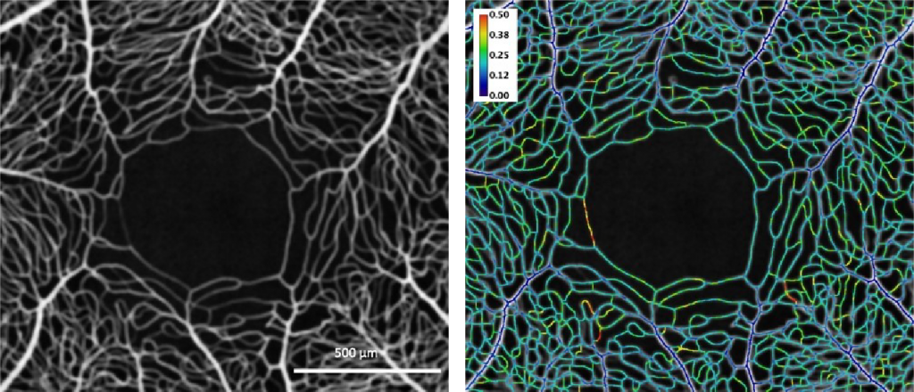
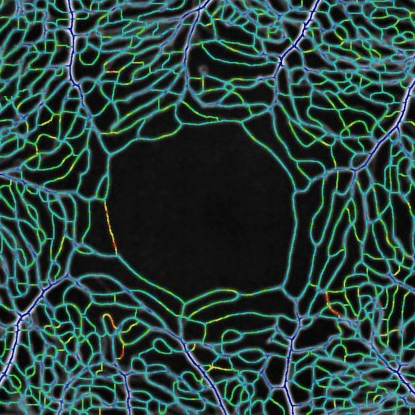
Left: En face mean intensity projection through 31 OCTA images from the macular region of a human subject. Right: Coefficient of variation along vessel centrelines showing spatial and temporal heterogeneity of capillary perfusion. The colour bar indicates the variation from dark blue (no variation) through to red (most variation).


The developed software tools have been used for characterising spatio-temporal variation of capillary perfusion in OCTA images.


Experimental results to date are both valuable and encouraging because they may be potentially useful for clinical diagnostic purposes and can be used to guide improvements in OCTA instruments and new algorithm development. These results move towards a non-invasive tool for the early detection of retinal vascular diseases in humans, and may also be used for other investigations of capillary perfusion where the retina is an appropriate model for microcirculation studies.


For further information, please contact Dr Andrew Mehnert.

This story was contributed by the University of Western Australia.

Messages inside Porites: Are corals exposed to repeated heatwaves coping?

Researchers at the Australian Research Council Centre of Excellence for Coral Reef Studies, the University of Western Australia, Curtin University, Aix-Marseille University in France, and the US National Oceanic and Atmospheric Administration wanted to know the history of coral bleaching events on the Great Barrier Reef and how corals are responding to climate change.


Hard long-lived corals, such as Porites, are the backbone of reef ecosystems such as the Great Barrier Reef. Such reef-building corals are sensitive to light levels and temperature. Reef-building corals are already reaching their limits with every heatwave. With ocean temperatures rising, can they hope to survive more frequent extreme temperature events?


Underwater photograph with two scubba divers insertingg a longg metal rod into a large coral bed

Dr. Thomas DeCarlo drilling a 2+ meter core from a massive coral on the Great Barrier Reef


The team of researchers collected Porite core samples across the northern Great Barrier Reef, the Coral Sea, and New Caledonia, bring them to the WA NIF Node at the University of Western Australia, where they were scanned on the Bruker Skyscan 1176 in vivo micro-CT. 3-dimensional image stacks of density variations revealed ‘bands’ within the coral skeletons, corresponding to age. Also seen were high/low density ‘stress bands’, corresponding to environmental stressors such as exceptionally high water temperature.


CT images showing the bands inside coral cores from 1815 - 2017

μCT scans (dark/light shading = low/high density) reveal high-density stress bands and partial mortality scars preserved within the skeletons of long-lived Porites corals.


Some of the oldest (and longest) core samples had over 200 annual bands, meaning they were a living coral that has been growing for two centuries. By comparing stress bands to age, change-induced bleaching episodes were mapped, providing a timeline of coral bleaching events. Three striking observations followed: First, the researchers found the first piece of evidence that coral bleaching has occurred prior to the 1980s. Second, a significant increase in the frequency of stress bands was seen over time, consistent with the effects of global warming sparking more frequent coral bleaching events. Finally, recent (within the past few years) acclimatization was seen, whereby corals became less sensitive to heat stress follow repeated exposure to marine heatwaves. These results, published in early 2019, offer hope that reef-building corals surviving heatwaves may be able to adapt for future heatwaves.


This story was contributed by the University of Western Australia. For enquiries, please contact Ms Diana Patalwala.


National Network of Trusted Data Repositories

During 2017 the National Imaging Facility (NIF) nodes at the University of Western Australia (UWA), University of Queensland (UQ), University of New South Wales (UNSW) and Monash University collaborated on a national project to enhance the quality, durability and reliability of data generated by NIF through the Trusted Data Repository project.

●        Quality pertains to a NIF user’s expectation that an animal, plant or material can be scanned and from that data reliable outcomes/characterisations can be obtained (e.g. signal, volume, morphology) over time and across NIF sites.

●        Durability refers to guaranteed long-term availability of the data.

●        Reliability means that the data is useful for future researchers, i.e. stored in one or more open data formats and with sufficient evidential metadata.

The Project, Delivering durable, reliable, high-quality image data, was jointly funded by the Australian National Data Service (ANDS) and Research Data Services (RDS). It was motivated both by NIF’s desire to enhance the quality of the data associated with the use of its facilities, and the desire of ANDS/RDS to facilitate the establishment of Trusted Data Repositories that enable access to data for at least 10 years and includes metadata that documents both the quality of the data and its provenance.

A trusted data repository service is essential for sharing data and ensures that project data created and used by researchers is “managed, curated, and archived in such a way to preserve the initial investment in collecting them” and that the data “remain useful and meaningful into the future” (https://www.coretrustseal.org).

The scope of the Project was limited to MRI data with the understanding that the developed requirements and trusted data repository services could be adapted to, or serve as a basis for other instruments/modalities.

The key outcomes from the Project include:

  1. The NIF agreed process for acquiring trusted data (NAP) – Lists the requirements that must be satisfied to obtain high-quality data, i.e. NIF-certified data, suitable for ingestion in a NIF trusted data repository service. They cover provisioning of a unique instrument identifier, instrument registration with Research Data Australia (https://researchdata.ands.org.au), quality control (QC), quality assurance measures, requisite metadata (including cross-reference to the QC data),  the process by which data is moved from the instrument to the digital repository service and the format(s) of the data.
  2. The NIF requirements for a trusted data repository service – Provides a platform-agnostic checklist of requirements that a basic NIF trusted data repository service should satisfy, including: identification of data by a unique Project identifier, ingestion of data from NIF-compliant instruments, authentication via the Australian Access Federation (https://aaf.edu.au), interoperability and easy deployment across NIF nodes.
  3. Implementations of trusted data repository services for two exemplars:
    1. Preclinical MRI data (with mouse brain data as an example) acquired across three NIF nodes—UNSW, UQ and UWA—using a Bruker BioSpec 9.4T MRI. The services have been implemented using the open source MyTardis/ImageTrove (https://www.mytardis.org) platform.
    2. Clinical ataxia MRI data acquired using a Siemens Skyra 3T MRI scanner in support of a Monash-proposed International Ataxia Imaging Repository (IAIR). The service has been Implemented using the open source XNAT (https://www.xnat.org) platform.

Software developed to support the implementation of the repository services includes: Docker (https://www.docker.com) Compose scripts to permit easy deployment at differents sites, client-side scripts for uploading NIF-certified data to ImageTrove/MyTardis and an XNAT plugin for uploading non-DICOM files.

  1. Assessments of the resulting trusted data repository services against a relevant international metric, the CoreTrustSeal (https://www.coretrustseal.org) Core Trustworthy Data Repositories Requirements.

For NIF users and the broader imaging research community the benefits and impact of this Project include:

  • Reliable and durable access to data
  • Improved reliability of research outputs and the provenance associated with it
  • Making NIF data more FAIR (Findable, Accessible, Interoperable, Reusable – https://www.ands.org.au/working-with-data/the-fair-data-principles)
  • Easier linkages between publications and data
  • Stronger research partnerships

For research institutions they include:

  • Enhanced reputation management
  • A means by which to comply with the Australian Code for the Responsible Conduct of Research
  • Enhanced ability to engage in multi-centre imaging research projects

For NIF they include

  • Improved data quality
  • Improved international reputation
  • The ability to run multi-centre trials

The transition plan post-funding includes: maintenance of existing services for 10 years; the integration of additional instruments; creation of a project web portal; planned new national and international service deployments; refinements and improvements; and CoreTrustSeal certification.

Project documents have been archived in the NIF Customer Relationship Management (CRM) system (accessible by NIF staff). Project software is hosted on GitHub and is freely available for download here: https://github.com/NIF-au/TDR. For further information please contact either the national Project Manager or NIF.

Project Manager and UWA lead: Andrew Mehnert (NIF Informatics Fellow, Centre for Microscopy, Characterisation and Analysis).
NIF lead – Graham Galloway (Chief Executive Officer, NIF)
UQ lead – Andrew Janke (NIF Informatics Fellow, Centre for Advanced Imaging)
UNSW lead – Marco Gruwel (Senior Research Associate, Mark Wainwright Analytical Centre)
Monash lead – Wojtek Goscinski (Associate Director, Monash eResearch Centre)
Privacy Settings
Youtube
Vimeo
Google Maps