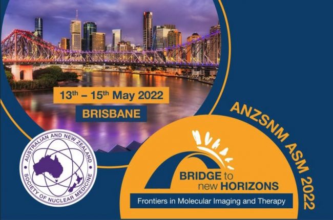
NIF Molecular Imaging and Radiochemistry Showcase to be presented at ANZSNM

National Imaging Facility enables access to imaging capabilities across the country and will present a Molecular Imaging and Radiochemistry Showcase at ANZSNM 2022, featuring presentations from a range of research leaders from Australia’s advanced imaging network.

See the full ANZSNM program here.

Register to attend ANZSNM 2022.

National Imaging Facility: Molecular Imaging and Radiochemistry Showcase
Saturday 14 May 2022, 3:15pm – 4:15pm
Session Chair: Prof Wojtek Goscinski, CEO National Imaging Facility

TimeSpeakerTopic
3:15 – 3:20Professor Wojtek Goscinski

Chief Executive Officer
National Imaging Facility

Introduction to NIF Molecular Imaging and
Radiochemistry Showcase

3:20 – 3:30Professor Steven Meikle

Head of the Imaging Physics Laboratory, Brain and Mind Research Institute, University of Sydney

Total Body PET
3:30 – 3:40Associate Professor Roslyn Francis

Head of Department of Nuclear Medicine and WA PET Service, Sir Charles Gairdner Hospital, University of Western Australia

Radiochemistry activities in Western Australia
3:40 – 3:50Professor Gary Egan

Professor and Foundation Director, Monash Biomedical Imaging

Director, ARC Centre of Excellence for Integrative Brain Function

Australian Precision Medicine Enterprise
3:50 – 4:00Prof Kristofer Thurecht

Acting Deputy Director (Research Technologies) and Group Leader – Principal Research Fellow,

Centre for Advanced Imaging, University of Queensland

Affiliate Principal Research Fellow and Group Leader,

Australian Institute for Bioengineering and Nanotechnology

Alpha therapies and activities
4:00 – 4:10Dr John Bennett

Research Infrastructure Platform Leader – Biosciences,
ANSTO

ANSTO’s new NIF Alpha Radioisotopes and
Radiopharmaceuticals Facility

NIF’s newest capability: Medical industry, manufacturers, and museums set to benefit from WA’s first high-power research-dedicated CT scanner

National Imaging Facility’s (NIF) University of Western Australia (UWA) node located at the Centre for Microscopy Characterisation and Analysis (CMCA) will grow its capacity with the arrival of a new computed tomography (CT) scanner to expand capabilities for industry, manufacturing and museums who require imaging of large samples.

The Nikon XT H 225 ST will increase NIF’s scope to cater for specimens that require a large field of view, including medical implants, additive manufacturing and industrial components, and environmental or historical artefacts.


Research applications of the new CT scanner will extend from medical material testing, industrial material including castings, turbine blades, plastics, packaging, dispensers, to precious palaeontology and archaeology articles.

Diana Patalwala, NIF’s Facility Fellow at UWA’s CMCA said the CT will enable engagement with the biomedical, agriculture, environmental, renewable resource, advanced manufacturing, electronics and defence industries.

“Our new CT capabilities will have increased applications in pre-clinical and clinical research involving medical prosthesis, dental implants, critical assemblies of medical devices and drug delivery systems,” she said.

“It is vital for components such as patient-specific medical implants manufactured through additive manufacturing technologies to be of outstanding quality, and an X-Ray CT can play an important role in this process from start to end.”

Other medical applications include verifying the dimensions of drug delivery systems’ inhaler chambers or dispenser mechanisms, syringes, stents, pacemakers and more.

“Industry will greatly benefit from the Nikon XT H 225 ST as it is the only CT technology of its kind to provide a 225kV (450W) rotating target X-ray source, this means we can image larger and denser samples with increased accuracy than previously possible,” Ms Patalwala said.  

“This makes it ideal for industry users involved in materials testing, inspection and quality control applications.

“This CT scanner would also be ideal for examining archaeological samples, museum specimens and fossils as well, enabling us to get the detailed inside picture without destroying these precious artefacts!” Ms Patalwala said.


With an X-ray source as powerful as 225kV/450W, it is the only high-power research-dedicated CT system in WA.

The unique and versatile scanner can examine specimens ranging in size from small rock cores, which are important for minimising the risk associated with the planned drilling operations in mining and increase the probability of meeting the target yield, to large industrial manufacturing components, such as casting moulds parts, batteries, fuel cells and electronic circuits.

The Nikon XT H 225 ST has an impressive maximum field of view (35cm x 35cm x 35cm), a sample height that can accommodate up to 65cm and a sample weight of 50kg – which will allow for greater capacity in imaging larger samples.


Its large field-of-view, makes it capable of CT scanning the internal tomography of an object non-destructively

The CT uses multiple axial scans to generate cross-sectional information or three-dimensional reconstructions. The X-ray CT has the typical mechanism for taking ‘slices’ which are then digitally reconstructed into 3D volumes.

The Nikon CT has an extremely high-powered X-ray source (450W) for penetrating geological, marine and industrial objects as well as the capability of producing lower energy X-rays (20W) for bio-medical applications.

With resolutions down to the 10um range, academia and industry will have access to 2D cross-sectional slices and 3D volume rendered models, as well as access to advanced quantitative analysis software packages capable of characterising material properties involving cracks, pores, and fibres – just to name a few.


The new Nikon XT H 225 ST CT scanner was delivered at the end of March, with installation commencing from April, and a view to opening to users in May.

This instrument has been funded by National Imaging Facility, enabled by the National Collaborative Research Infrastructure Strategy, with the Government of Western Australia and supporters of the Western Australia National Imaging Facility.

For further information about the instrument, contact NIF Facility Fellow, Diana Patalwala diana.patalwala@uwa.edu.au.

#WorldHealthDay: Imaging unlocking research to keep people healthy

#WorldHealthDay: As Australia’s advanced imaging network, we’re focused on addressing national science and research priorities to help keep people healthy. Our expertise, equipment and services are critical to Australia’s ability to translate health discoveries, undertake clinical trials and commercialise medical products.

The importance of protecting Australians from health threats is critical, as is Australia’s strong medical research capability and reputation for quality and standards.

The National Imaging Facility is unlocking solutions to the world’s biggest imaging challenges across commercial, clinical and research fields. We have helped Australians innovate in fields such as bioengineering, clinical science, biology, medical technology, pharmaceutical and non-pharmaceutical therapies.

Thousands of scientists, doctors, and professionals across hundreds of Australian institutions, companies and research organisations use our work to help answer their medical research questions. We also work with engaged volunteers and patients who make a valuable contribution to health and discovery by being part of research.

We’ve included some examples of the medical projects we’re proud to have partnered with to keep people healthy below:

Dr Ciara Duffy from Western Australia’s Harry Perkins Institute of Medical Research imaging the investigation of honeybee venom to treat breast cancer cells at the University of Western Australia’s Centre for Microscopy, Characterisation and Analysis in collaboration with Microscopy Australia

Associate Professor David Parsons and Dr Martin Donnelly performing preclinical testing of a ground-breaking and simple to use ‘field ventilator’ that can be locally produced at a low cost from easily acquired parts at SAHMRI, in collaboration with 4DMedical, and the University of Adelaide

Supporting Australian trials of Biogen’s Aducanumab (Aduhelm), the first disease modifying therapy for Alzheimer’s disease approved by the United States Food and Drug Administration (FDA) with the University of Melbourne, Herston Imaging Research Facility, the Hunter Medical Research Institute, Australian Imaging Biomarkers and Lifestyle Study of Ageing at The Florey Institute of Neuroscience and Mental Health and Austin Health

#WomenInScience: A conversation with Diana Patalwala

#WomenInScience: A conversation with Diana Patalwala, Research Officer, Preclinical and Materials Imaging and National Imaging Facility Fellow at the Centre for Microscopy Characterisation and Analysis, University of Western Australia  

11 February is the United Nations’ International Day of Women and Girls in Science, highlighting the importance of full and equal access and participation. 

We’re proud to create powerful collaborations across the research and innovation sector, building teams with world-class expertise, who manage our state-of-the-art equipment, and partner with experts in other fields.  

Our mission is to make cutting-edge imaging capabilities accessible to Australian researchers, and we envision a society that provides equal opportunity for people of all genders to learn, work and engage in science. 

As we look to the future of research, it’s clear Australia’s success depends on us developing and encouraging the next generation of scientists, problem solvers and leaders – regardless of their gender, background or any other factor. 

Today we highlight the exceptional work of women leading the way in science and thank them for their work to deliver the impacts of life-changing research. 


Diana Patalwala has worked with the National Imaging Facility (NIF) as a Facility Fellow at the University of Western Australia (UWA) for the best part of a decade, dedicating her time to enabling research translation to real-world benefits.  

The breadth of impact that advanced imaging techniques has on research outcomes is what drives her to come to work every day. 

“We have researchers working on projects spanning in scope from investigating the anti-tumour effects of honeybee venom to treat breast cancer, the most common cancer in women worldwide, all the way to studying the acclimatisation of reef-building corals to consecutive heatwaves, contributing to the understanding of how different coral species are responding to climate change,” she says. 

“This sort of research is contributing to society, it’s giving back, it’s impactful!” 

Image: Coral stress band imaging, as part of Diana’s work assisting researchers with the study of acclimatisation of reef-building corals

Diana oversees the operations and development of research projects, providing user training and support at the Centre for Microscopy Characterisation and Analysis (CMCA) Bio-Imaging Facility (BIF), which supports interdisciplinary and multimodal imaging of small animals and materials using X-ray CT, High Frequency Ultrasounds and Photoacoustic Imaging, Fluorescence Multispectral and Bioluminescence Imaging.  

Her valuable skills and experience in imaging methodologies enable her to assist researchers with data collection, reconstruction, analysis and visualisation. 

When asked what led her to this career path, Diana says her post-graduate studies piqued her interest – but not in the way you might expect. 

“My postgrad degree in Medical Biotechnology had a few units which involved data analysis from preclinical imaging instruments,” she explains. 

“Although we were taught the theoretical principles on which these pre-clinical instruments worked, we were never allowed to operate them ourselves, which was disappointing because the science behind the instruments was really fascinating to me!” 

“Seeing my professors at the university working with these instruments motivated me to envision my career in a pre-clinical imaging facility,” she says. 

Now, Diana’s work allows her to have a hands-on role in imaging, enabling potentially life-changing research in medical biotechnology. 

Before new medical treatments and drugs reach the clinical trial phase (when research studies are performed on people for evaluation), they undergo pre-clinical testing and development. 

Diana says this is where pre-clinical imaging comes into the picture to provide invaluable data.  

“High resolution and high throughput pre-clinical imaging equipment such as pre-clinical CT scanners, high frequency ultrasounds, photoacoustics, Invivo bioluminescence and fluorescence imaging techniques better facilitate the development of these treatments and drugs during their pre-clinical phase,” she says. 

“As a NIF Facility Fellow, I operate and train researchers to use these instruments in a way in which we can get the maximum output from them and analyse the data they generate.” 

Talking to Diana, it is clear she is extremely passionate about her job and how her work can benefit the research community.  

At the end of last year, she presented her work on In vivo MicroCT and In vivo Fluorescence Imaging to an international audience at NIF’s webinar series in partnership with Global BioImaging, which for most people would be a career highlight – but for Diana, it’s quite a competitive ranking. 

“EVERYDAY is a career highlight!” she says. 

“Every day, researchers come to us with questions that have never been answered before, and we at NIF help them design experiments that give them access to world-class, cutting-edge pre-clinical and clinical imaging technologies.” 

“We provide them a better insight into their research needs, and ultimately aim to generate answers to some of the biggest challenges facing society!” 

When asked what advice she would give to someone who is considering working with a NIF capability, Diana says collaboration is at the heart of her work. 

“Come and have a chat with us – we are here for you!” 

“No one knows our instruments better than us – so talk to us before you design your experiments. We can put these instruments to use in ways you might not have thought of, and we will help you get the maximum output from them.” she says. 

For more information on NIF’s UWA Node, or to chat about how NIF’s capabilities could be used in your research project, contact Diana here. 

#ImagingTheFuture Week: Unlocking solutions to major health challenges

#ImagingTheFuture Week: Unlocking solutions to major health challenges


Chan Zuckerberg Initiative’s (CZI) Imaging the Future Week puts a spotlight on the significance of imaging science in biomedicine, and the importance of building a vibrant imaging community across the world to tackle these challenges at scale.

Imaging science and the highly skilled researchers behind it are vital to addressing global health challenges, and driving innovation in disease management, prevention, and cure.

The National Imaging Facility (NIF) invests in state-of-the-art equipment and partners with world-class experts to process and interpret data and apply imaging to solve challenging health problems.

CEO Prof Wojtek Goscinski said he was proud of the NIF’s partnerships which enable the translation of discoveries through to real world applications to improve the health of the population.

“Advanced imaging techniques make it possible to deepen our understanding of health and disease in the human body through visualisation,” Prof Goscinski said.

“Imaging already plays a critical role in healthcare, and the acceleration of its advancements in biomedicine are positioning us, and our colleagues world-wide to continue this work well into the future.”

“We are supportive of the efforts of CZI and I’m excited for NIF to work alongside them and our other international imaging colleagues, building a cutting-edge imaging community at the forefront of global imaging research,” Prof Goscinski said.

You can find out more about Imaging the Future Week here.

Keep scrolling to check out some of the impressive imaging work from a few of the Australian National Imaging Facility’s Nodes.

Time-of-flight angiography of the human brain using 7 Tesla MRI – courtesy of the Centre for Advanced Imaging, University of Queensland

Human Tooth CT scan – courtesy of Diana Patalwala, University of Western Australia

Angiogram scanned on the Siemens 3T Skyra magnet – courtesy of the Large Animal Research and Imaging Facility, South Australian Health and Medical Research Institute

Tractography template image of a sham rat – courtesy of David Wright, The Florey Institute of Neuroscience and Mental Health

Cancer cells killed by honeybee venom

Breast cancer, the most common cancer in women worldwide, may one day be treated using the venom of the European honeybee.

Dr Ciara Duffy from Western Australia’s Harry Perkins Institute of Medical Research has found that venom from honeybees can rapidly kill aggressive and hard-to-treat breast cancer cells. NCRIS-enabled facilities at the Centre for Microscopy, Characterization and Analysis (CMCA), UWA, were integral to the research, published recently in the journal Nature Precision Oncology.

Read More

Understanding Feto-Placental Vasculature

Proper vascular development of the human placenta is crucial for meeting the metabolic needs of the developing fetus during pregnancy. Maternal environmental stressors such as malnutrition disrupt the elaboration of the feto-placental vasculature that, in turn, impacts on placental function and results in reduced fetal growth. The ramifications of this are not only on short-term foetal health but also on long-term health outcomes. Indeed, distortion in placental shape and size strongly associate with later adult health outcomes such as cardiovascular disease, obesity and cancer.

Read More

Micro-CT of re-regeneration in lizard tails

Re-regeneration to reduce adverse effects associated with tail loss

Caudal autotomy, the ability to drop and regenerate a portion of the tail, is a widely used anti-predation strategy in many lizard species. Intra-vertebral autotomy planes within a series of the lizard’s caudal vertebrae allow individuals to autotomise a portion of their tail to escape a threat, such as a predator’s grasp. Once autotomised, the tail regenerates with a rigid cartilage rod in place of the original bony vertebrae. Although an effective anti-predation strategy, it has both short and long-term costs to the individual associated with physical tail loss, as well as the energy required for regeneration. Additionally, a regenerated tail lacks autotomy planes, where subsequent autotomy events having to at a more proximal position at a caudal vertebra with an intact autotomy plane.

Read More
Privacy Settings
Youtube
Vimeo
Google Maps