Detecting retinal vascular disease
One of the leading causes of blindness in the Western world are vascular diseases affecting the retina. The arteries and veins inside the light-sensitive layer in the back of the eye – the retinal vasculature – are an intricate network supplying the inner retina with crucial nutrients while removing metabolic waste. The heterogeneity in space and time of blood flow in this microvasculature is critical as the retina has one of the highest metabolic demands of the central nervous system.
Pathological retinal neovascularisation and oedema – that is swelling, thickening, or unusual growth in the retinal vasculature – are commonly responsible for certified visual loss. Therefore, understanding the spatial and temporal heterogeneity and active regulation of retinal blood flow in the retina is critical for the diagnosis of retinal vascular disease. Furthermore, these blood vessels are affected by systemic vascular diseases and, as such, evidence of these conditions may be observed through the microvasculature inside the eye.
Recently, a technique known as optical coherence tomography angiography (OCTA) has been developed as a non-invasive approach to visualising blood vessels. This method uses the reflectance of light on the surface of moving blood vessels to map the vasculature of the retina.
A multi-disciplinary team, headed by Prof Dao-Yi Yu at the Lions Eye Institute (affiliated with the UWA Centre for Ophthalmology and Visual Science) are using OCTA to reveal remarkable spatial and temporal heterogeneity of retinal capillary perfusion. The project aims to use OCTA as a non-invasive tool for the early detection of retinal vascular diseases.
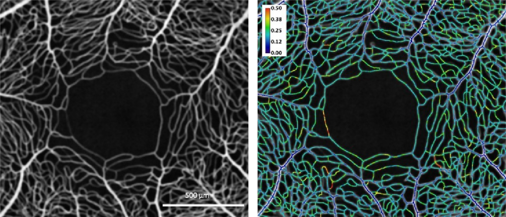
NIF Informatics Fellow, Dr Andrew Mehnert, is contributing his analysis expertise to guide improvements in OCTA instrumentation and algorithms. Image analyses, undertaken in the Characterisation Virtual Laboratory (CVL) using both MATLAB and FIJI/ImageJ, showed remarkable resolution of capillary perfusion in the macular region of human and porcine subjects.

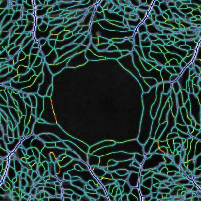
Left: En face mean intensity projection through 31 OCTA images from the macular region of a human subject. Right: Coefficient of variation along vessel centrelines showing spatial and temporal heterogeneity of capillary perfusion. The colour bar indicates the variation from dark blue (no variation) through to red (most variation).
The developed software tools have been used for characterising spatio-temporal variation of capillary perfusion in OCTA images.
Experimental results to date are both valuable and encouraging because they may be potentially useful for clinical diagnostic purposes and can be used to guide improvements in OCTA instruments and new algorithm development. These results move towards a non-invasive tool for the early detection of retinal vascular diseases in humans, and may also be used for other investigations of capillary perfusion where the retina is an appropriate model for microcirculation studies.
For further information, please contact Dr Andrew Mehnert.
This story was contributed by the University of Western Australia.


