#ImagingTheFuture Week: Unlocking solutions to major health challenges

#ImagingTheFuture Week: Unlocking solutions to major health challenges


Chan Zuckerberg Initiative’s (CZI) Imaging the Future Week puts a spotlight on the significance of imaging science in biomedicine, and the importance of building a vibrant imaging community across the world to tackle these challenges at scale.

Imaging science and the highly skilled researchers behind it are vital to addressing global health challenges, and driving innovation in disease management, prevention, and cure.

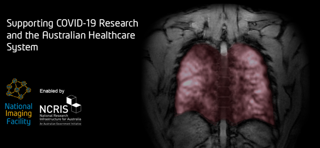
The National Imaging Facility (NIF) invests in state-of-the-art equipment and partners with world-class experts to process and interpret data and apply imaging to solve challenging health problems.

CEO Prof Wojtek Goscinski said he was proud of the NIF’s partnerships which enable the translation of discoveries through to real world applications to improve the health of the population.

“Advanced imaging techniques make it possible to deepen our understanding of health and disease in the human body through visualisation,” Prof Goscinski said.

“Imaging already plays a critical role in healthcare, and the acceleration of its advancements in biomedicine are positioning us, and our colleagues world-wide to continue this work well into the future.”

“We are supportive of the efforts of CZI and I’m excited for NIF to work alongside them and our other international imaging colleagues, building a cutting-edge imaging community at the forefront of global imaging research,” Prof Goscinski said.

You can find out more about Imaging the Future Week here.

Keep scrolling to check out some of the impressive imaging work from a few of the Australian National Imaging Facility’s Nodes.

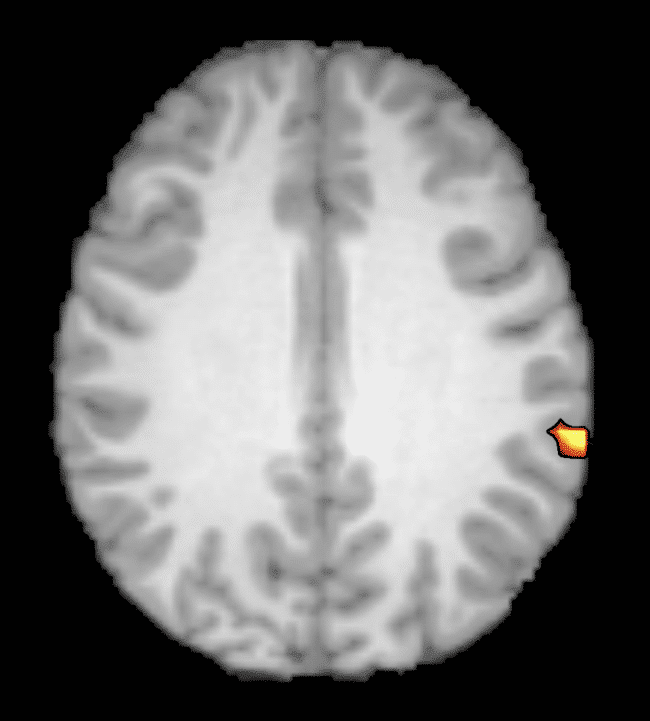
Time-of-flight angiography of the human brain using 7 Tesla MRI – courtesy of the Centre for Advanced Imaging, University of Queensland

Human Tooth CT scan – courtesy of Diana Patalwala, University of Western Australia

Angiogram scanned on the Siemens 3T Skyra magnet – courtesy of the Large Animal Research and Imaging Facility, South Australian Health and Medical Research Institute

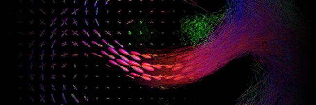
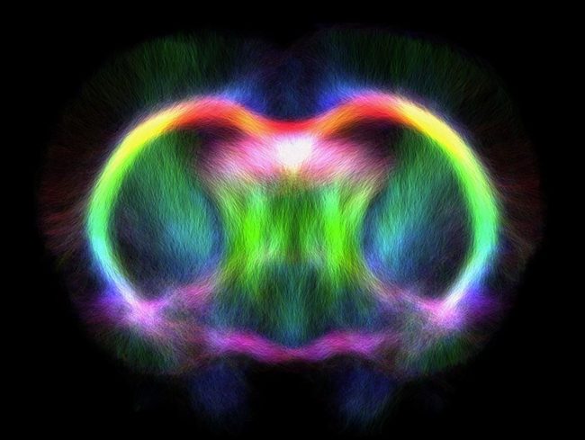
Tractography template image of a sham rat – courtesy of David Wright, The Florey Institute of Neuroscience and Mental Health

The Australian Epilepsy Project

The Australian Epilepsy Project (AEP) will change the lives of people living with epilepsy by reducing uncertainty surrounding diagnosis and fast-tracking the path to optimal treatment using the combination of advanced imaging, genetics, cognition, and artificial intelligence. Such improvements will result in better outcome prediction at disease onset, a higher rate of seizure freedom, reduced economic burden of disease and will increase life-participation of people with epilepsy. 

Read More

MRtrix3

MRtrix3: Advanced tools for the analysis of diffusion MRI data

Diffusion-weighted MRI (dMRI) is a commonly-used medical imaging modality for the investigation of tissue microstructure, exploiting the local hindrance and restriction of water diffusion as indirect probes. The neuroimaging research community utilises this technology extensively for the study of brain white matter in particular, reconstructing structural connectivity pathways and analysing estimated tissue properties.

Read More

Local Connectivity Networks Disrupted by Sports-Related Concussion

From https://journals.sagepub.com/doi/10.1177/2059700219861200
Functional local connectivity is decreased in acutely concussed players compared to controls. (a) Statistically significant brain regions at family-wise error rate p < 0.05; 5000 permutations using threshold-free cluster enhancement. Cool-scaled color bar denotes the magnitude of voxel t-values. (b) Individual-level z-normalized fMRI local connectivity values where each grey circle denotes an individual subject; red line is the group mean; red shaded area is the 95th percentile of the mean value; and blue shaded area is one standard deviation from the mean.

Head injuries, including concussion, are taken very seriously in sporting professions. To date, making an accurate diagnosis of acute concussion has been made difficult by the lack of a reliable direct biomarker for injury and recovery. This diagnostic gap can lead to unknown recovery periods and potentially long-term impacts for athletes.


Researchers at the Florey Institute of Neuroscience and Mental Health set out to understand functional brain changes in professional players in the Australian Football League who had been diagnosed with acute sport-related concussion.


The world-first study, published in the Journal of Concussion, utilised NIF infrastructure, the 3T Trio and Skyra MRI scanners, and NIF expertise, Facility Fellows Shawna Farquharson and David Abbott.

 Functional MRI (fMRI) was undertaken to assess functional connectivity alongside anatomical imaging. Although no anatomical damage was observed, the authors described a decreased intrinsic fMRI connectivity within the right frontoparietal regions in acutely concussed footballers. In other words, all 20 concussed athletes showed reduced activity in parts of the brain responsible for executive function, working memory and switching tasks.


“By looking at how the different parts of the brain talk to each other, we can see how these three brain networks are affected, and these changes may help explain the symptoms we see in concussed players.” – Dr Mangor Pedersen, study co-author, from the Florey Institute of Neuroscience and Mental Health.


One interesting finding in this study is that concussion appears to affect particular networks in the brain. These findings are in agreement with some of the typical clinical features of concussion; however, they are based on trends seen as a group. In future, the authors intend to investigate individual brain networks and develop guidelines for personalised treatment and recovery.


This story was contributed by the Florey Institute of Neuroscience and Mental Health NIF Node. For more information, please contact Shawna Farquharson or David Abbott.


More information about concussion is available here, or by speaking to your GP.

New SMRT President appointed

Profile photo of Shawna facing camera and smiling
NIF Facility Fellow and Florey Chief Radiographer, Shawna Farquharson. Photo attributed to S. Farquharson

NIF Facility Fellow Shawna Farquharson is leading the way to empower, connect, educate and inform MRI Radiographers and Technologists worldwide. Following their 2019 Annual Meeting, the Society for MR Radiographers & Technologists (SMRT), a Section of the International Society for Magnetic Resonance in Medicine (ISMRM), appointed Shawna Farquharson as its new President of SMRT. SMRT is the leading non-profit organisation that provides an international forum for education, information and research in magnetic resonance for radiographers and technologists throughout the world.

Read More

Imaging shows Alzheimer’s decline

Researchers at the Florey have invented a breakthrough imaging technique to describe in micro-detail the brain degeneration occurring in people with early Alzheimer’s and the full-blown disease.

Using the Siemens 3T Trio scanner at the Florey node of the National Imaging Facility (NIF), researchers have identified the precise locations of brain degeneration in a cohort of living Alzheimer’s patients. The work is important as it sheds new light on the underlying cognitive degeneration in Alzheimer’s, helping us focus our efforts to slow the decline.

To develop the technique, the team analysed brain scans from 177 Australians as part of the Australian Imaging, Biomarkers and Lifestyle study, who were either healthy, had an early form of Alzheimer’s or had the full-blown disease.

The brain pathways identified by the team have all been implicated in Alzheimer’s disease previously; those known to be crucial for memory formation, emotion and reasoning.

Alzheimer’s disease is usually thought to be caused by abnormal production and buildup of a peptide called amyloid beta.

Professor Alan Connelly, who led the study, said, “Interestingly, the mildly affected patients with low amyloid had more fibre degeneration in particular brain regions than those with high amyloid levels. This suggests that firstly, specific degeneration of certain brain areas will not necessarily be useful in predicting which mildly impaired individuals will progress to Alzheimer’s disease, and secondly that degeneration of this pathway is related to cognitive impairment, regardless of the buildup of the amyloid peptide.

“This is an important advance for a field still struggling to come to grips with what exactly causes Alzheimer’s. Our study shows we still have a way to go in interrogating the natural history of this insidious disease,” Alan says.

Lead author Remika Mito says, “This study was conducted by comparing the averages of each group of patients against each other, in order to give us the most statistically, and biologically, relevant results. In the future, we want to be able to compare an individual patient against a normal, healthy standard, to see how far along the disease trajectory they are. Or we could compare back to their previous scans to determine what effect a new medication is having as part of a clinical trial for example.”

Remika recently explained her results in an online abstract for Brain. If you would like to more about the details of the study, head over to Youtube to view Remika explain her work.


This story was contributed by the Florey Institute of Neuroscience and Mental Health. 

Privacy Settings
Youtube
Vimeo
Google Maps