NIF’s newest capability: Medical industry, manufacturers, and museums set to benefit from WA’s first high-power research-dedicated CT scanner
National Imaging Facility’s (NIF) University of Western Australia (UWA) node located at the Centre for Microscopy Characterisation and Analysis (CMCA) will grow its capacity with the arrival of a new computed tomography (CT) scanner to expand capabilities for industry, manufacturing and museums who require imaging of large samples.
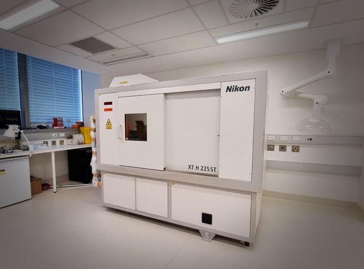
The Nikon XT H 225 ST will increase NIF’s scope to cater for specimens that require a large field of view, including medical implants, additive manufacturing and industrial components, and environmental or historical artefacts.
Research applications of the new CT scanner will extend from medical material testing, industrial material including castings, turbine blades, plastics, packaging, dispensers, to precious palaeontology and archaeology articles.
Diana Patalwala, NIF’s Facility Fellow at UWA’s CMCA said the CT will enable engagement with the biomedical, agriculture, environmental, renewable resource, advanced manufacturing, electronics and defence industries.
“Our new CT capabilities will have increased applications in pre-clinical and clinical research involving medical prosthesis, dental implants, critical assemblies of medical devices and drug delivery systems,” she said.
“It is vital for components such as patient-specific medical implants manufactured through additive manufacturing technologies to be of outstanding quality, and an X-Ray CT can play an important role in this process from start to end.”
Other medical applications include verifying the dimensions of drug delivery systems’ inhaler chambers or dispenser mechanisms, syringes, stents, pacemakers and more.
“Industry will greatly benefit from the Nikon XT H 225 ST as it is the only CT technology of its kind to provide a 225kV (450W) rotating target X-ray source, this means we can image larger and denser samples with increased accuracy than previously possible,” Ms Patalwala said.
“This makes it ideal for industry users involved in materials testing, inspection and quality control applications.
“This CT scanner would also be ideal for examining archaeological samples, museum specimens and fossils as well, enabling us to get the detailed inside picture without destroying these precious artefacts!” Ms Patalwala said.
With an X-ray source as powerful as 225kV/450W, it is the only high-power research-dedicated CT system in WA.
The unique and versatile scanner can examine specimens ranging in size from small rock cores, which are important for minimising the risk associated with the planned drilling operations in mining and increase the probability of meeting the target yield, to large industrial manufacturing components, such as casting moulds parts, batteries, fuel cells and electronic circuits.
The Nikon XT H 225 ST has an impressive maximum field of view (35cm x 35cm x 35cm), a sample height that can accommodate up to 65cm and a sample weight of 50kg – which will allow for greater capacity in imaging larger samples.

Its large field-of-view, makes it capable of CT scanning the internal tomography of an object non-destructively
The CT uses multiple axial scans to generate cross-sectional information or three-dimensional reconstructions. The X-ray CT has the typical mechanism for taking ‘slices’ which are then digitally reconstructed into 3D volumes.
The Nikon CT has an extremely high-powered X-ray source (450W) for penetrating geological, marine and industrial objects as well as the capability of producing lower energy X-rays (20W) for bio-medical applications.
With resolutions down to the 10um range, academia and industry will have access to 2D cross-sectional slices and 3D volume rendered models, as well as access to advanced quantitative analysis software packages capable of characterising material properties involving cracks, pores, and fibres – just to name a few.
The new Nikon XT H 225 ST CT scanner was delivered at the end of March, with installation commencing from April, and a view to opening to users in May.
This instrument has been funded by National Imaging Facility, enabled by the National Collaborative Research Infrastructure Strategy, with the Government of Western Australia and supporters of the Western Australia National Imaging Facility.
For further information about the instrument, contact NIF Facility Fellow, Diana Patalwala diana.patalwala@uwa.edu.au.


