International partnership launches Monash University-Helmholtz lab to solve global oncology, cardiology and infectious disease challenges
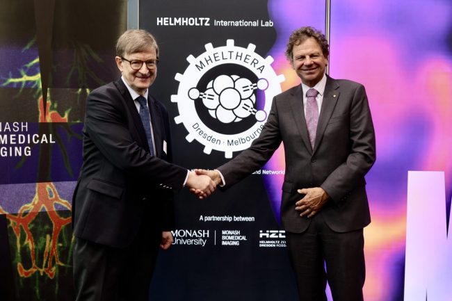
Monash University has partnered with the Helmholtz-Zentrum Dresden-Rossendorf (HZDR), a member of the Helmholtz Association of German Research Centres to establish the Monash-Helmholtz Laboratory for Radio-Immuno-Theranostics (MHELTHERA) to enable clinical translation at a launch event in Melbourne today.
The MHELTHERA Lab is a collaboration to optimise non-invasive imaging techniques and personalised therapy, drawing on diagnostic and therapeutic expertise in cancer, infectious and cardiac disease.
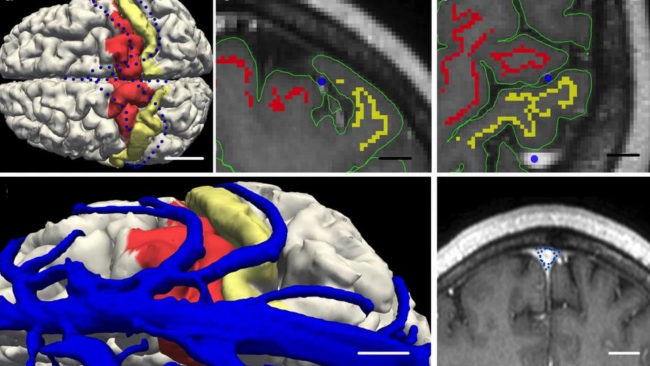
HZDR and Monash are developing next-generation biomedical imaging platforms and the MHELTHERA Lab will specialise in molecular imaging through nuclear techniques such as Positron Emission Tomography (PET) and Single Photon Emission Computed Tomography (SPECT), as well as optical techniques including Fluorescence Imaging (FLI).
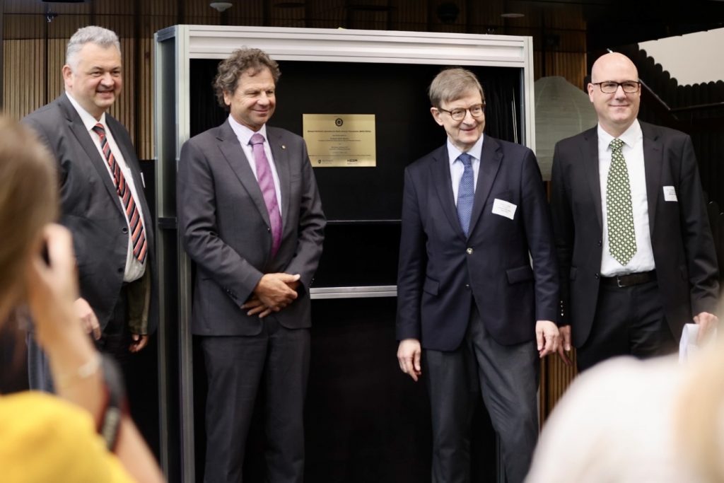
Prof Sebastian M. Schmidt, Scientific Director of HZDR, Mr Simon McKeon AO, Chancellor of Monash University, Prof Otmar Wiestler, President of Helmholtz Association of German Research Centres and Prof Christoph Hagemeyer, National Imaging Facility Monash Biomedical Imaging Node Director opened the MHELTHERA Lab.
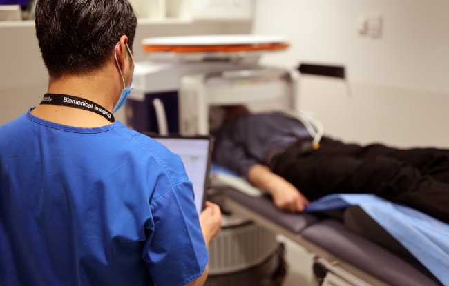
The MHELTHERA Lab was officially opened at its Australian site, Monash Biomedical Imaging (MBI), a node of the National Imaging Facility (NIF) by Mr Simon McKeon AO, Chancellor of Monash University, Prof Otmar Wiestler, President of Helmholtz Association of German Research Centres, and Prof Sebastian M. Schmidt, Scientific Director of HZDR.
Mr McKeon said the partnership is an excellent example of the power of international collaborations.
“No single institution or nation has all the answers when it comes to addressing the most pressing health challenges of our time. By partnering with institutions such as Helmholtz, we can bring together the best talent, knowledge, and resources to tackle these challenges together,” Mr McKeon said.

Prof Wiestler said the Helmholtz Association is Germany’s largest research organisation, developing solutions and innovative technologies to preserve the foundations of human life.
“By supporting MHELTHERA, we are bringing together leading international researchers to develop a highly original class of radiopharmaceuticals that will provide new treatment prospects for cancer patients in particular,” Prof Wiestler said”.

Prof Schmidt said MHELTERA will offer excellent opportunities, especially for young researchers.
“The cooperation is particularly attractive for PhD students, who will have the opportunity to improve their skills through joint research training programs of HZDR and Monash University,” Prof Schmidt said.

MHELTHERA builds upon a successful long-standing collaboration between the two organisations and is led by Prof Christoph Hagemeyer from Monash University, who is also the NIF MBI Node Director, and Prof Michael Bachmann from the HZDR Institute of Radiopharmaceutical Cancer Research.

“The joint laboratory will leverage the research infrastructure capabilities at both sites to accelerate the development of promising technologies in the field of precision medicine,” Prof Hagemeyer said.
NIF CEO Prof Wojtek Goscinski said there are ten Helmholtz International Labs dedicated to innovative research located around the world, and it was a privilege for NIF co-funded infrastructure and staff to support the first of its kind in Australia.
“NIF has invested in world-class radiochemistry expertise and capabilities at MBI and this is an exciting opportunity for us to support a unique Helmholtz lab at the global cutting-edge of cancer research,” Prof Goscinski said.
“The application of radiopharmaceuticals and nuclear theranostics through PET imaging is revolutionising cancer treatment, and NIF is committed to improving health outcomes by supporting these innovative medical products,” Prof Goscinski said.
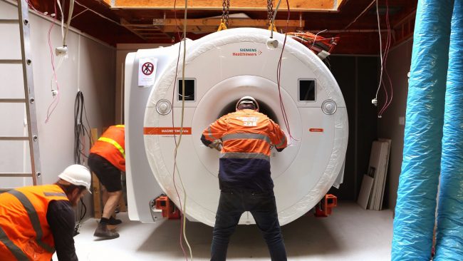
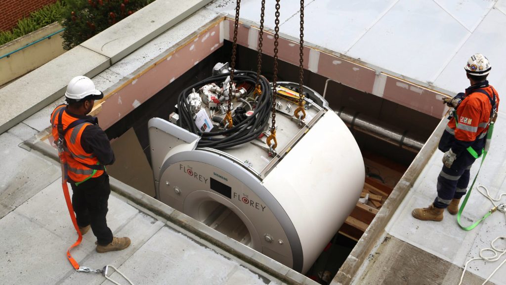
NIF has committed $4.95 million to a new cyclotron, enabling critical radioisotope production, and an expanded radiochemistry facility at MBI which is central to the infrastructure supporting the collaboration.
This critical infrastructure will complement the lab, providing a platform for manufacturing capability, drug innovation, treatment and advanced training.