Victorian collaboration raises over $50m investment in critical imaging capabilities

[Image: La Trobe University – Olivia Newton-John Cancer Research Institute NIF Fellow, Dr Ingrid Burvenich with Minister Tierney]

The Victorian Government has invested $14.83m in National Imaging Facility’s (NIF) research infrastructure in Victoria, in partnership with the Victorian Biomedical Imaging Capability (VBIC), equating to a boost of just over $50m through collaborative co-investment. 

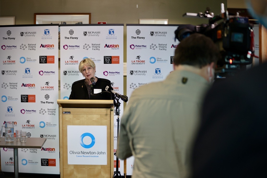
[Image: Victorian Minister for Higher Education, the Hon Gayle Tierney MP]

Victorian Minister for Higher Education, the Hon Gayle Tierney MP visited the Olivia Newton-John Cancer Research Institute on Tuesday, to highlight the impact of the collaboration and the State Government’s investment of through the Victorian Higher Education State Investment Fund (VHESIF) initiative.  

“Collaborative projects such as this demonstrate how our government is supporting higher education and industry to become international leaders in their field,” Minister Tierney said.

[Image: Victorian Minister for Higher Education, the Hon Gayle Tierney MP toured the facilities at the Olivia Newton-John Cancer Research Institute and the Austin hospital]

The funding is supporting the upgrade and expansion of imaging capabilities across NIF’s research facilities in Victoria, including The Florey, La Trobe University and the Olivia Newton-John Cancer Research Institute, Monash University, Swinburne University of Technology, and University of Melbourne in partnership with Peter MacCallum Cancer Centre and the Austin Hospital.  

Critical medical research in areas of national priority such as dementia cancer and epilepsy, as well as agriculture research will be enabled by the co-investment, which includes $26.7m from NIF through the Australian Government’s National Collaborative Research Infrastructure Strategy (NCRIS) funding scheme. 

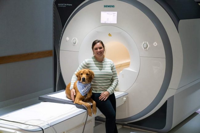
Infrastructure funded under the collaboration includes human magnetic resonance imaging (MRI) capabilities at Swinburne University of Technology and The Florey, where state-of-the-art high-intensity focused ultrasound will support the development of new treatments for essential tremor and tremor-dominant Parkinson’s disease. 

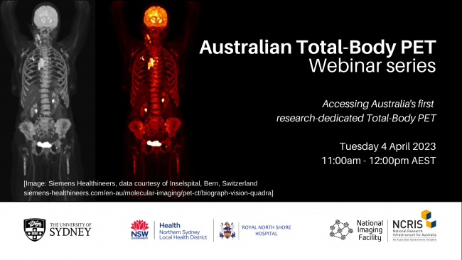
In addition to this, the University of Melbourne upgraded their ultra-high-field 7T MRI (one of only two in Australia), and acquired a new human PET-CT. 

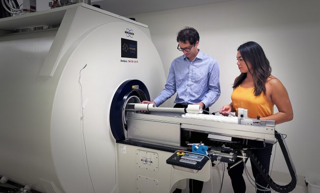
Preclinical capabilities including PET/MRI and PET/CT to support important drug discovery and testing have been installed at the Olivia Newton-John Cancer Research Institute/La Trobe University and Monash University. 

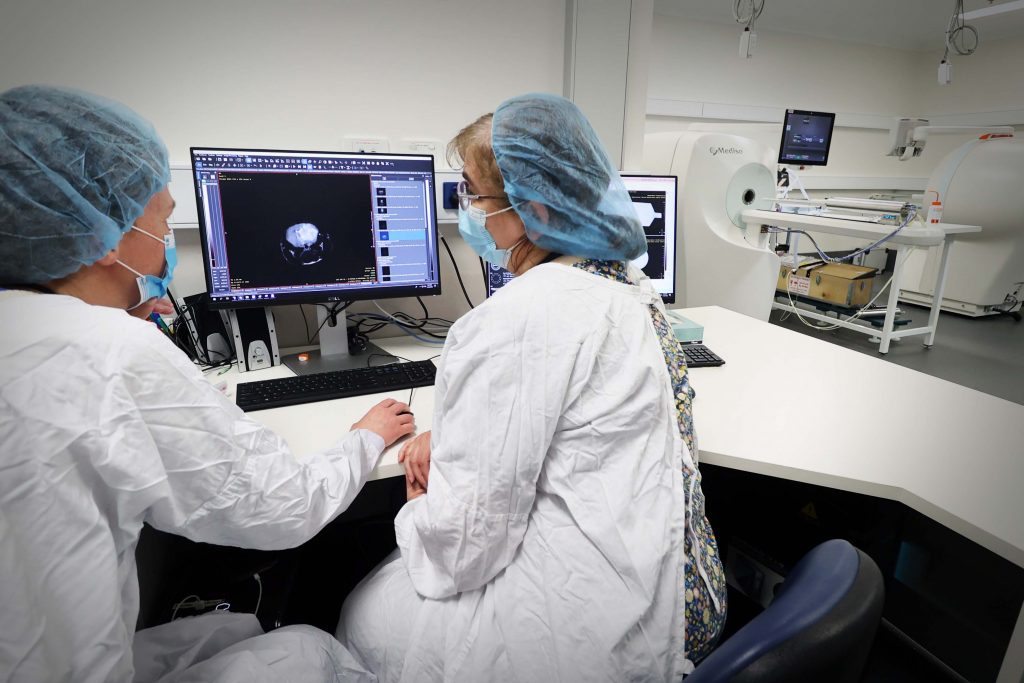
[Image: NIF preclinical capabilities at the Olivia Newton-John Cancer Research Institute]

A new research cyclotron at Monash (the Australian Precision Radiopharmaceutical Facility APRF) will enable the production of radioisotopes under GMP standards, and enhance Australia’s sovereign capability to produce therapeutics and diagnostics. Complementary to this, radiochemistry hotcell infrastructure upgrades at Monash, the Austin Hospital, and Peter MacCallum Cancer Centre will support the design and development of novel cancer treatments.  

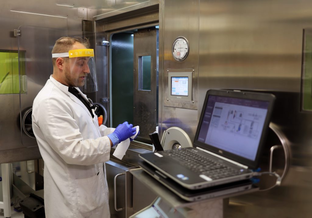
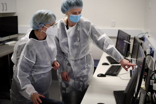
[NIF radiochemistry capabilities at the Austin hospital/Olivia Newton-John Cancer Research Institute]

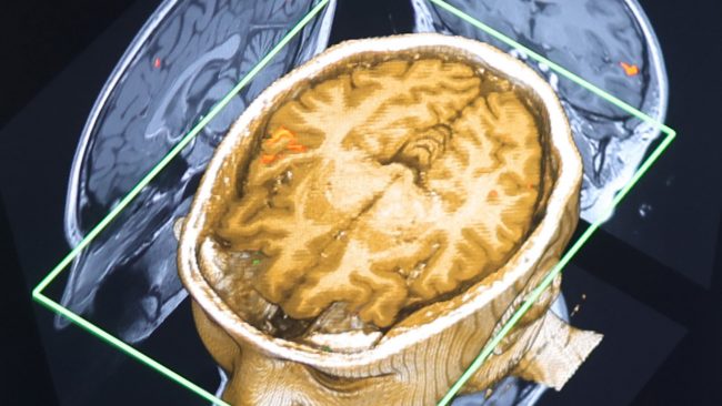
The funding also enables upgrades to the magnetoencephalography (MEG) at Swinburne, one of only two systems in Australia, supporting the study of brain function. 

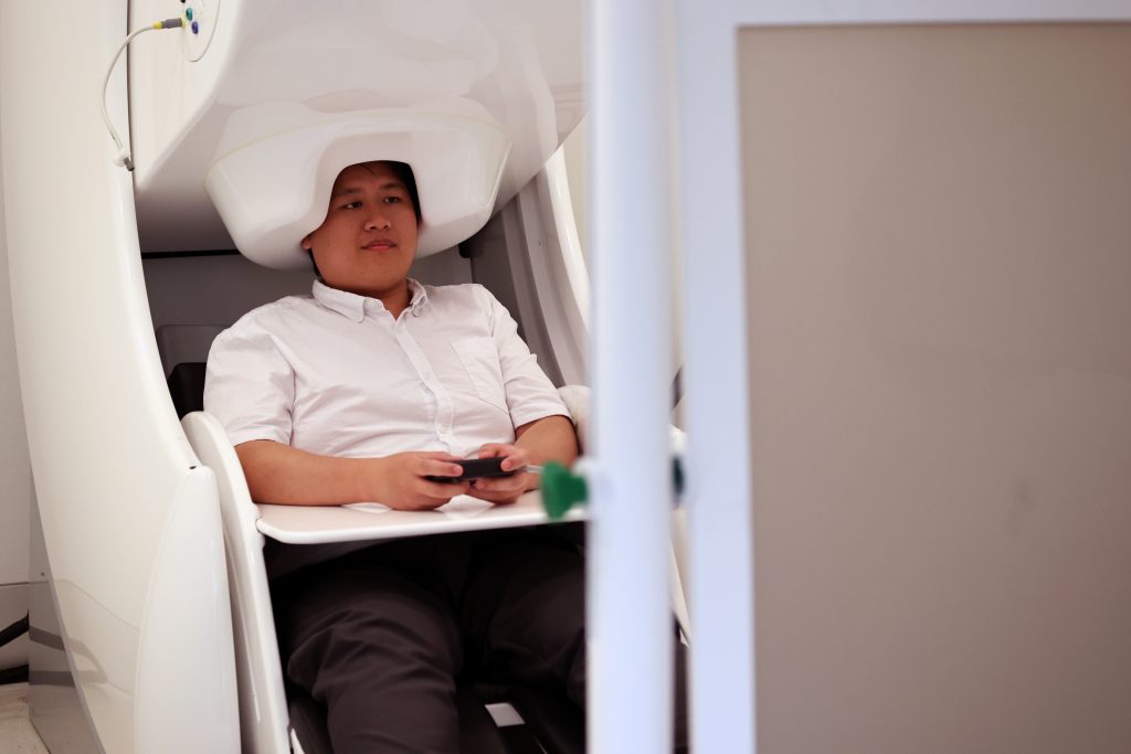
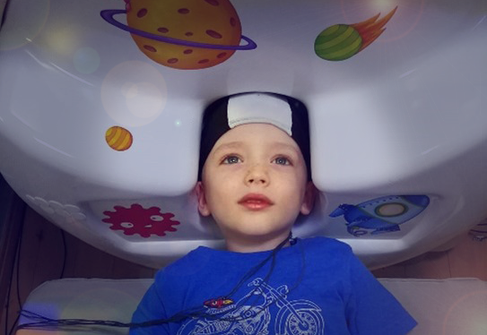
[Image: NIF MEG at Swinburne University of Technology]

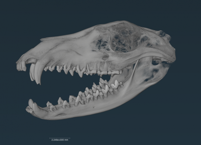
Development of specialised plant imaging capabilities at the University of Melbourne will underpin research into the effect of climate change on crops and soil, and strategies and applications for agricultural improvements to support Australia’s standing as a world leader in food and beverage production. 

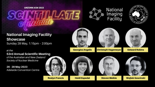
NIF Chief Executive Officer, Prof Wojtek Goscinski said the co-contributed investment underpins transformational initiatives in a number of national priority areas including precision medicine, molecular imaging, drug discovery, diagnostics and plant soil imaging. 

“It’s a privilege for NIF to partner with the Victorian Government and VBIC to support Australia’s strategic science and research priorities” Prof Goscinski said. 

“These capabilities will support Australia as a world-leader in applying advanced imaging technology, resulting in better healthcare, better products, and important discoveries.”

Read the Victorian Government’s announcement here. 

23 June: Take Your Dog to Work Day – even if you work at a research institute!

[photo credit: Mick Ross]

How do you reduce stress when you’re preparing people for medical imaging? Simply add a dog. That’s what PhD candidate Caroline Faucher at the Hunter Medical Research Institute (HMRI) has done, training golden retriever Maple to be a calming influence for the young, old and nervous.

Caroline and Maple are part of the Systems Neuroscience Group, under the leadership of Michael Breakspear, NIF’s University of Newcastle Node (UON) Co-Director.

The group brings together mathematicians, neuroscientists, psychologists, clinicians, social scientists and physicists to solve important research and healthcare questions.

Mathematical models, advanced statistics and behavioural sciences are combined with NIF imaging and used to improve understanding in areas such as dementia, mental health, sleep, hearing loss and ageing.

NIF’s MRI technology at Newcastle produces detailed 3D images of soft tissue such as the brain, spinal cord, nerves, muscles, ligaments and tendons.

Caroline uses MRI to study the brain of adults in mid to late life.

“My PhD focuses on the influence of sleep on cognition and brain health through a combination of clinical and neuroscience approaches,” she says.

“Using the MRI means I can study how sleep quality is related to brain activity and structure.”

The research is important in ensuring people can sleep well so their mind and body can recharge – which in turn can help decrease the risk of developing conditions such as dementia, cardiovascular disease and mental health disorders such as depression.

“My research specifically looks at the links between sleep, cognitive abilities, brain structure and activity in older adults, both with and without a genetic risk of developing dementia.”

It aligns with Federal Government priorities for ensuring better health for the young and older Australians, leveraging imaging technology to understand and promote healthy development and ageing.

Being at UON also enables researchers to undertake imaging studies across a representative selection of the Australian population, including people from regional, rural and metro areas – another important national priority.

UON is NIF’s first regional node, providing an opportunity to leverage its unique links to regional, rural and remote communities through the Hunter and New England districts of NSW.

Caroline says participants undergoing MRI scans may be tired or nervous – making Maple’s role as a therapy dog invaluable.

“It’s still early days, but the hope is that Maple can assist participants coming for research. I’m also hoping to research this topic in the near future, looking at how animal-assisted therapy can help with scanner anxiety. 

“Of course, Maple also helps with the general wellbeing of the team.

“Positive interactions between people and therapy animals are known to bring emotional and physiological benefits.

“Therapy dogs are increasingly used across Australian hospitals and health clinics, aged care and mental health facilities, schools and workplaces.”

Recent figures show that therapy dogs bring positive interactions to about 20,000 people around the country each week.

Maple also enjoys the interactions.

“My favourite part of my job is getting all of the pats and hearing that I am a good boy.”

Caroline and Maple graduated as an animal-assisted therapy human-canine clinical team from Therapy Dogs Australia.

National Imaging Facility Showcase at ANZSNM 2023

National Imaging Facility will host a Showcase at ANZSNM 2023, featuring presentations from Australia’s advanced imaging network.

See the full ANZSNM Program and register here.

Sunday 28 May, 1:15pm – 2:00pm
City Room 3/4
Adelaide Convention Centre

Chair:   Prof Steven Meikle
Head, Imaging Physics Laboratory
Brain and Mind Centre, University of Sydney

Time

1:15pm

Topic

Introduction and session open

Speaker

Prof Wojtek Goscinski
Chief Executive Officer
National Imaging Facility

1:20pm

Australian National Total Body PET Facility

Dr Georgios Angelis
NIF Total Body PET Fellow
University of Sydney

1:30pm

Monash MHELTHERA Lab and cyclotron capabilities

Prof Christoph Hagemeyer
Acting Director, NIF Node Director
Monash Biomedical Imaging

1:40pm

SAHMRI Radiopharmaceutical Chemistry

Dr Edward Robins
Head, Radiopharmaceutical Research and Development
Molecular Imaging and Therapy Research Unit
SAHMRI

1:50pm

New capabilities of the Western Australia Node

A/Prof Roslyn Francis
Head, Department of Nuclear Medicine and the WA PET Service, Sir Charles Gairdner Hospital
NIF Deputy Node Director
University of Western Australia

Dr Heidi Espedal
NIF Facility Fellow
University of Western Australia

MS Awareness Month: Australia’s advanced imaging technology takes aim at multiple sclerosis

[Image: NIF Fellow Dr Tim Rosenow and researcher Dr Virginie Lam with NIF’s preclinical MRI at the University of Western Australia.]


Multiple sclerosis affects more than 30,000 Australians. Most have relapse-remitting MS (RRMS), known for having flare-ups and recovery that in turn damage and repair insulating layers protecting nerves in the brain and spinal cord. Researchers are collaborating with NIF to study the cycles of attack and remission – and determine if a novel therapy can help.


RRMS flare-ups involve demyelination, a process in which immune cells attack myelin, the protective coating around nerve fibres. Damaged myelin slows or prevents signals travelling between the brain, spinal cord, organs and limbs. 

Curtin University’s Dr Virginie Lam leads research into promoting the flipside to that process, remyelination – repair that is part of the RRMS remission phase and can halt disease progression. 

A focus is protecting fatty molecules called lipids that are abundant in myelin. 

Dr Lam is testing a lipid-based therapy with antioxidant, anti-inflammatory and cell-controlling properties that shows promise in enhancing remyelination. 

The Western Australia NIF node supports the research with in vivo MRI to monitor the amount and health of myelin levels in preclinical MS models for up to 16 weeks. 

The research is a collaboration with Professor John Mamo, Director of the Curtin Health Innovation Research Institute, Dr Michael Bynevelt, a neuroradiologist at WA Health and the University of Western Australia, and NIF Fellows Dr Tim Rosenow and Dr Sjoerd Vos.  

It involves use of some of Australia’s most advanced MRI technology, an ultra-high field preclinical research MRI scanner at the University of Western Australia. 

It is important national infrastructure supporting preclinical research for modelling disease mechanisms and testing early drug candidates, ahead of plans to move into human imaging and clinical trials. 

The NIF platform can determine the structure and health of myelin in the specific brain regions that are most important in MS. Critically, this can be done non-invasively, allowing repeated measurements over time. 

This enables Dr Lam’s team to monitor the health of myelin and brain function, study pathways needed for healthy brain function, and track the effects of the therapy. 

“Imaging is important for determining the efficacy of the therapy we are researching,” Dr Lam says. 

“The therapy we are using is a purified form of a lipid which makes up a large portion of myelin.  

“The myelin lipid in the therapy is not found in high quantities in the foods we eat, so we need to boost that through a purified supplement form. 

“There is no cure for RRMS and little is known about the mechanism for myelin repair.  

“We want to better understand the underlying processes – and at the same time determine our therapy’s success – in restoring myelin function, enhancing remyelination and preventing or slowing MS progression.” 

Four weeks into the study, interim results have already shown increased myelin in the brain and reduced demyelination in an important part of the brain that is prone to demyelination in MS, the corpus callosum, Dr Lam says. 

The findings come as peak body MS Australia publishes research in February 2023 showing the number of Australians living with MS is increasing at a significant and accelerating rate – up from 25,607 in 2017 to 33,335 in 2021. 

Total costs for all people with MS in Australia have increased substantially in the same period, from $1.75 billion to $2.5 billion, with an annual per-person cost that is $20,000 above the next highest comparable complex chronic disease, Parkinson’s. 

People with RRMS are typically diagnosed in their 20s and 30s – earlier than other forms of MS. 

Common RRMS symptoms include fatigue, numbness, vision problems, spasticity or stiffness, bowel and bladder issues, and problems with learning, memory or information processing. 

Australian health research infrastructure underpins world-first brain cancer collaboration

The NCRIS Health Group will support Australian brain cancer research through Brain Cancer Biobanking Australia’s project to develop an integrated network of research platforms to improve patient outcomes, test new treatments and work toward a cure, with the announcement of a Medical Research Future Fund (MRFF) grant worth just under $6M.

The project brings together clinicians, researchers and healthcare specialists from institutions across Australia to establish three essential interlinked platforms:

  • An Australian Brain Cancer Registry to systematically collect treatment and outcome data of Australians living with brain cancer, enabling researchers to identify and address variations in clinical practice and outcomes, immediately increasing the quality of care that every patient experiences.
  • Registry Clinical Trials and Patient Enrolment Platform to connect researchers with people living with brain cancer and enable rapid, cost-effective clinical trials and patient donations of data and specimens of research for testing novel ideas designed to improve patient survival.
  • Biobanking and Organoid Platform to establish national standards and protocols for brain cancer biobanking and the creation of Australian brain cancer organoids (cutting-edge brain tumour models). This will assist in providing the resources Australia needs to drive the innovative genomic research that will deliver new treatments in brain cancer.

The NCRIS Health Group’s research infrastructure will enhance collaborative opportunities for this important national initiative through the support of critical capabilities in biobanking, imaging, modeling and providing access to linked data.

Around 2,000 Australians develop brain cancer every year, and it takes the lives of more children and adults under 40 than any other cancer, with no improvement in survival rate for over 40 years.

Brain Cancer Biobanking Australia Chair A/Prof Lindy Jeffree will lead the important national collaboration to improve the lives of Australians living with brain cancer.

“The significance of this grant cannot be overstated. These funds will enable our national team to establish an integrated network of research platforms that will not just be an Australian-first, but a world-first in brain cancer,” A/Prof Jeffree said.


About the NCRIS Health Group

The NCRIS Health Group includes Bioplatforms Australia (BPA), National Imaging Facility (NIF), Phenomics Australia (PA), Population Health Research Network (PHRN) and Therapeutic Innovation Australia (TIA), enabled by the Australian Government Department of Education’s National Collaborative Research Infrastructure Strategy (NCRIS).

The NCRIS Health Group supports cross-disciplinary research, giving Australian researchers access to world-leading facilities for more impactful outcomes across the whole translation cycle. Contact any of the affiliated organisations for more information about open access to capabilities.

Australian Total-Body PET webinar series: Accessing Australia’s first research-dedicated Total-Body PET

Register: How to access Australia’s first research-dedicated Total-Body PET

Imaging to understand whole-body processes such as novel drug interactions during therapeutic development will soon be accessible through Australia’s first research-dedicated Total Body PET (TB-PET).

The Sydney Imaging Core Research Facility at The University of Sydney, in partnership with Northern Sydney Local Health District (NSLHD) and the National Imaging Facility (NIF), is establishing a Total Body PET facility in the Department of Nuclear Medicine at Royal North Shore Hospital.

TB-PET can be used to capture molecular processes from all organs simultaneously. The high-performance instrument can be used to study diseases that affect the entire body and build a better picture of complex processes such as ageing, metabolism, brain signalling and drug interactions.

Due to its exquisite sensitivity, it also has the potential to use much lower radiation doses compared to conventional PET scanners, making it safe to scan children, healthy volunteers, and to scan patients repeatedly to better understand disease progression and treatment effects.

TB-PET is accessible for research studies through Sydney Imaging Core Research Facility, the University of Sydney Node of the National Imaging Facility. For more information, contact Dr Georgios Angelis.

Webinar details

11:00 AM – 12:00 PM AEST
Tuesday 4 April 2023 
Register via Eventbrite here.

In this first webinar:

  • The Director of Sydney Imaging, Prof Fernando Calamante, will introduce the new facility, the operational model and the available resources to all researchers across Australia
  • Prof Steve Meikle will provide an overview of Total Body PET technology and explain how the new facility will fit within the existing clinical research imaging landscape
  • Amanda Hammond, Molecular Imaging Product Manager at Siemens Healthcare, will present an overview of the technical specifications and capabilities of the new Biograph Vision Quadra TB-PET system set to be installed.
  • The presentations will be followed by a short Q&A session.

NIF investment builds understanding of Australia’s unique paleontological collections

A 105-million-year-old shark vertebrae fossil and a 4,000-year-old thylacine skull are amongst the first items to be scanned on NIF’s materials investment at the University of Western Australia (UWA), building a valuable digital collection of unique national artefacts with the Museum of Western Australia.

UWA NIF Fellow, Diana Patalwala said computed tomography was an invaluable tool for imaging materials providing high resolution 3D data, and preserving accurate anatomical information on the relative shape, size and location of different structures that would not be attainable by physical dissection.

“For each specimen, our materials-dedicated CT scanner takes approximately 1800–3000 x-ray images as the sample is rotated in the x-ray beam, at a level of resolution 100 times that of a typical medical CT scanner used on humans,” Ms Patalwala said.

“These images are then used to create a 3D model of the entire specimen, which is in essence a stack of virtual dissection slices that can be manipulated, rotated, and studied from every angle, revealing unprecedented details of the internal structure of the specimen.”

NIF is supporting the capture of this valuable data to create permanent digital records of specimen collections and enabling its reuse by multiple researchers to minimise duplication of efforts and resources.

Western Australian Museum Curator of Entomology, Collections and Research, Dr Nikolai Tartanic said the data captured could serve several different purposes.

“We’ve used scans to generate 3D models of items that can be printed and put on display. Sometimes these are scale models of minute organisms that are otherwise too small to observe and appreciate, other times we use 3D models to replace fragile or rare specimens,” Dr Tartanic said.

“The 3D datasets can also be shared with colleagues electronically, which in some cases replaces the need to send the physical specimen without putting the specimen at risk.”

Western Australian Museum Head of Earth and Planetary Sciences Dr Mikael Siversson said prior to the availability of this technology, some information about specimens could only be found by physical dissection, which could result in damage.

“In the old days, palaeontologists studying internal structures of vertebrate fossils would sometimes cut up the fossil slice-by-slice, and build a 3D model using clay,” Dr Siversson said.

“Printed 3D models enable palaeontologists to hands-on examine the morphology of primary type specimen without risking damaging the actual specimen.”

Scroll on to see some of the scans captured for the Museum’s collection.


Nullarbor Thylacine Skull sub-fossil: (Scan resolution: 105um; 185kV; 100uA)
Thylacinus cynocephalus (Tasmanian Tiger)
Murra-El-Elevyn Cave, Nullarbor

This subfossil skull from the Nullarbor was scanned in addition to a modern skull from Tasmania, enabling the team to capture data that can be used for morphometric (shape variation and comparison) studies.

This fossil is about 4,000 years old (carbon dated – 3,885 carbon years) and there’s a hole in the jaw where they took a sample for the carbon dating. It is thought to be a female, based on the size of the skull. 


Shark vertebrae fossil: (Scan resolution: 56um; 200kV; 100uA)
Anacoracidae sp. (undescribed species of anacoracid shark)
Toolebuc Formation, Richmond, Queensland

This shark vertebra fossil is about 105 million years old (Albian Stage, Cretaceous Period), and belongs to a group of lamniform sharks called the anacoracids which were the Cretaceous ecological equivalents to modern whaler sharks.

Dr Siversson said the fossil represents a new species.

“It is exceptionally well preserved – anacoracid vertebrae are notoriously fragile – and this particular vertebra is surprisingly large considering the small size of anacoracid teeth in the same geological formation,” Dr Siversson said.

The team suspects this species was a plankton feeder, and based on its size, the animal would have been about four metres in length.


Aplysinopsis sponge: (Scan resolution: 91um; 850V; 250uA)

This species of sponge belongs to the order Dictyoceratida, which are sponges with spongin fibres. Some of them incorporate sand granules in their fibres, and small crustanceans are often found inside the sponge cavities.

Scanning revealed this sponge had several brittle stars living within it.


Echinodictyum Clathroides: (Scan resolution: 121um; 180kV; 100uA)

This species of sponge was first discovered in Shark Bay area, making its location a type locality. The sponge has three different spicules found in its fibres, some densely covered with spines.


Amber resin (Scan resolution: 78um; 190kV; 100uA)

These are specimens of amber-like natural resin of paleobotanical origin collected from WA beaches. The ultimate origin of this amber-like resin is likely South East Asia and it is thought these samples may have floated down to Rottnest Island where they were collected all the way from Indonesia.

Chemical analysis of similar amber found on Cape York in Queensland identified that it was produced by the Dipterocarpaceae, a family of lowland tropical rainforest trees.

Dr Tatarnic said the research team were looking for trapped insects using the CT scanning, but unfortunately did not find any.

“Scans of amber can detect the presence of now extinct insects. These may be new to science, or they may help us reconstruct past ecosystems, or identify from where the piece of amber originates,” Dr Tatarnic said.


The Nikon XT H 225 ST CT scanner was delivered to UWA in 2022 and is funded by National Imaging Facility, enabled by the National Collaborative Research Infrastructure Strategy, with the Government of Western Australia and supporters of the Western Australia National Imaging Facility.

For further information, contact NIF Facility Fellow, Diana Patalwala diana.patalwala@uwa.edu.au.

NIF’s SAHMRI Node becomes second Hub of the Australian Epilepsy Project

[Image: Prof Graeme Jackson, AEP Chief Investigator, Dr Michelle Kiley AM, Director of Epilepsy Services, CALHN and Lead Epileptologist AEP South Australia, Martin Adams, Chair of the Florey Board, The Florey Institute of Neuroscience and Mental Health and Prof Steve Wesselingh, Executive Director SAHMRI]

ICYMI, an Australian Epilepsy Project (AEP) Hub has opened at SAHMRI – South Australia’s independent not-for-profit health and medical research institute, and National Imaging Facility’s (NIF) second node to join the national multidisciplinary collaboration to improve epilepsy outcomes.

AEP’s first Hub was established at NIF’s Node at the Florey Institute of Neuroscience and Mental Health in Melbourne and the SAHMRI Hub will provide magnetic resonance imaging (MRI) scanning facilities and expertise to support this important initiative in Adelaide.

Epilepsy affects over 150,000 Australians, and its expenditure burden on the national health system is around $333M each year.

[Image: Ned Travers, AEP Lived Experience Ambassador South Australia, Dr Michelle Kiley AM, Director of Epilepsy Services CALHN and Lead Epileptologist AEP South Australia, Amanda Anderson, AEP Lived Experience Ambassador and Participant Lead and Carolyn Travers, AEP Lived Experience Ambassador South Australia]

The AEP aims to develop a critical resource to progress epilepsy research to reduce diagnosis uncertainty and facilitate fast-tracking of optimal treatment by combining advanced imaging, genetics, cognition, and artificial intelligence (AI).

AEP’s Chief Investigator, Professor Graeme Jackson said the ultimate aim of the AEP is to improve the standard of care and change the lives of people with epilepsy.

“Epilepsy is life-long condition and we need life-long solutions. Using algorithms, imaging and rich data we can extract insights to predict patterns in epilepsy and create individualised treatment plans for patients. This is an exciting new standard of care that we’ll be able to offer people living with epilepsy,” Prof Jackson said.

The AEP program has been developed using novel advanced imaging techniques with AI and machine learning, supported by NIF’s world-class infrastructure, which will provide the highest quality of data to epilepsy research.

NIF CEO Prof Wojtek Goscinski said the geographical expansion of the project will drive major advances in decision support tools to guide diagnosis and highlight opportunities for precision treatment for epilepsy, while addressing the disparity in epilepsy research in Australia’s diverse population.

“NIF’s national network of world-class human MR expertise and infrastructure will enable scanning across the country, in alignment with our impact goals addressing health equity for all Australians” Prof Goscinski said.

“It’s a privilege for NIF to support this life-changing project at our node at the Florey for patients in Victoria, and now SAHMRI for patients in South Australia,” Prof Goscinski said.

[Image: Dr David Vaughan, AEP Imaging Lead and Clinician, Paul Lightfoot, AEP Operations Lead, Jemima Gore, Operations Officer, SAHMRI Clinical Trials Platform, Lisa Carne, SAHMRI Operations Manager and Dr Karen Best, Director, SAHMRI Clinicial Trials Platform]

SAHMRI Clinical Trials Platform Director Dr Karen Best said enabling Adelaide’s medical research sector to engage with national initiatives like the AEP is a key reason that SAHMRI’s Clinical Trials Platform exists.

“We’re proud to be able to help at all stages of the project’s SA-based activities, from coordinating patient enrolment to making connections for diagnostic testing at facilities like the SAHMRI Clinical Research Imaging Centre.”

In addition to the AEP’s expansion to South Australia, Hubs in Queensland and New South Wales are set to launch in mid-2023.


Want to join the Australian Epilepsy Project? Ask your neurologist for a referral.

People in South Australia as well as Victoria living with epilepsy can be referred into the hub for advanced testing, free of charge, as part of their participation in the AEP. Find out more at epilepsyproject.org.au

NIF imaging to underpin research building complete picture of concussion

Concussion is a form of mild traumatic brain injury (mTBI). Across Australia each year, an estimated 35,000 people with reported mTBI experience symptoms that persist for monthsor even years. There is a pressing need to develop improved clinical and imaging tools to aid early diagnosis and better monitor ongoing recovery for patients. NIF advanced imaging technology will be used to establish a national imaging data resource of people experiencing mTBI across Australia. 

NIF is partnering with AUS-mTBI, a national consortium of clinicians, researchers, industry partners and decision-makers working to build Australia’s first clinical and imaging data resource of people experiencing mTBI. 

The world-leading initiative aims to help provide a better understanding of normal patterns of recovery and to identify the risk factors associated with delays or persistent post-concussion symptoms. 

Curtin University neuroscientist Professor Melinda Fitzgerald leads AUS-mTBI, with funding from the Federal Government’s Medical Research Future Fund, in collaboration with experts in brain biology, trauma, human behaviour, risk assessment, software design and development, support and patient care. 

NIF imaging will play an important role in helping the consortium in realising its aims of better understanding – and ultimately treating – concussion. 

NIF’s MRI technology will be used to scan the brains of people to be recruited to the research project in coming months. 

This will improve understanding of each person’s brain biology to accurate predict their outcomes and guide personalised treatment. 

Professor Fitzgerald says the imaging will validate the complex picture of concussion that AUS-mTBI is building, including each person’s unique biology, background and behaviour. 

She says AUS-mTBI will build this complex picture of concussion as it expands upon the HeadCheck app to gather a range of information including demographic data and factors that will have a bearing on a person’s outcome. 

The database will provide the information to people with concussion and clinicians to improve treatment recommendations. 

“We aim to have an evidence-based resource for everyone who may come across someone with a concussion, especially GPs, physiotherapists and trainers,” Professor Fitzgerald says. 

“The resource will also be for people with concussion who may not have sought access to clinical care.” 

Professor Fitzgerald says concussion is complex and a detailed picture is needed to predict a patient’s outcome, ahead of personalising the treatment. 

“In order to predict whether people will have continuing symptoms following a concussion, it’s important to have information about the type of injury, such as if the person had amnesia after the initial trauma, their mental health, previous concussions and even social factors such as family support and access to healthcare. 

“The research will find out whether all this information helps with predicting which people may be likely to have long-lasting negative impacts or a delayed recovery.” 

Up to 200,000 TBIs are reported each year in Australia, typically resulting from traffic accidents, falls, contact sports or acts of violence. 

While about 180,000 are considered mild, an estimated 35,000 people can have long-lasting symptoms, such as headaches, dizziness, fatigue, irritability, anxiety, trouble sleeping, ringing in the ears and loss of concentration and memory. 

AUS-mTBI will also develop programs designed with Aboriginal and Torres Strait Islander people and those in who live in rural and remote areas. 

The research is starting with recruitment of people who will undergo brain scans using NIF capabilities at the University of Western Australia and University of Queensland Nodes. 

Consortium members include Curtin, Monash, Edith Cowan, Griffith, Macquarie and Deakin universities, software company Curve Tomorrow, the Queensland Brain Institute, Poche Centre for Indigenous Health and support organisation Synapse Australia. 

“This research is valuable because it will determine the best information to collect to predict the outcome of mild TBI, analyse that information to guide treatment, make treatment more consistent across Australia – and possibly the world – and provide personalised care plans,” Professor Fitzgerald says.   

“We have an opportunity, through better healthcare, to improve quality of life for people with mild TBI and reduce the impacts on their families, our society and the healthcare system.” 


The NCRIS Health Group 

This cross-disciplinary research project is supported by National Imaging Facility and the Population Health Research Network as part of the NCRIS Health Group, assisting Australian researchers to leverage access to world-leading facilities for impactful outcomes.  

The NCRIS Health Group enhances collaborative opportunities between infrastructure capabilities, enabling support across the whole research translation cycle. It includes Bioplatforms Australia (BPA), National Imaging Facility (NIF), Phenomics Australia, Population Health Research Network (PHRN) and Therapeutic Innovation Australia (TIA). Click here for more information. 

Researchers gain unique insights, setting aim for the stars through southern hemisphere’s only paediatric MEG

National Imaging Facility’s (NIF) node at Macquarie University is home to Australia’s only paediatric magnetoencephalography (MEG) system, allowing children to undergo scans to discover how the brain develops normally as well as what might be different in epilepsy and autism.  

MEG enables highly precise, real-time study of brain activity and it is one of the most advanced methods of recording and evaluating the brain while it is actively functioning.  

It non-invasively measures the magnetic fields produced by the brain’s electrical currents through sensors in a helmet to help identify sources of activity in the brain. 

NIF’s Macquarie University Node Co-Node Director Professor Paul Sowman oversees the KIT-Macquarie Brain Research Laboratory, where he has worked since 2009 after being awarded National Health and Medical Research Council training fellowship to join the lab in its early development. 

At the time, the lab was the only MEG facility in the southern hemisphere and had the only whole-head child MEG system in the world. Over ten years later, it is still the only paediatric MEG facility in the southern hemisphere.  

He led the first investigation to longitudinally track the changes in young children’s auditory function as they grow older; providing much needed insight into typical brain development and an important first step towards understanding neurodevelopmental disorders such as autism.  

Prof Sowman said the preschool years are a time of huge change in children’s cognitive abilities although little is known about the corresponding changes in brain function. 

“Using MEG, we’ve been able to show that normal cognitive development is characterised by an increasing tendency for the brain to make predictions about its sensory environment,” Prof Sowman said.  

“This ‘predictive-coding’ theory of cognition is thought to be a key site of cognitive difference in schizophrenia and autism, and hence developing objective brain-based measures of this might be key to a better understanding of those neurodiverse states,” he said. 


Brain scans can be scary, especially if you’re a preschooler, but the team at Macquarie have created an environment to put kids at ease with the MEG set up as an outer space adventure.  

Intrepid mini astronauts (the kids) are put through a fun space cadet preparation, including taking measurements, training and simulation to make them comfortable and confident about their ultimate mission. 

This astronaut training process prepares the cadets so that when it’s time to launch the real MEG spaceship, the kids are well versed and ready to follow instructions from ground control (the scanner control room), ensuring accurate results. 

NIF Macquarie Node Facility Fellow Dr Judy Zhu supports research projects at the MEG laboratory and is the first point of contact for potential new users.  

“I love the opportunity to work with researchers on different projects,” Dr Zhu said. 

“We are very excited to be part of NIF so we can enable more collaborations and make the MEG facility available to a broader research community.” 

For more information, and to access the paediatric MEG as well as other NIF imaging capabilities at Macquarie University, contact us. 

Privacy Settings
Youtube
Vimeo
Google Maps