World-first: Cancer Council supports Western Sydney Uni’s AI enhanced cancer research

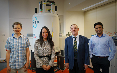
[Pictured above: Dr Tim Stait-Gardner, Dr Trang Pham (UNSW/Liverpool Hospital), Professor Bill Price and Dr Abhishek Gupta]


Cancer Council NSW has awarded a grant for over $430K to Western Sydney University researchers and an expert multi-institutional team to investigate the use of artificial intelligence (AI) to improve the effectiveness of radiation therapy for people living with cancer.

The world-first study will support the MRI-Linac, a next-gen radiotherapy technology developed by the NSW-based Ingham Institute for Applied Medical Research, combining an MRI scanner and a radiotherapy linear accelerator (Linac) into one integrated system.

In typical radiotherapy treatment, still images of the patient and their cancerous tumour taken prior to treatment are used to help plan and guide the direction of the radiation beam, but this radiation process can also damage normal tissues that are subjected to the radiation beam during treatment.

The MRI-Linac combines the technology of a Linear Accelerator and an MRI scanner, which can display real-time images enabling the monitoring of movement in tumour locations caused by normal functions like breathing or swallowing during treatment. The MRI-Linac can pinpoint parts within the tumour that are most active and aggressive, so a higher dose of radiation can be delivered to those areas.

In this study, MRI will be used to characterise cancer heterogeneity (differences among tumours and cancer cells), which can lead to cancer recurrence.

The multi-institutional research team is led by NIF Node Director Prof Bill Price and includes NIF Facility Fellow, Dr Tim Stait-Gardner and Research Fellow, Dr Abhishek Gupta from Western Sydney University; Dr Trang Pham, A/Prof Lois Holloway and Prof Erik Meijering from UNSW, as well as Prof Daniel Moses from the Prince of Wales Hospital. The research team also includes members from the Ingham Institute of Applied Medical Research, Auckland Bioengineering Institute and the University of Queensland.

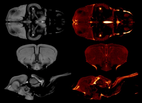
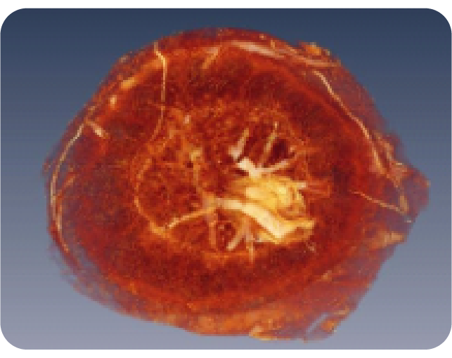
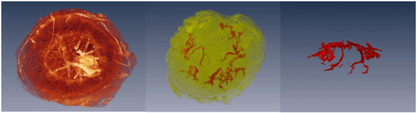
The team will use ultra-high-strength MRI scanners to produce microscopic resolution images of tumour samples. These highly detailed images will allow them to characterise the biological differences between tumours.

The team will then use deep learning, a specialised form of AI, to transfer this knowledge into clinical MRI scanners to enhance the resolution of imagery in MRI-Linac.

Prof Price said the research will allow clinicians to better predict the effectiveness of treatment and enable personalised care, with half of all cancer patients requiring radiotherapy.

“Radiotherapy is an important part of treatment for many cancer patients, however, in current practice it offers little capacity for personalised care,” Prof Price said.

“We have identified an opportunity to further enhance treatment by considering biological characteristics of an individual’s tumour with the help of AI.”

Prof Price said the implementation of this new enhanced imaging technology along with the precision of the MRI-Linac has the potential to greatly improve treatment outcomes and patient survival rates.


The study, ‘Targeting cancer heterogeneity with ultrahigh field MRI and radiotherapy using deep learning’, will be a collaboration between Western Sydney University, UNSW, Liverpool Hospital, Prince of Wales Hospital, Ingham Institute, University of Queensland, and Auckland Bioengineering Institute.

For more information on this study, contact NIF Node Director, Prof Bill Price.

Read Western Sydney University’s media release here.

Creating a Lizard Brain Atlas

Until recently, reptilian evolutionary studies lacked an important resource – a lizard brain atlas. As the subject of numerous ecological and behavioural studies, the Australian tawny dragon (Agamidae: Ctenophorus decresii) was an ideal candidate for creating a high-resolution MRI atlas of a representative scaled reptile (squamata). Such data is not only a resource for studies of the genus but also informs environmental decision making through an improved understanding of animal adaptation and evolution.

Read More

MRI investigations of placental structure and function

Preeclampsia is a medical condition affecting up to 3% of pregnant women in Australia. Characterised by high blood pressure and protein in the urine, it is a leading cause of morbidity and mortality in both mothers and infants. Furthermore, preeclampsia has been linked to long-term health consequences for both mother and child.

3-D reconstructions of the placenta from MRI images. (left) Foetal surface view of the placenta. (middle) Maternal surface view of the placenta with an overlay showing maternal vasculature. (right) Side view showing maternal vasculature alone

Hampering early diagnosis, prevention, and treatment efforts is a lack of understanding of preeclampsia pathophysiology.  Currently, the cause of this condition is unknown. Prof Annemarie Hennessy and a team of researchers at Western Sydney University are utilising the WSU NIF Node, in collaboration with NIF Fellow Dr Timothy Stait-Gardner, to learn more about this serious condition.

In this project, high-resolution magnetic resonance imaging is being used to examine placental changes in vivo in mouse models of preeclampsia. In addition to the in vivo studies, high-resolution scans of fixed mouse placentas, normal and abnormal, have been used to create a placental atlas. The creation of a placental atlas and a number of publications have provided important information on mouse models of preeclampsia, including its characterisation and how to differentiate between different models of preeclampsia from T2 maps of the mouse placentas. These works have provided some of the basis for investigations of new treatments of preeclampsia.

Publications:

This story was contributed by the Western Sydney University NIF Node. For further information, please contact Dr Timothy Stait-Gardner.

Grape split imaging to examine physical changes before and after splitting using diffusion magnetic resonance

Berry split is a condition in which the grape epidermis splits which often occurs during periods of high rainfall and is a significant cause of grape crop loss. In damp conditions there is increased uptake of water via osmosis and decreased water loss from transpiration. The pre-dawn turgor pressure of table grape cultivars lies in the vicinity of 5-38 kPa but prior to berry split can be as high as 1.5-3.7 MPa.
In order to examine and characterise the immediate effect of fruit split on grape, National Wine & Grape Industry and Western Sydney University node of National Imaging Facility have been collaborating on an ongoing project, which investigates the physical changes within the grape berry both before and after splitting using diffusion magnetic resonance imaging. Thirty-six table grapes of the Thompson Seedless variety were involved in the study and assigned to three groups: a control group (12 berries), a group in which they were wrapped in damp tissue (12 berries), and a total immersion group (12 berries). Five axial images (including diffusion tensor images) spaced evenly apart along the length of each berry were acquired simultaneously every hour to create a time-course study of each grape.
For each grape that split within the MRI during the study it was observed that there was an immediate change in the diffusion coefficient in the region of the wound. This region increased in volume over the course of the subsequent scans and correlated with regions of non-vital cells (as determined by fluorescence microscopy). It was determined from the study that grape berries left exposed to standing water after splitting exhibit greater cell death within the vicinity of the split. Therefore, the surface of split berries should be kept dry if possible to reduce further damage.


Collaborators

Nanoscale Organisation and Dynamics Group, University of Western Sydney
School of Medicine, University of Western Sydney
National Wine & Grape Industry Centre
NSW Department of Primary Industries

Sources

Dean, R.J., Bobek, G., Stait-Gardner, T., Clarke, S.J., Rogiers, S.Y. and Price, W.S. (2015), Time-course study of grape berry split using diffusion magnetic resonance imaging. Aust. J. Grape Wine R. doi: 10.1111/ajgw.12184
Dean R.J., Stait-Gardner, T., Clarke, S.J., Rogiers, S.Y., Bobek, G. and Price, W.S. (2014) Use of diffusion magnetic resonance imaging to correlate the developmental changes in grape berry tissue structure with water diffusion patterns. Plant methods 10(1):35


Privacy Settings
Youtube
Vimeo
Google Maps