Brain research supercharged by new ‘control panel’ accessible worldwide

The flood of data from brain research worldwide has now been channelled into an easy-to-use, open-access processing tool built to supercharge our understanding of the human brain.  

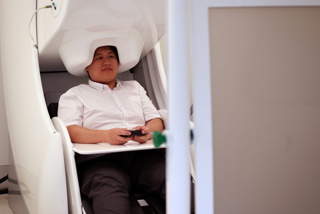
Neurodesk – a platform for processing, analysing and sharing massive datasets – has solved critical issues in the search for answers about brain disorders and diseases such as epilepsy, dementia, schizophrenia and traumatic brain injury. Neuroimaging data comes from modalities such as MRI, PET and MEG/EEG scans. 

Neurodesk was proposed at, led by and supported by National Imaging Facility partners including the University of Queensland, Swinburne University of Technology, and the Australian Imaging Service through University of Sydney.  

“It has removed a significant bottleneck that has hindered our ability to explore the complexities of the human brain, streamlining the processing of vast amounts of neuroimaging data,” says Dr Steffen Bollmann, project lead at the University of Queensland. 

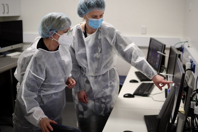
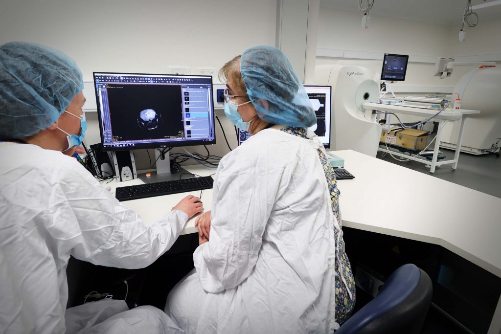
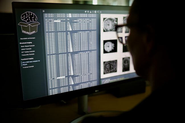
[Image: Members of the Neurodesk team, Aswin Narayanan, Thuy Dao, Dr Steffen Bollmann. Image credit: Dr Nicholas Hamilton.]

Both individual people and society stand to benefit: unravelling the brain’s mysteries helps us find cures for debilitating brain disorders, relieve people of the pain of untreated mental health issues and its societal cost, develop personalised medical treatments, inform public health policies, and understand human behaviour. 

Neurodesk is like the ultimate, seamless sci-fi control panel. Accessible through a user-friendly browser interface, it works across operating systems, pulling together different neuroimaging programs that run in all different ways, and is able to run giant research projects or small ones with its seamless access to computing power. 

Dr Bollmann sees Neurodesk’s accessibility as a leap forward in democratising neuroscience tools: “It empowers researchers worldwide to use open-source neuroimaging tools effectively and focus on their research questions, rather than grappling with technical obstacles.” 

“It is difficult to overstate how much benefit Neurodesk provides to our facility,” says Dr Tim Rosenow, NIF Facility Fellow and Neurodesk user at UWA’s Centre for Microscopy Characterisation and Analysis. “Researchers no longer need to be experienced in IT to perform their analyses, because software, dependencies, and conflicts are already handled.” 

However, the most important aim of Neurodesk’s creators was reproducibility: the process of other researchers replicating studies to check results and build a foundation of reliable scientific knowledge. The platform contains techniques that enable researchers to accurately replicate other teams’ analyses.  

Scientists can even share and publish their analyses, which can significantly reduce time and effort for collaboration and creating new knowledge. They can then tackle bigger neuroscience questions and analyse larger datasets. 

The ability to handle huge amounts of data is crucial, moving forward in brain research. Neurodesk gives researchers access to supercomputers and cloud computing to process neuroimaging data efficiently, letting them work with ‘biobanks’ of data (for instance, the UK Biobank holds imaging data from 50,000+ people). 

“By simplifying neuroimaging, fostering collaboration and empowering scientists to handle huge datasets, this breakthrough not only advances our understanding of the healthy human brain, but also sheds light on the intricacies of various brain disorders,” says Dr Bollmann, “promising improved diagnostics and treatments in the future.” 

Read the new paper published in Nature Methods here. 

Victorian collaboration raises over $50m investment in critical imaging capabilities

[Image: La Trobe University – Olivia Newton-John Cancer Research Institute NIF Fellow, Dr Ingrid Burvenich with Minister Tierney]

The Victorian Government has invested $14.83m in National Imaging Facility’s (NIF) research infrastructure in Victoria, in partnership with the Victorian Biomedical Imaging Capability (VBIC), equating to a boost of just over $50m through collaborative co-investment. 

[Image: Victorian Minister for Higher Education, the Hon Gayle Tierney MP]

Victorian Minister for Higher Education, the Hon Gayle Tierney MP visited the Olivia Newton-John Cancer Research Institute on Tuesday, to highlight the impact of the collaboration and the State Government’s investment of through the Victorian Higher Education State Investment Fund (VHESIF) initiative.  

“Collaborative projects such as this demonstrate how our government is supporting higher education and industry to become international leaders in their field,” Minister Tierney said.

[Image: Victorian Minister for Higher Education, the Hon Gayle Tierney MP toured the facilities at the Olivia Newton-John Cancer Research Institute and the Austin hospital]

The funding is supporting the upgrade and expansion of imaging capabilities across NIF’s research facilities in Victoria, including The Florey, La Trobe University and the Olivia Newton-John Cancer Research Institute, Monash University, Swinburne University of Technology, and University of Melbourne in partnership with Peter MacCallum Cancer Centre and the Austin Hospital.  

Critical medical research in areas of national priority such as dementia cancer and epilepsy, as well as agriculture research will be enabled by the co-investment, which includes $26.7m from NIF through the Australian Government’s National Collaborative Research Infrastructure Strategy (NCRIS) funding scheme. 

Infrastructure funded under the collaboration includes human magnetic resonance imaging (MRI) capabilities at Swinburne University of Technology and The Florey, where state-of-the-art high-intensity focused ultrasound will support the development of new treatments for essential tremor and tremor-dominant Parkinson’s disease. 

In addition to this, the University of Melbourne upgraded their ultra-high-field 7T MRI (one of only two in Australia), and acquired a new human PET-CT. 

Preclinical capabilities including PET/MRI and PET/CT to support important drug discovery and testing have been installed at the Olivia Newton-John Cancer Research Institute/La Trobe University and Monash University. 

[Image: NIF preclinical capabilities at the Olivia Newton-John Cancer Research Institute]

A new research cyclotron at Monash (the Australian Precision Radiopharmaceutical Facility APRF) will enable the production of radioisotopes under GMP standards, and enhance Australia’s sovereign capability to produce therapeutics and diagnostics. Complementary to this, radiochemistry hotcell infrastructure upgrades at Monash, the Austin Hospital, and Peter MacCallum Cancer Centre will support the design and development of novel cancer treatments.  

[NIF radiochemistry capabilities at the Austin hospital/Olivia Newton-John Cancer Research Institute]

The funding also enables upgrades to the magnetoencephalography (MEG) at Swinburne, one of only two systems in Australia, supporting the study of brain function. 

[Image: NIF MEG at Swinburne University of Technology]

Development of specialised plant imaging capabilities at the University of Melbourne will underpin research into the effect of climate change on crops and soil, and strategies and applications for agricultural improvements to support Australia’s standing as a world leader in food and beverage production. 

NIF Chief Executive Officer, Prof Wojtek Goscinski said the co-contributed investment underpins transformational initiatives in a number of national priority areas including precision medicine, molecular imaging, drug discovery, diagnostics and plant soil imaging. 

“It’s a privilege for NIF to partner with the Victorian Government and VBIC to support Australia’s strategic science and research priorities” Prof Goscinski said. 

“These capabilities will support Australia as a world-leader in applying advanced imaging technology, resulting in better healthcare, better products, and important discoveries.”

Read the Victorian Government’s announcement here. 

Victorian imaging network meets to map out innovative future

[Pictured: VBIC Annual Network Meeting guest speakers Prof Lindy Fitzgerald and Prof Amy Brodtmann] 

Innovation, industry partnerships and commercialisation will be among the topics discussed at a meeting bringing together NIF’s capabilities from around Victoria on November 24. 

The annual Victorian Biomedical Imaging Capability (VBIC) network meeting will attract researchers, clinicians and industry partners to Monash Biomedical Imaging in Melbourne. 

Participants will represent NIF nodes at the University of Melbourne, Monash, Swinburne, the Florey, Olivia Newton John Cancer Research Institute and La Trobe University. 

Neurologist Prof Amy Brodtmann will draw on her interests in imaging, stroke and dementia to present research findings from the Cognitive Health Initiative at Monash and Alfred Health  

Guest speaker Prof Melinda Fitzgerald from Curtin University and the Perron Institute will present on a national initiative she heads as CEO, called Connectivity, the Mission for Traumatic Brain Injury.  The initiative includes use of MRI and clinical biomarkers in a national trial to improve the diagnosis and prognosis of traumatic brain injury. 

Sessions at the meeting will also cover the work of imaging experts in research programs employing ultra-high field MRI, CT and PET; as well as presentations from experts in industry partnering and commercialisation. 

NIF Chief Executive Officer Prof Wojtek Goscinski said the meeting would provide a platform for early career researchers and emerging leaders, a showcase for new-generation imaging, and opportunities for important in-person networking. 

It would also enable discussion on the NIF Imaging Roadmap, including supporting innovation and ensuring Australia’s international comparative advantage, Prof Goscinski said. 

The roadmap will add to the substantial impact and jobs that VBIC and NIF have already delivered, with a recent report estimating more than $350 million in economic activity for Victoria. 

VBIC nodes have grown to employ more than 150 FTE imaging staff, and partner with more than 90 organisations, including Austin Health, CSIRO, Melbourne Health, Mental Health Research Institute and the Peter MacCallum Cancer Centre. 

Capital investments have reached $37 million, providing access to human and preclinical MRI and PET-CT, preclinical DEXA scanners and confocal endomicroscopy, as well as magnetoencephalography and nuclear scintigraphy. 

A massive $235 million in major grants has been secured, enabling new research projects in cancer, infection and inflammation, brain function, epilepsy, dementia and even long-term aspirin use. 

Click here for more information about the VBIC network meeting and full programme.

The Australian Epilepsy Project

The Australian Epilepsy Project (AEP) will change the lives of people living with epilepsy by reducing uncertainty surrounding diagnosis and fast-tracking the path to optimal treatment using the combination of advanced imaging, genetics, cognition, and artificial intelligence. Such improvements will result in better outcome prediction at disease onset, a higher rate of seizure freedom, reduced economic burden of disease and will increase life-participation of people with epilepsy. 

Read More

Catching the wave

Catching the wave: non-invasive MEG/EEG for epilepsy seizure localisation

Over 250,000 Australians currently live with epilepsy, a chronic disorder of the brain. In addition to significant social, physical and psychological consequences, epilepsy is linked with an increased risk of death compared to the general population. Up to 70% of epilepsy cases may be controlled through lifestyle and medication; however, many patients are unable to be treated using conservative measures. A major clinical problem, drug-refractory focal epilepsy may require aggressive treatments, including surgery, to resect a portion of the brain.

The localisation of the epileptogenic zone (the portion of the brain requiring resection to reduce seizures) is typically carried out clinically using magnetic resonance imaging (MRI). In many cases, no abnormalities are detected, requiring further investigation. Surgeons may also use invasive surgically implanted electrodes (intracranial electrode depth or grid, or ICEEG) for seizure source localisation, increasing the risk to the patient. A lower risk option is further functional imaging with positron emission tomography (PET), single-photon emission computed tomography (SPECT), and simultaneous electroencephalography functional MRI (EEG-fMRI). Unfortunately, many of these modalities lack temporal resolution to distinguish seizure onset.

“Catching the earliest wave, the earliest is key”
Representation of MEG (green) and EEG (blue) overlays.

Magnetoencephalography (MEG) and electroencephalography (EEG) offer a non-invasive method to detect rapidly evolving brain wave spikes and seizures at millisecond temporal resolution.  A/Prof Chris Plummer and team, Mr Simon J. Vogrin, Dr William P. Woods, A/Prof Michael A. Murphy, Prof Mark J. Cook, and Prof David T.J. Liley, have developed a world-class multimodal technique for non-invasive source localisation and epileptogenic zone characterisation. The method (using co-acquired MEG and high-density EEG) has already improved the surgical treatment of medically refractory epilepsy, with clinical validity demonstrated in a key 2019 publication.

The team utilised the world-class MEG at the SUT NIF Node together with the expertise of NIF Facility Fellow Dr Will Woods to look at the efficacy of concurrent HDEEG (High-Density EEG > 64 electrodes) and MEG for identifying the epileptogenic zone. Ninety-six patients have been involved in the long-term prospective study. Seventy-one patients were MRI negative and twenty-five MRI positive with either complex lesions or multiple lesions. More than five years’ data collection in collaboration with neurologists and medical scientists found that the combined method was able to detect interictal spike discharges (between seizures) at early, mid and late-phase as well as ictal discharges (during seizures).  The earliest resolvable localisation by either EEG or MEG was determined to be the most suitable site for surgical resection.

“We find that for most cases, modelling either the mid-upswing or the later peak (still common practice) results in mislocalization of the cortical generator (and the putative Epileptogenic Zone) due to the effects of discharge propagation across the cortical surface. We look for the earliest feature from the discharge; sometimes it appears first in the Magnetometer, sometimes it is first in one of the Gradiometers, and sometimes it is first in the HDEEG. For a given patient, there is typically a dominant leading modality, but the modality that leads in time will vary from patient to patient. This is fundamentally the reason for recording both. Otherwise, you risk having half the story before diving into invasive investigations.”  – A/Prof Plummer

To date, successful source localisation has guided successful surgical outcomes in 25 patients, 8 of whom were spared invasive intracranial monitoring due to this method. Many of the over 100 studies performed are yet to progress to surgery, underpinning the latent potential of yet to be followed complete surgical workup and seizure-freedom outcomes following surgical therapy.

“Accurate, non-invasive localisation with this method will reduce the medical burden of invasive intracranial electrode monitoring by reducing the number of invasive electrodes required and, in some cases, remove the need for these in the first place to allow the patient to proceed directly to potentially curative surgery.” – A/Prof Plummer

This work intends to revolutionise epilepsy management in Australia, with data from a larger and more diverse cohort expected to provide evidence for a Medicare rebate to facilitate the accessibility of this technique for Australians.

Success stories:

From 500 seizures per month to none; MEG/EEG guided surgical resection success

A young man with a five-year history of disabling seizures was suffering up to 500 seizures per month; his schooling was disrupted, and all the standard non-invasive tests gave conflicting results. Surgical resection was performed correlating with the earliest EMSL result. The patient is now seizure-free and on minimal medication following surgery.

A 10-year old child had a four-year history of left leg motor-sensory events, up to 200 times per day, leading to falls. PET and MRI suggested localisation of seizures in the right superior frontal gyrus and the right paracentral lobule, while EMSL results showed seizure onset further to the rear at the precuneus. Surgical resection of the paracentral lobule and superior frontal gyrus did not relieve the seizures seen by ICEEG during surgery. Further exploration revealed a second pocket of dysplasia in line with the EMSL result, resection of which normalised the ICEEG and relieved the patient of seizures.

A 36-year-old patient presented with speech-arresting complex partial turns with a typical MRI result. Their surgical plan was altered to include a hippocampal depth electrode, where seizures were found to start by MEG. Standard anteromesial temporal lobectomy revealed a cortical dysplasia involving the entorhinal cortex. The patient is now seizure-free and works as a speech therapist.

Recovering day to day activities without disturbing the complex dysplasia

A 36-year old mother of two young children suffered a 33-year history of multiple-daily disabling motor seizures. She was denied surgery due to a complex lesion seen by MRI (frontoparietal dysplasia).  EMSL captured the seizure onset, targeting surgical resection without interfering with the bulk of the lesion. The patient is now on reduced medication and experiences only brief non-disabling focal seizures. She states that she “has her life back”.

You can learn more about epilepsy here or by speaking to your GP. This work was contributed by St Vincent’s Hospital, Swinburne University of Technology, and the University of Melbourne. For more information, please contact Dr Will Woods.

Privacy Settings
Youtube
Vimeo
Google Maps