NIF – ACRF collaboration launches Australian-first facility for new cancer therapies

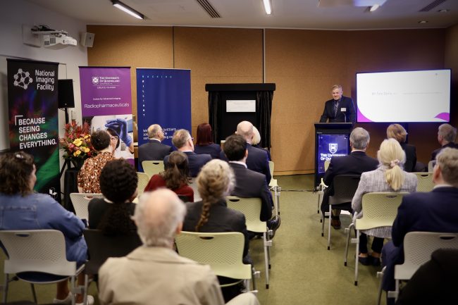
Queensland’s Assistant Minister for Health and Regional Health Infrastructure, the Hon Brittany Lauga MP joined the University of Queensland’s Vice-Chancellor and President Prof Deborah Terry AO and Chair of the Australian Cancer Research Foundation (ACRF), Mr Tom Dery AO to officially open the ACRF Facility for Targeted Radiometals in Cancer (AFTRiC) at the Centre for Advanced Imaging (CAI) in Brisbane today.

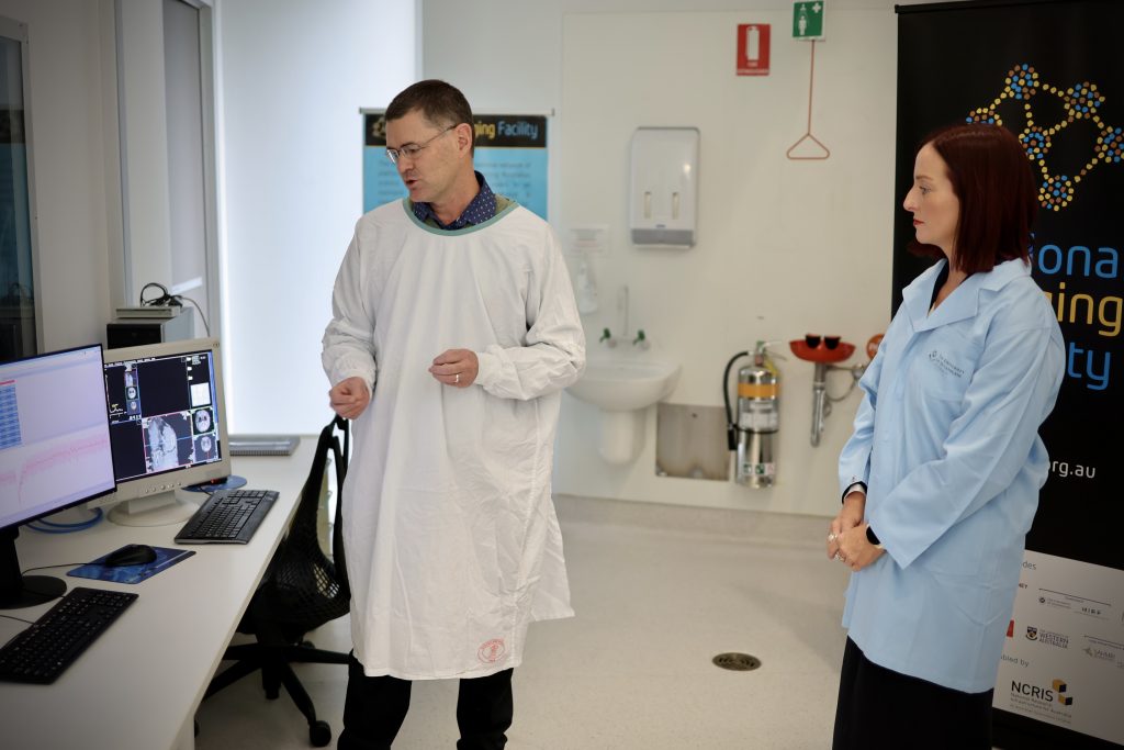
[NIF Fellow Dr Gary Cowin demonstrating the capabilities of the new Facility, with Assistant Minister for Health and Regional Health Infrastructure, the Hon Brittany Lauga MP]

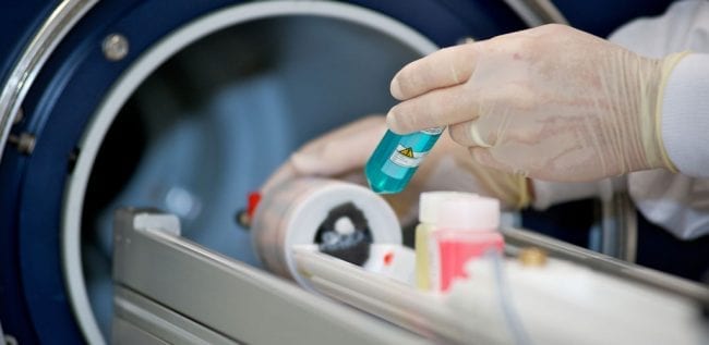
AFTRiC will be Australia’s first facility for the discovery, development and clinical application of novel alpha particle-based cancer therapeutics, as well as other radiometal and molecular imaging agents.

The Facility has been co-funded by the National Imaging Facility (NIF) ($1.2m) in partnership with the ACRF ($1.2m), the Ian Potter Foundation ($180k) and the University of Queensland.

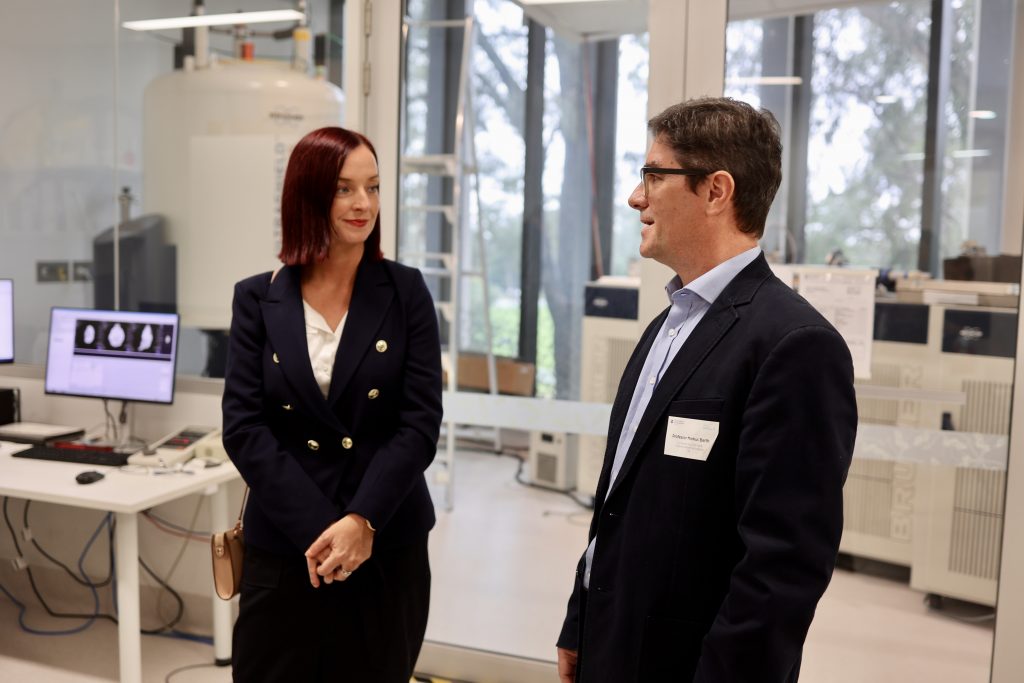
[Assistant Minister for Health and Regional Health Infrastructure, the Hon Brittany Lauga MP with UQ NIF Node Director Prof Markus Barth]

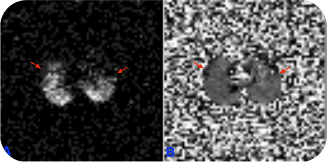
AFTRiC will support the advancement of cancer theranostics (combined therapy and diagnostics in imaging), using cancer-seeking molecules attached to alpha particles to deposit high-energy radiation to cancer cells, without impacting healthy tissue.

NIF Chief Executive Officer, Prof Wojtek Goscinski said it was a privilege to partner with the University of Queensland, the ACRF and the Ian Potter Foundation to boost Australia’s open-access alpha particle research capabilities.

“Our investment in AFTRiC aligns with NIF’s commitment to grow our capability to help researchers and industry use alpha particles to produce and test new-generation theranostics,” Prof Goscinski said.

“The development of new theranostics through AFTRiC will support this fast-growing area of significant healthcare innovation, allowing doctors to ‘see what they treat’ by combining diagnosis and treatment to improve cancer therapy and outcomes.”

CAI Deputy Director (Research) Prof Kris Thurecht said several pieces of AFTRiC equipment had already been installed at CAI and used to evaluate first-in-class radiopharmaceuticals, building a strong case for the development and clinical translation of new cancer drugs.

“Radiopharmaceuticals and theranostics have been identified by all levels of government as a next-generation research priority, and AFTRiC firmly positions us as one of the country’s leading capabilities in this space,” Prof Thurecht said.

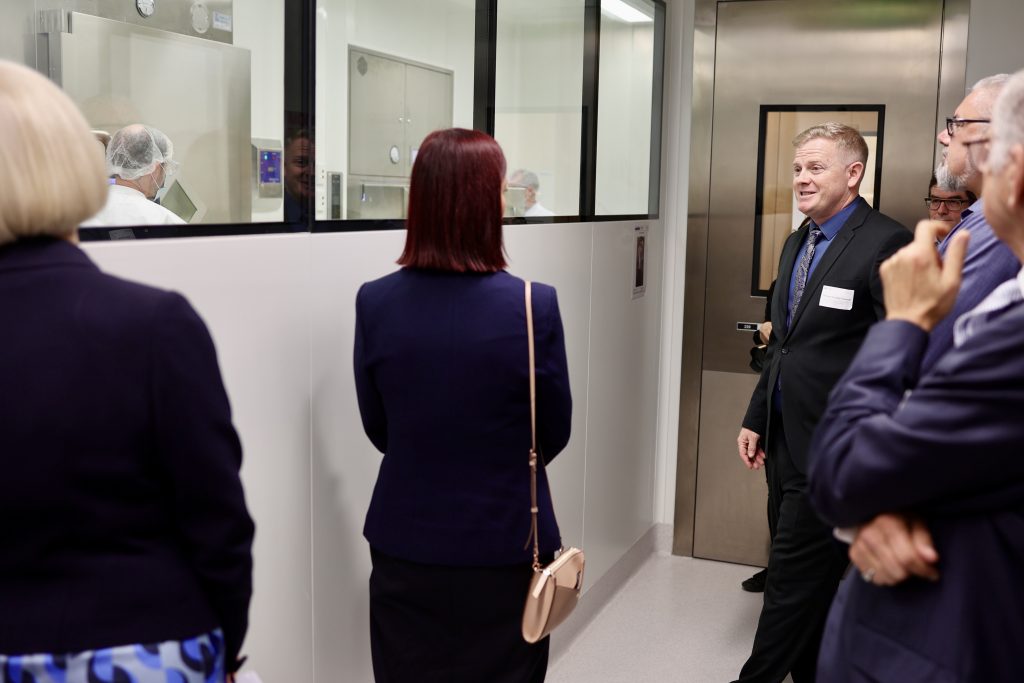
[CAI Deputy Director (Research) Prof Kris Thurecht lead a tour of the new Facility]

“We will be one of the few places in the country that can produce these specialised radiopharmaceuticals and, in collaboration with our industry partners, we will evaluate and hopefully develop clinical grade product for clinical trials.”

AFTRiC is one of two NIF investments underpinning a national capability for targeted alpha particle therapeutics. The other is establishing a new facility at ANSTO’s Lucas Heights campus to enable the development and translation of alpha-emitting radiopharmaceuticals, having access to radioisotopes produced in the OPAL research reactor.

NIF Molecular Imaging and Radiochemistry Showcase to be presented at ANZSNM

National Imaging Facility enables access to imaging capabilities across the country and will present a Molecular Imaging and Radiochemistry Showcase at ANZSNM 2022, featuring presentations from a range of research leaders from Australia’s advanced imaging network.

See the full ANZSNM program here.

Register to attend ANZSNM 2022.

National Imaging Facility: Molecular Imaging and Radiochemistry Showcase
Saturday 14 May 2022, 3:15pm – 4:15pm
Session Chair: Prof Wojtek Goscinski, CEO National Imaging Facility

TimeSpeakerTopic
3:15 – 3:20Professor Wojtek Goscinski

Chief Executive Officer
National Imaging Facility

Introduction to NIF Molecular Imaging and
Radiochemistry Showcase

3:20 – 3:30Professor Steven Meikle

Head of the Imaging Physics Laboratory, Brain and Mind Research Institute, University of Sydney

Total Body PET
3:30 – 3:40Associate Professor Roslyn Francis

Head of Department of Nuclear Medicine and WA PET Service, Sir Charles Gairdner Hospital, University of Western Australia

Radiochemistry activities in Western Australia
3:40 – 3:50Professor Gary Egan

Professor and Foundation Director, Monash Biomedical Imaging

Director, ARC Centre of Excellence for Integrative Brain Function

Australian Precision Medicine Enterprise
3:50 – 4:00Prof Kristofer Thurecht

Acting Deputy Director (Research Technologies) and Group Leader – Principal Research Fellow,

Centre for Advanced Imaging, University of Queensland

Affiliate Principal Research Fellow and Group Leader,

Australian Institute for Bioengineering and Nanotechnology

Alpha therapies and activities
4:00 – 4:10Dr John Bennett

Research Infrastructure Platform Leader – Biosciences,
ANSTO

ANSTO’s new NIF Alpha Radioisotopes and
Radiopharmaceuticals Facility

NIF’s Professor Fernando Calamante elected President of the International Society for Magnetic Resonance in Medicine

National Facility of Imaging’s (NIF) Co-Director of the University of Sydney/ANSTO joint node, Professor Fernando Calamante, has been elected President of the International Society for Magnetic Resonance in Medicine (ISMRM). ISMRM draws on a multidisciplinary membership of over 9,000 clinicians, physicists, engineers, biochemists, and technologists who contribute to discovery, innovation and clinical translation in magnetic resonance.

Prof Fernando Calamante

Professor Calamante’s research is at the forefront of his field and includes the development of novel methods for Diffusion MRI, Perfusion MRI and brain connectivity, and their applications to neurology and neuroscience. He has gained international recognition for his MRI methods including his contribution to the MRtrix software for Diffusion MRI analysis, which is considered one of the most widely adopted tools in the field.

“I feel greatly honoured to have been elected to this role. The ISMRM has played such an important part of my research career and has been the source of so many collaborations and inspiration to my research; it is a real privilege to be able to play my part in contributing to ensuring the ISMRM continues to deliver its vision. These are challenging times, and it will be interesting to see how Societies such as ours react, adapt and evolve in the face of the challenges COVID has given us.” Professor Calamante said.

Professor Calamante is the first researcher from outside Europe or North America to be elected as ISMRM President.

 “It is a great milestone for the ISMRM, and testimony of its international nature and diversity. Having come originally from Argentina and now living in Australia for the last 16 years, I feel I can fly the international flag as ISMRM President. As the first ‘rest of the world’ President, I will do my best to increase the presence of the ISMRM in under-represented regions,” Professor Calamante said.

Professor Calamante is the Director of Sydney Imaging, Core Research Facility at the University of Sydney and Professor in the School of Biomedical Engineering, Faculty of Engineering.

NIF Chief Executive Officer, Professor Graham Galloway said he was delighted that Professor Calamante was elected ISMRM president and has been recognised for his expertise in magnetic resonance. ISMRM is recognised by the MR community as the premiere organisation for sharing of discovery and applications and driving the development of MR technology. It brings together basic scientists, engineers, clinicians and industry.  Professor Calamante’s election demonstrates that NIF is at the forefront of leading-edge imaging instrumentation and expertise in imaging technology.

National Preclinical PET QA

The NIF Molecular Imaging & Radiochemistry (MIR) Theme is a group of NIF Fellows, Directors, and users of NIF facilities that focus on state-of-the-art radiochemistry and molecular imaging applications using PET, SPECT, and MRI.

Integrating preclinical PET systems into a national resource requires the development of defined QA programs to monitor and integrate the data from individual systems. Hence, the MIR Theme initiated a national quality assurance (QA) program for the NIF preclinical PET instruments.

Read More

Hyperpolarized 129Xe MR imaging of lung

Magnetic Resonance Imaging (MRI) has a range of applications in medical diagnosis, and more than 25,000 scanners are estimated to be in use worldwide. However, lung imaging suffers from some technical challenges, limiting its application in pulmonary disease diagnosis and treatment.


Due to low proton density, movement and high susceptibility difference between air and tissue, conventional proton MRI struggles to image lung tissue and function. These can be partially overcome by the introduction of contrast agents into the lung. Hyperpolarised gases are promising contrast agents for imaging lung structure and function. The two most common gasses are helium (3He) and xenon (129Xe) isotopes. Helium isotopes are challenging and expensive to obtain, and do not provide significant functional readouts. Xenon can be challenging to work with, but promises novel physiological measurements not previously feasible. This resulting technique, termed hyperpolarized 129Xe MR imaging, has revolutionised the field of functional lung imaging.


Five years ago, an Australian Research Council (ARC) grant was awarded to researchers at Monash Biomedical Imaging (MBI) and ANSTO to design and build a machine that could reliably produce hyperpolarized xenon. In this project, Dr Wai Tung Lee and NIF Facility Fellow Dr Gang Zheng have teamed up to develop a new multimodal technology capable of high-quality investigations of the human lungs. While Dr Lee, of ANSTO, was responsible for the construction of the polarizer and production of polarized 129Xe for MR imaging, Dr Zheng, of Monash University, designed the MR experiments and adapted imaging protocols.

Figure 1. Schematic diagram of the polarizer system. Note: Some details omitted for clarity.

Hyperpolarized 129Xe (HP-Xe) gas was generated in a custom designed and constructed SEOP system at Monash Biomedical Imaging (Figure 1 and Figure 2). The system consists of a gas-polarizing unit and a gas injection and recycle unit. The core component of the gas-polarizing unit is an optical pumping cell (OPC) where the gas is polarized. The OPC is placed in an oven to regulate the amount of Rb vapour by maintaining a constant temperature between 60ºC-90ºC. The optical pumping process uses two 240W narrow-bandwidth lasers tuned to the Rb D1 transition wavelength of 794.7nm. Additional optics condition the laser light to circular polarization and shape the laser profile to fully illuminate the OPC. HP-Xe is produced in batches rather than using a continuous-flow process.

Figure 2. The spin-exchange-optical pumping system. A. Gas polarizing unit in Room 1; B. Gas injection and recycling unit in Room 2.

MR experiments were performed on a clinical 3T whole-body system (Skyra, Siemens Medical Solutions, Erlangen, Germany) with a broadband RF amplifier. HP-129Xe MRI was enabled using a bird-cage transmit/receive chest coil (RAPID Biomedical GmbH, Wuerzburg, Germany). A 2D-GRE sequence for X-nuclei MRI was used to image HP-Xe gas in lung. Proton channel images were acquired for the localization of the lung. The resonance frequency of 129Xe was set to 34.09 MHz on the 3T scanner. The human subject first quickly flushed his lung with pure nitrogen, and then immediately inhaled HP-Xe in the Tedlar bag. After inhalation of HP-Xe, the subject held their breath during the scan. The total imaging time was less than 10s.


Dr Zheng outlines the results to date, “We have successfully imaged gas-phase HP-129Xe in a range of phantoms, such as the Tedlar bag, syringe and the glass cell (Figure 3). We repeated the gas-phase imaging in both ex-vivo (Figure 4) and in-vivo lamb lungs (Figure 5). Imaging results supported that our polarizer can provide sufficient polarization for lung imaging. In 2019, we successfully performed a human experiment and a gas signal from the lungs was clearly identified (Figure 6). In addition, all studied showed a clear Rf peak of gaseous and dissolved xenon which should allow us to develop methods to assess lung function. We plan to study human pulmonary disease in the near future and hope to find additional collaborators to help translate this modality into clinical use.”

Gallery Notice : Images have either not been selected or couldn't be found

Construction of the hyperpolarizer is complete; it can routinely produce HP-129Xe for research use. The technique has successfully imaged HP-129Xe in phantoms, ex-vivo and in-vivo lamb lungs and a human lung. If you are considering lung research, please see more on the Monash website, or get in touch with Dr Gang Zheng to discuss your project needs.


Publications:

Zheng G, Lee WT, Tong X, et al., A SEOP Filling Station at the Monash Biomedical Imaging Centre. Conference on polarization in Noble gases (PiNG), 2017.


Zheng G, Lee WT, Tong X, et al., Phase imaging of hyperpolarized 129Xe gas in a human lung. ISMRM 2020, submitted on 6 Nov 2019.


Acknowledgements:

National Imaging Facility, ARC (Grant LE130100035); NHMRC (Grant APP606944); CASS Foundation; Monash Biomedical Imaging, ANSTO


This story was contributed by Dr Gang Zheng of the Monash University NIF Node.


Privacy Settings
Youtube
Vimeo
Google Maps