4D MRI to measure fetal blood flow
The fetus is remarkable in that it grows and develops in an environment of physiological hypoxaemia and hypoglycaemia relative to the mother.
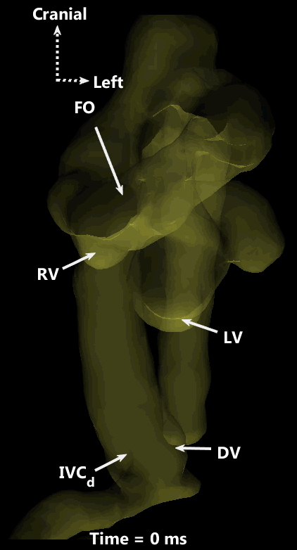
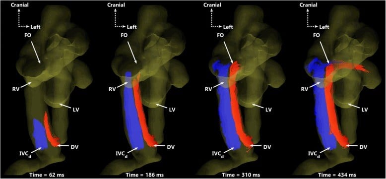
However, the fetus thrives in this environment because the sharp concentration gradient between the mother and the fetus ensures that both oxygen and glucose diffuse across the placental membranes and into the blood of the fetal umbilical vein, through the fetal liver to the heart and brain. While some of this oxygen and glucose-rich blood perfuses the liver, more than half flows directly to the heart through the ductus venosus. Remarkably, this oxygen and glucose-rich blood does not mix with blood returning from the lower body of the fetus but is directed in the posterior left side of the inferior vena cava in a phenomenon known as streaming. As a consequence of this streaming effect, and despite the fetus being hypoxemic, there is a relatively high oxygen and glucose delivery to the brain, which is essential for healthy brain development.
Thus, understanding how the tone (degree of constriction) of the ductus venosus regulates streaming is vital in understanding how the fetus maintains the delivery of oxygen and glucose to the brain where these substrates are in short supply.
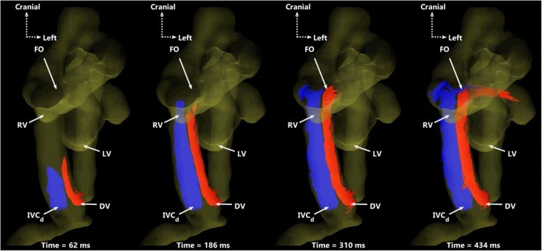
Professor Janna Morrison and the Early Origins of Adult Health Research Group (University of South Australia) have, in collaboration with researchers at Sick Kids Hospital (Toronto, Canada), validated the use of 4D MRI to measure fetal blood flow, even in the smallest and most intricate parts of the fetal circulation. This technique has led to a better understanding of the cardiac streaming of oxygen and nutrient-rich blood toward the brain during fetal life (Schrauben et al., 2019). Professor Morrison and her team are now using this technique in combination with pharmacological agents to understand better how the tone of ductus venosus is regulated as part of an ARC Discovery Project. Understanding the intricate mechanisms behind ductal tone may lead to intervention strategies and more targeted therapies in situations where fetal substrate supply is restricted such as intrauterine growth restriction.
This story was contributed by Dr Jack Darby of the University of South Australia. For inquiries, please contact Mr Raj Perumal or NIF Central.
For more details and to see additional animations, please check:
Schrauben EM, Saini BS, Darby JRT, Soo JY, Lock MC, Stirrat E, Stortz G, Sled JG, Morrison JL, Seed M & Macgowan CK. (2019). Fetal hemodynamics and cardiac streaming assessed by 4D flow cardiovascular magnetic resonance in fetal sheep. Journal of cardiovascular magnetic resonance: official journal of the Society for Cardiovascular Magnetic Resonance 21, 8.


