Michael de Veer

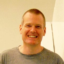
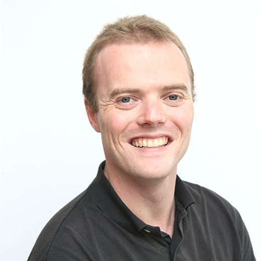
Dr de Veer obtained his PhD in Biochemistry at Monash University and has extensive post-doctoral experience in signalling pathways, linking tracers and mapping cell migration and immune modulation within the lymphatic system. A large portion of his research is conducted with industry partners and he understands the interface between academic and translational research. He is currently leading the pre-clinical imaging team at Monash Biomedical Imaging and brings extensive experience with the large animal and rodent models that our users want to image. His own imaging research is aimed at developing MR and PET-MR imaging agents to delineate the lymphatic system in pre-clinical models of oedema and disease. He is an advocate for the benefits and impact of incorporating multi-modal imaging into projects to produce excellent research outcomes. A large part of his role is to facilitate industry engagement with the facility and drive translational research.

Will Woods

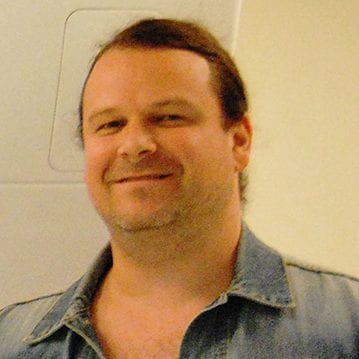
Dr Will Woods received his degree in Applied Mathematics in 1991 and his PhD in 1995 in Chemical Engineering, both from the University of Newcastle upon Tyne, UK. From 1995 to 1997 he held an EU/JSPS Fellowship in the Department of Applied Mathematics at Kyoto University, Japan. He then worked on applications of minimal realisations of bilinear models to networks of neurons in the Department of Physiology in Newcastle upon Tyne, before moving to the University of York, UK in 2004 to facilitate the setup and management of only the second MEG lab in the UK. Since 2010 he has held the position of Senior Lecturer in Imaging at Swinburne University, Melbourne. His main research interests are applications of graph theoretic methods to neuroimaging data and inverse problems in MEG

Matthew Hughes

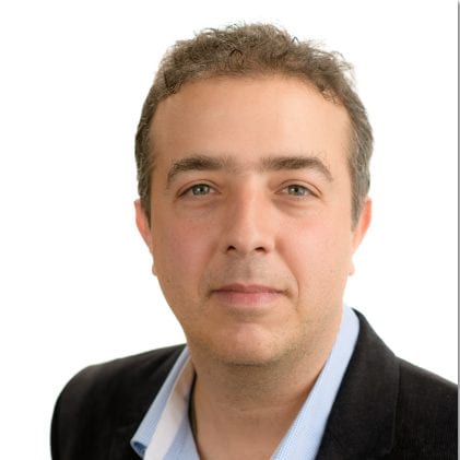
Dr Matthew Hughes supports users on the 3T MRI. Dr Hughes provides support for a number of projects within Swinburne as well as collaborative projects.

Karine Mardon

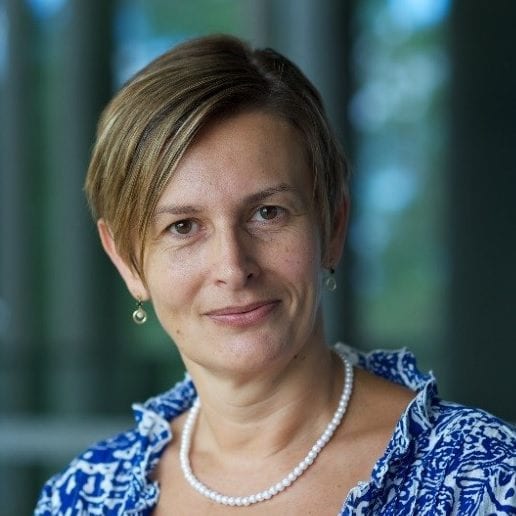
Dr Karine Mardon obtained her PhD in radiopharmacology from University Paris XII in 1994; she then moved to Australia to pursue postdoctoral studies at ANSTO in the Radiopharmaceutical Division and worked there for 5 years in the development and characterisation of radiopharmaceuticals for SPECT and PET. She has extensive experience with in vitro and in vivo preclinical research, particularly in the evaluation of drugs developed for the study of movement disorders as well as in the evaluation of radiolabelled peripheral benzodiazepine receptor ligands as markers of neurodegeneration and tumour occurrence. She moved to Brisbane in 2000 and joined the University of Queensland where she developed further experience working in the field of preclinical drug development in the ADME division of TetraQ. She joined the Centre for Advanced Imaging in October 2010 as NIF Facility Fellow for preclinical PET/CT.

Gary Cowin

Dr Cowin develops new MR based projects on Bruker (9.4T large animal, 16.4T small animal micro-imaging system and 7T ClinScan systems) and Siemens human MRI systems (1.5T and 3T clinical and 7T research systems) for Human and Animal studies. He is also developing simultaneous PET and MRI imaging on the world’s first commercial prototype of a preclinical combined PET/MRI system. He has extensive experience investigating fat localisation and mobilisation, extending image resolution on ultra-high field Animal (16.4T) and Human (7T) MRI systems, prostate, liver and spinal cord research including spectroscopy, diffusion and anatomical imaging. Dr Cowin is experienced in various aspects of MRI QA, including effects of gradient non-linearity on image quality. He is developing hyperpolarised gas lung imaging in both human and animals, obtaining the first Helium images in Australia.

Oren Civier

Dr Oren Civier received his PhD from Boston University, where he used computational modelling to study cortical and subcortical mechanisms of speech production. After his PhD, Oren investigated white matter anomalies in speech disorders using diffusion MRI (DTI), and the neural control of oral and hand movements using MEG. Leveraging those qualifications and industry experience in Computer Science, Oren’s focus is on developing multimodal neuroimaging analysis tools based, in part, on the MRtrix software, including diffusion MRI combined with fMRI (continuing from his previous position at the University of Sydney), and diffusion MRI combined with MEG (current position). These efforts parallel his research interest in elucidating the brain’s connectivity network on multiple dimensions (conduction speed, throughput, redundancy, etc.) in order to develop more accurate diagnoses and more targeted/personalised treatment of brain disorders. Some additional research methodologies and techniques Dr Civier has used throughout his career are tDCS, electromagnetic/camera-based motion tracking, and auditory/visual feedback manipulation.

Rob Smith

Dr Robert Smith completed Bachelors degrees in Applied Physics and Electronic Engineering at RMIT University in 2008 and has since completed a PhD and subsequent post-doctoral research at the Florey Institute of Neuroscience and Mental Health. His research focuses on the development of advanced processing and reconstruction methods for the analysis of diffusion MRI data, which enable robust inference of brain white matter connectivity and derivation of / inference on quantitative measures in the presence of complex white matter fibre configurations. These developments, in addition to other advanced data processing tools, are made freely available to the research community as part of the software package MRtrix3, for which extensive community support is provided via online forum and software teaching workshops. Dr Smith additionally provides support for the two 3T human MRI systems installed at the Melbourne Brain Centre in Heidelberg, which includes the recent Siemens Prisma scanner upgrade.

Tom Close

Dr Thomas Close specialises in neuroinformatics and computational neuroscience. He obtained a PhD investigating advanced techniques in diffusion MRI tractography of cerebral white matter and has completed a post-doctoral position in the Computational Neuroscience Unit of Okinawa Institute of Science and Technology Graduate University in Japan. Tom provides support for the imaging operations at Sydney Imaging. Tom is actively collaborating with other NIF Informatics Fellows on the national Trusted Data Repositories Project and has developed a deployed exemplar utilizing the XNAT platform.

Andrew Mehnert

Dr Andrew Mehnert is Senior Lecturer in Data Management, Analysis and Visualisation at the Centre for Microscopy, Characterisation and Analysis (CMCA) at the University of Western Australia (UWA). His position is jointly funded by NIF and Microscopy Australia. He provides informatics support (data management, analysis and visualisation) to, and collaborates with, CMCA researchers across various disciplines and microscopy and imaging techniques. Andrew’s research interests focus on the development of image analysis methods for biomedical imaging applications involving MRI, CT and optical microscopy. Such methods include visualisation, denoising, spatial co-registration, segmentation, parametric modelling of contrast enhancement (DCE-MRI), computational diffusion MRI and classification (feature extraction, feature selection and classifier design, training and validation).

David Abbott

Professor David Abbott is a physicist-neuroscientist with over twenty years’ experience in neuroimaging informatics. He heads the Neuroinformatics Laboratory established in the Epilepsy and Imaging Divisions of the Florey Institute of Neuroscience and Mental Health. David develops enhanced national informatics capability in the processing, analysis and interpretation of neuroimaging data. Scope includes research, development, implementation and application of advanced analysis procedures; automated processing pipelines; image data management; policy development and education. David’s research has largely focused on clinically relevant advances in human brain mapping. This includes pioneering work in electronic medical image distribution and neuroimaging methodology including structural and functional MRI analysis and simultaneous EEG/fMRI. David has authored over 100 papers in well-regarded international journals, with a cumulative citation count exceeding 8,000 according to Google Scholar. Portions of his research work are also embodied in publicly released software, including iBrain and the iBrain Analysis Toolbox for SPM (for image processing, analysis and visualisation), SOCK (for fully automated noise classification and filtering of fMRI data), and the ERICA Toolbox (for data-driven analysis of event-related functional MRI).

Privacy Settings
Youtube
Vimeo
Google Maps