Simon Cherry and Ramsey Badawi’s visionary work has redefined the boundaries of medical imaging, enhancing the precision of detecting disease, and opening new avenues for research and treatment.
Simon Cherry and Ramsey Badawi’s invention of the total-body PET scanner represents a significant leap forward in medical research – with profound implications for patient care.
The innovative imaging technology allows for comprehensive, whole-body scans in a single session, providing unprecedented detail and accuracy. By enabling earlier and more precise detection of diseases, the total-body PET scanner opens new avenues for research, ultimately leading to more effective treatments and better patient outcomes.
National Imaging Facility, the University of Sydney and the Northern Sydney Local Health District are proud to host Australia’s first research-dedicated total-body PET scanner, making this cutting-edge technology accessible to local researchers and clinicians. The Australian National Total Body PET Facility is set to accelerate medical research and improve patient care across the country.
“I think you’re poised to make huge contributions: in Australia you have some of the leading PET imaging scientists and methodology in the world, and are well known for translational science and medicine. What better place to have this national facility?” says Distinguished Research Professor Simon Cherry.
“And that was the good news! The bad news is that the eyes of the world are upon you: we’re expecting great things from this facility and from all of you in the future,” laughs Prof Cherry.
Distinguished Research Professor Simon Cherry and Professor Ramsey Badawi (University of California, Davis) are pioneers in biomedical imaging technology. Professor Cherry leads the Cherry Lab‘s efforts in molecular imaging, focusing on advancing positron emission tomography (PET). He is also a key member of the Australian National Total Body PET Facility’s Research Advisory Committee.
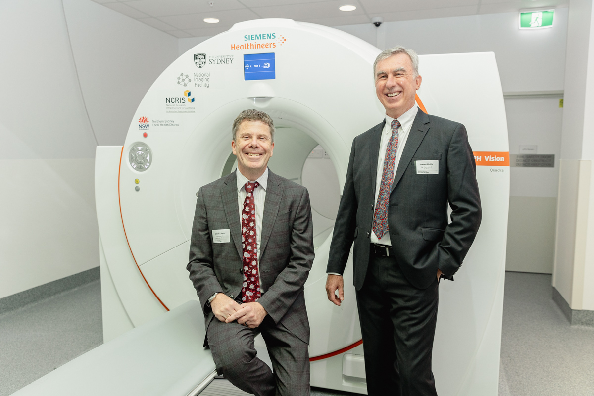
Professor Badawi, a medical physicist and co-director of the EXPLORER Molecular Imaging Center at UC Davis Health, specialises in developing novel PET imaging instruments and translational imaging applications. Together, they co-led the EXPLORER project, creating the world’s first total-body PET scanner.
Their team’s work ultimately means faster and more sensitive detection technologies – with wide-ranging benefits for diagnosis, patient treatment categorisation, and treatment response assessment.
Luckily, it’s the polar-opposite of bad news. In the nine months since the first open-access research-dedicated total body PET (TB-PET) facility opened in Australia, the excitement hasn’t abated.
TB-PET has been called an “extraordinary leap forward in medical imaging research”. The step change from traditional PET scanners encompasses:
- imaging of all organs and tissues of the body simultaneously
- opportunities to see whole-body processes and signalling between different organs
- higher sensitivity for single organ and total-body scans
- better resolution (referred to as signal-to-noise ratios)
- shorter scanning times
- lower radioactive injection doses.
“This is such an exciting advance,” says Prof Cherry, “because now for the first time ever we’ve got all the major organs and tissues in the field of view of the scanner at once, and we can follow our radioactively labelled tracer as it moves through the body.”
Prof Badawi adds that “the high sensitivity of TB-PET is a game changer, especially for imaging techniques like immunoPET. It allows us to obtain clear, dynamic images and perform kinetic modelling, which was previously unimaginable.”
The early challenges in developing Total Body PET
“I came from a small-animal background where we were imaging the whole body of a mouse,” Prof Cherry explains. “Ramsey has co-led and directed this project with me from the from the very outset, and was interested what happens to PET scanner performance as you make the scanner longer and longer.”
[Cherry also acknowledges “our fantastic team” at UC Davis, and the many researchers at United Imaging “who believed in us when nobody else did, and helped us build the very first system which has now spawned this whole field of total body imaging”.]
Although Cherry and Badawi struggled for years to secure funding, they generated preliminary data with funding from NIH, UC Davis RISE, and Siemens, then created their prototype TB-PET.
Prof Badawi adds, “It took a lot of elevator pitch conversations and bringing this up whenever we got a chance to talk in public. People must have got a bit fed up with us going on about the same idea year after year, but it was necessary to get the funding we needed.”
“We have a regular PET scanner, but replicate it 7 times to get 8 rings in total that covers the whole body. There are half a million detectors in the device, measuring a billion different lines through the human body simultaneously,” Prof Cherry says.
“When I think about what’s so exciting about the new system installed 9 months ago in Sydney, it’s that it’s really thrown wide open the protocols for clinical imaging – we can choose image acquisition time, dose, trading off throughput and image quality – so many options!”
Nearly a silver bullet: gains in sensitivity, timing, dose reduction
PET imaging is the most sensitive and non-invasive technique that can quantitatively measure physiology, metabolism and molecular pathways.
Previously, its use has been limited by worries about patients’ radiation dose (from the injectable tracers required), long imaging times (where patients must remain very still or risk the image quality), and the difficulty catching the image-making photon pairs with its narrow detecting ring (resulting in what’s called a ‘low signal-to-noise ratio’).
However, the TB-PET’s drastically increased sensitivity provides an answer for all those issues. Clinicians will be able to detect smaller or lower-contrast tumours or lesions, and measure them more accurately, acquire sharper images, scan more ‘sensitive’ patients (e.g. foetuses, infants, children), and track the radiotracers for longer.
“TB-PET is a beginning, not an end,” says Prof Cherry. “We suspect many impactful applications remain to be uncovered and developed.”
Prof Badawi adds, “The high sensitivity of TB-PET allows us to perform dynamic imaging and kinetic modelling, which opens up new possibilities for understanding disease processes and developing new treatments.”
He also notes that “one of the most exciting aspects of TB-PET is its potential to revolutionise how we approach chronic diseases. By making PET scanning easier and more accessible, we can start to address major health issues like fatty liver disease and autoimmune disorders.”
The imaging platform for this facility is the Siemens Biograph Vision Quadra. It is flagship infrastructure within Sydney Imaging and the National Imaging Facility network.
“I see that we have a huge opportunity before us; this facility will be a major player in integrating PET into a systems approach to medicine,” Prof Cherry says.
Unparallelled new applications of TB-PET
TB-PET broadens and deepens the possibilities of current PET scans, and opens the door to many previously inaccessible applications.
Its use will help generate new knowledge, inform therapies, and improve disease detection, staging and prognosis. TB-PET will also likely come to be used in studies of personalised precision medicine, development, ageing, nutrition and exercise.
TB-PET holds innumerable possibilities for patients, clinicians and researchers, such as:
- improving how we detect and stage cancer and other whole-body (‘systemic’) conditions (e.g. inflammation, vascular disease, sepsis or infectious diseases)
- allowing low-risk investigations of diseases where multiple organs interact (e.g. the gut–brain connection for irritable bowel syndrome, schizophrenia, autism, and Parkinson’s disease)
- developing and checking toxicity of new drugs and where, how, and how fast these drugs move and are absorbed in/excreted from the body
- tracking and checking new therapies that use cells or nanoparticles, such as cancer immunotherapy
- potentially no longer avoiding PET scans for pregnant people and foetuses because of the very low radiation doses possible with TB-PET
- studying how nutrition and exercise affect metabolism, inflammation, immunity, the heart, the endocrine system, and the microbiome
- improving care for current patients by reducing the time for a scan by 24 times.
Australia’s first open-access facility with dedicated research capacity
The TB-PET facility is a flagship capacity for Australia to study many medical fields and is the first step toward a national network for cancer research and pharmaceutical development. It provides a unique national capability for whole-body, ultra-high sensitivity PET/CT imaging.
Its capacity is shared equally between clinical use and open-access research use.
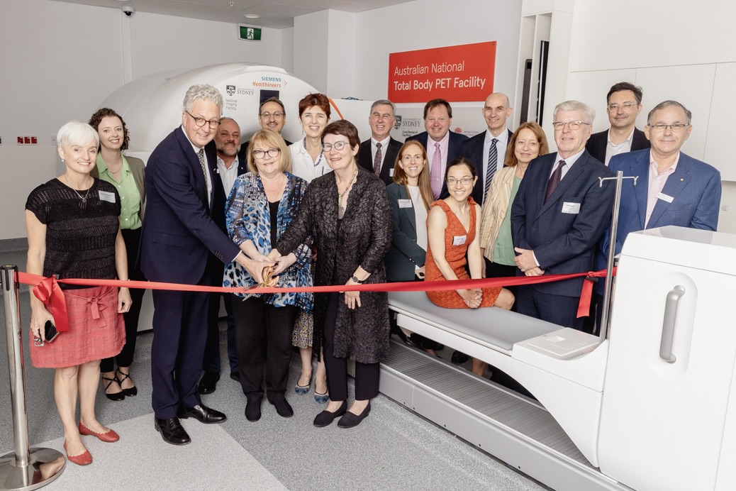
Opened in late 2023, the Australian National Total Body PET Facility was established through a $15-million partnership between the National Imaging Facility, the University of Sydney, Northern Sydney Local Health District (NSLHD) – supported by the Australian Government’s National Collaborative Research Infrastructure Strategy (NCRIS).
The expert staff working to support its operation range from NSLHD technical staff to operate the scanner, NIF and University jointly funded staff to help with research operations and to provide technical support and advice to researchers.
Webinars give window into TB-PET experts’ work
NIF has partnered with the University of Sydney to deliver an ongoing webinar series about the world of the first research-dedicated TB-PET.
Watch the most recent webinar, A conversation with Simon Cherry and Ramsey Badawi: Origins, current progress and the future of Total Body PET:
Watch a brilliant and accessible talk by Simon Cherry about the potential of TB-PET
Register for future events and access previous webinars in the series here
Opportunities for researchers to use the TB-PET
Because the facility operates using an arrangement for equal time-share between clinical use and academic research, researchers can apply to do a project at any time during the year.
Projects to use the TB-PET are assessed individually on the basis of their scientific merit, according to NIF/NCRIS principles.
After consulting with a NIF Fellow, researchers may submit a project proposal, after which they are assessed by a Scientific Review Panel for scientific merit and technical feasibility. Successful projects are then endorsed by the Facility.
The National Imaging Facility, The Sydney Imaging Core Research Facility at The University of Sydney, and the Northern Sydney Local Health District (NSLHD) operate in partnership to operate the TB-PET facility in the Department of Nuclear Medicine at Royal North Shore Hospital.
Read more in these peer-reviewed articles: Total-Body PET: Maximizing Sensitivity to Create New Opportunities for Clinical Research and Patient Care; First Human Imaging Studies with the EXPLORER Total-Body PET Scanner