Our NIF imaging community has delivered again: last year, we put a call out for spectacular images that you have captured or reconstructed. Our imaging communities will have seen many of these submissions featured in the 2025 NIF Impact Report.
NIF is excited to announce this year’s competition, which will illustrate your incredible work and source innovative captures for the next NIF Impact Report. We also feature the three winners of last year’s competition.
Enter the 2026 competition
We are ready to celebrate the year’s outstanding research images that you have generated through NIF expertise or instruments. There is a wealth of diversity, creativity and scientifically impactful imaging occurring every day across Australia.
We invite NIF Scientists, researchers, users, students, clinicians, technical and communication staff to share striking research images created using NIF capabilities.
From revealing the architecture of cells and tissues to visualising whole-body structures and processes, NIF-supported imaging technologies allow researchers and clinicians to see, measure and understand what would otherwise remain hidden.
Every image has the potential to reveal something unexpected, demonstrate the power of an imaging technique, or provide a new perspective on an important research question.
Entries close at 11.59 pm AEST on Sunday, 22 November. If you didn’t receive an email directly, your associated Node staff have the guidelines that tell you about the prizes, themes, where we’d showcase your image, and AI and processing caveats. Or email us!
Congratulations 2025 winners
Sincere thanks go out to everyone who submitted their images, or communication and facility folks who submitted for users or staff. We appreciate the time and effort you took to share your work with us.
All of the images shared with us were high quality, and showcase the outstanding research and imaging capabilities supported by the NIF community.
Our three winners were (in alphabetical order):
- Dr Nyoman Kurniawan, a Senior Research Fellow for UHF Preclinical MRI at The University of Queensland
- Prof John Magnussen, a Professor of Radiology from Macquarie University’s Faculty of Medicine, Health and Human Sciences
- A/Prof Brad (Bradford) Moffat, a Principal Research Fellow in MRI Science from the University of Melbourne’s Department of Radiology.
The winners’ standout submissions are featured below in much more detail, plus other submissions to the competition. We hope you enjoy seeing them as much as we did receiving them.
Unexpected fruit fibre information could guide producers
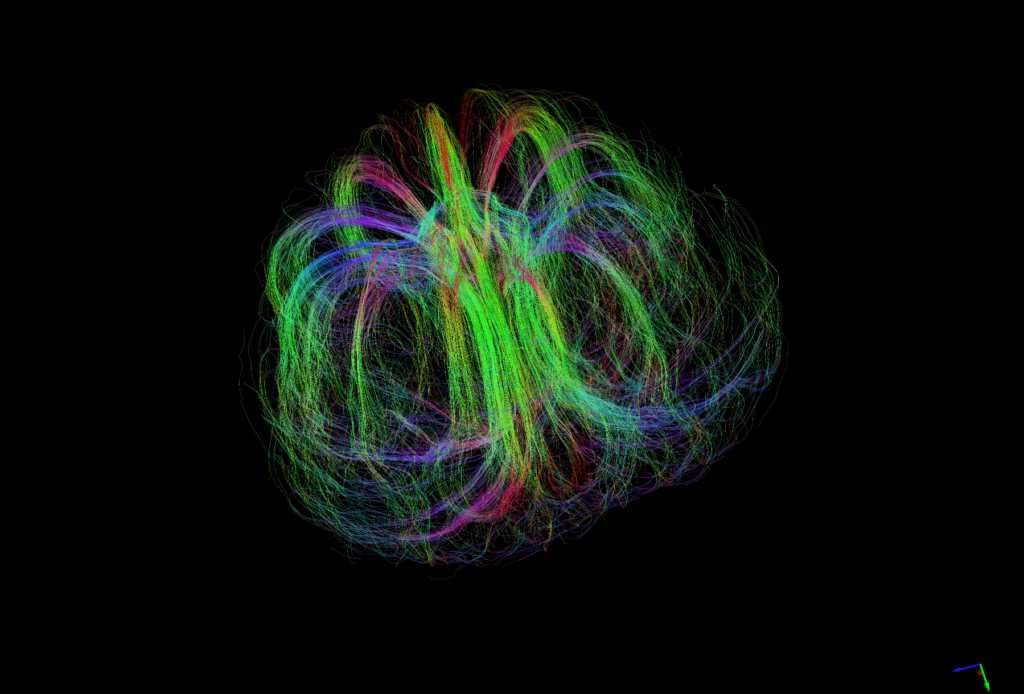
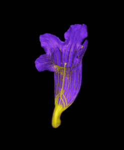
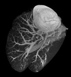
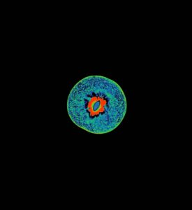
“When I created the image, I wanted to turn an abstract measurement into something that people can see and ask questions about,” said image creator Dr Nyoman Kurniawan (UQ, Senior Research Fellow for UHF Preclinical MRI).
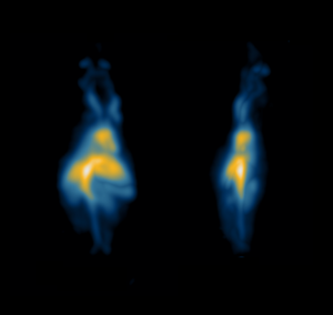
This project explored how a native Australian rainforest fruit – the Burdekin plum – gets water and nutrients to its flesh along vascular bundles during development, while its centre hardens into a woody stone.
The team used a 14.5-hour diffusion-weighted MRI scan at the AIBN – “brain scientists use the same principle to trace the wiring of the brain” – to see if the method “could also be used to map the hidden plumbing of fresh fruit without cutting it open at each stage of development,” Dr Kurniawan said.
This is because in biological tissue, water more easily moves along along vascular fibres than across them.
Dr Kurniawan described the team’s surprise at the shape of the plum’s fruit fibre bundle. In most fruits, fibers run like spokes from the centre towards the skin, but the Burdekin plum fibres were bundled tightly along the central axis and fanned out only towards the outer part (the flesh) at the top and bottom of the fruit.
He explained that long MRI scans like these can inform horticultural research into “features of the fruit that can help selecting good varieties and design harvesting, transport and commercialisation.”
A related study on green plum combined MRI scans with chemical and physical measurements to describe size, sweetness-to-acidity balance and ripening across four regions in the Northern Territory. “That kind of information can guide harvest timing, handling and site selection,” said Dr Kurniawan.
Not an eagle ray, but a unicorn ray with extra pelvic fins
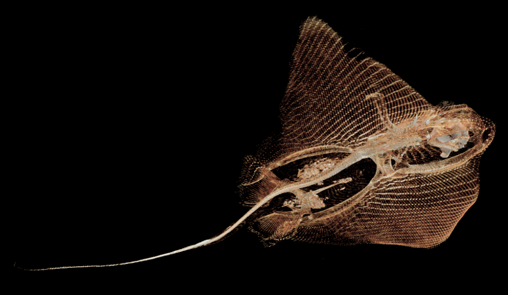
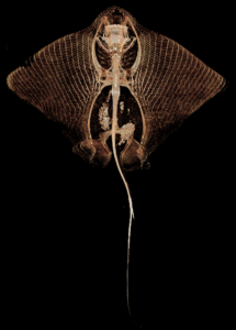
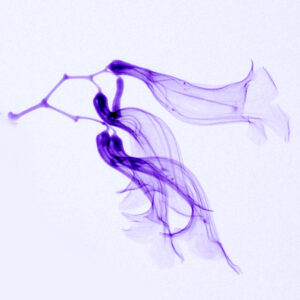
“Every now and then, a researcher will approach us with something weird and wonderful, asking if we can help them scan it,” said Dr Marg Pardey (Macquarie Medical Imaging’s Translational Research Imaging Manager), who submitted the image on Prof John Magnussen‘s behalf.
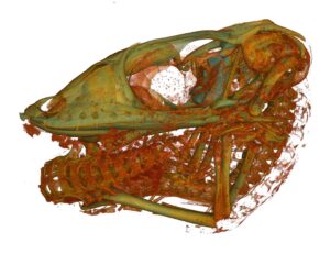
In April 2024, a biology PhD student did just that. They had found an unusual growth on the top of the head of a free-swimming stingray caught in the Port Kembla region.
After the ray was scanned at the Macquarie University NIF node on a GE Revolution CT scanner, Prof Magnussen used GE AW software to complete the reconstructions for the final image.
“For a facility that normally focuses on imaging humans who are alive and usually quite well, it’s amazing to see what mysteries we can find inside. Being able to look below the surface of the world, without breaking it, is a beautiful adventure,” said Prof Magnussen.
Although the team has scanned many specimens, from ancient Egyptian mummies, to clay tablets, viking swords, and sharks, this was the first ray, noted Prof Magnussen.
“The process of reconstruction for rays is certainly not well established! It’s designed around imaging live humans. So every one of these studies is a journey of discovery: no two non-human studies is ever really alike. How we scan and reconstruct is tailored to the individual item.”
The fine lines of the image are the ray’s cartilage, Prof Magnussen explained. “To be able to pull that level of detail is quite a treat. People might take it for granted now that we can actually look inside things without having to cut or break them open, but the ability to do so is just remarkable.”
7T MR brain scan that can reveal tiny abnormalities from disease

A/Prof Brad Moffat – a medical imaging physicist and chemist who (deputy) directs the Melbourne Brain Imaging Capability, The University of Melbourne NIF node – sent us this incredible image.
It’s an axial 7T susceptibility-weighted MR image of a brain at a resolution of 0.3 x 0.3 mm.
Scans like these are used to find tiny brain abnormalities in diseases such as epilepsy, multiple sclerosis, stroke and traumatic brain injury.
The image also represents A/Prof Moffat’s work in areas such as quantitative ultra high field (7T) MR imaging of human subjects, molecular imaging biomarker research, fMRI, diffusion MRI, functional diffusion mapping, MR perfusion, MRS, voxel-based morphometry, PET and nano-theranostics.
Thanks to our other submitters